Mamatova Muborak Nurpulatovna
Doctor of Biological Sciences, Action Professor, Samarkand State Medical University, Samarkand, Uzbekistan
Correspondence to: Mamatova Muborak Nurpulatovna, Doctor of Biological Sciences, Action Professor, Samarkand State Medical University, Samarkand, Uzbekistan.
| Email: |  |
Copyright © 2023 The Author(s). Published by Scientific & Academic Publishing.
This work is licensed under the Creative Commons Attribution International License (CC BY).
http://creativecommons.org/licenses/by/4.0/

Abstract
Rabies is an infectious disease of humans and animals caused by members of the family Rhabdoviridae of the genus Lyssavirus and leads to death in 100% of cases [2,3]. Rabies, which causes the greatest economic damage worldwide, occupies an extremely important place in the infectious pathology of humans and animals. But despite the successes of modern science, a huge area of the globe is not free from the rabies virus [13,14,15]. The world range of rabies does not yet tend to decrease due to natural focal disease. In the spread of rabies among people, stray dogs are of great importance, and among farm and domestic animals, wild carnivores play a major role [7,9,11].
Keywords:
Rabies, Pathogenesis, Infected, Inoculation, Intracerebral, Intraplantar
Cite this paper: Mamatova Muborak Nurpulatovna, Study of the Biological Properties of Rabies by the Method of Diagnosis of the "Gold Standard", American Journal of Medicine and Medical Sciences, Vol. 13 No. 6, 2023, pp. 838-844. doi: 10.5923/j.ajmms.20231306.13.
1. Relevance of the Topic
Natural foci of rabies have also become more active in our Republic. In order to clarify the nature of natural epizootics and to successfully solve the problem of rabies disease, as well as to develop means of preventing and combating rabies, not only the study of the ecology of the host but also data on the study of the biological characteristics of the rabies virus isolated in a certain geographical zone are of great importance. Due to the decisive importance of the antigenic and pathogenic properties of the virus, we were tasked with studying isolates isolated from various geographical zones of Uzbekistan [1,4,8,10].And also, when carrying out anti-epizootic measures, as well as prescribing a course of anti-rabies vaccinations to victims of animal bites, timely and accurate diagnosis is of great importance. The need for constant monitoring of the state of natural foci of rabies and the expansion of work in recent years on oral vaccination of wild carnivores require the improvement of express methods for isolating and indicating the virus. The investigation of hydrophobia of animals in different areas of the republic which investigated at Uzbekistan scientific investigation of Veterinary Institute. The results of this during the last years shows that spreading of this disease among agricultural animals and also plays a great role of wild raft toxic animals as foxes, jackals and wolfs, and also in other wild types animals. For the sake of that hydrophobia of domestic animals are often infected in country-side, especially during pasturable period.The speed of spreading virus of hydrophobia into central nervous system from the bits determines pathogenesis of disease and affectivity of treating and prophylaxis. Last times in literatures we came across different opinions about the question [1,10].We studied the time of penetration of virus of hydrophobia into central nervous system on the base of location of tamp on the body of different types animals.
2. Materials and Methods
In the course of the work, comparative tests of the "gold standard" in the diagnosis of rabies were carried out - a method of biological testing on white mice, followed by a study of IPA and ELISA.Legislative acts regulate different methods of laboratory diagnosis of rabies: the method of fluorescent antibodies (IPA), the method of isolation of the rabies virus in the culture of cells of the mouse neuroblastoma CCL-131 (or neuromas of the Gasser node of the rat - NGUK-1), a biological test on white mice, the method of enzyme immunoassay (ELISA), the reaction of diffusion precipitation (RDP). The effectiveness of anti-epizootic measures and the success of emergency vaccine prophylaxis largely depends on the timely and accurate identification of the causative agent of the disease [6], therefore, in some cases, it is critically important to be able to identify the rabies virus in the test material as soon as possible. At the moment, the "gold standard" in the diagnosis of rabies infection is the method of setting a biological test on white mice with the subsequent study of the IPA [12], although this approach is not without significant drawbacks - the duration of the analysis is up to 30 days (a negative diagnosis can be given only after 30 days of observation, provided); To study the results of the IPA, high qualification of a specialist is necessary; to conduct a study, it is necessary to have a vivarium.Improvement of laboratory diagnostics of anti-rabid infection in the territory of Uzbekistan despite the rapid development of molecular genetic methods for diagnosing infectious diseases, at the time of the beginning of the work presented in the territory of the republic there were no standardized sets of reagents that allow identifying rabies by setting UP RT-PCR in real-time [4,5].
3. Results
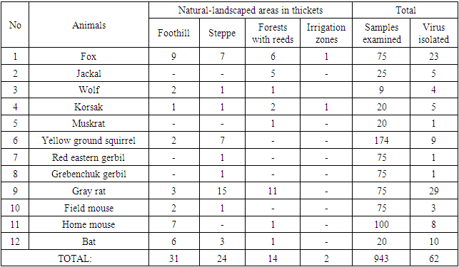
An analytical review of modern methods of laboratory diagnosis of rabies, as well as the results of its own research, indicate the high sensitivity and specificity of such express methods for identifying the causative agent of rabies, as a method for detecting the rabies virus antigen by labeled Fluorescein isothiocyanate anti-rabies antibodies, with the formation of characteristic luminous complexes-inclusions detected in the field of view of a luminescent microscope, which will provide early diagnosing rabies and in turn reducing people's risk of disease. In the last 10 years, 1787 samples of material from animals suspected of rabies have been examined using complex diagnostics, i.e. luminescence microscopy methods, as well as bioassay on mice. At the same time, the rabies virus was detected in 157 samples. Of the total number, 10 isolates of the street rabies virus from different species of animals living in various natural and climatic zones of Uzbekistan were selected for further study.Table 1. The results of the isolation of the rabies virus from wild carnivores and rodents settled in different natural zones of Uzbekistan
 |
| |
|
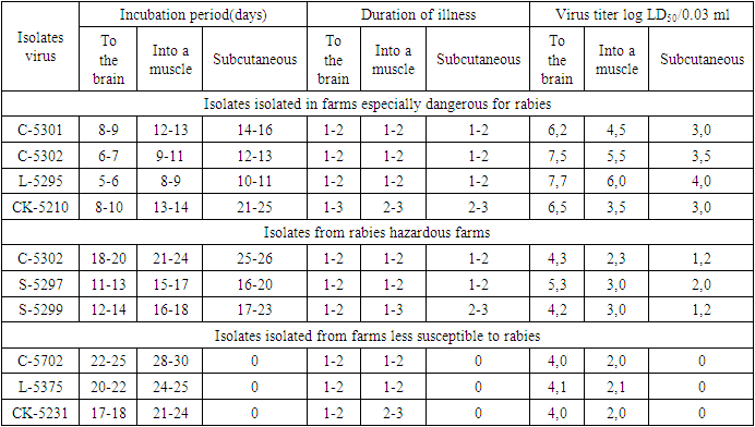
From the data presented in the table, it can be seen that in different natural zones of Uzbekistan, the rabies virus circulates among wild animals - often in foxes and gray rats. In recent years, it has been established that wild animals are the main carriers of the virus, thereby creating natural foci of rabies, i.e. especially dangerous areas for rabies. Isolation of the rabies virus from rodents, in particular, from gray rats living in livestock buildings, and yards, indicate that they are the main potential source of this infection.According to morphological, antigenic, and other indicators, the identified isolates of the virus are assigned to the family of rhabdoviruses, the genus of lyssavirus, and the species – of rabies virus.When determining the titer of the studying viruses on white mice and its calculation according to the Reed and Mench method, high titers of 6.2-7.7 logs of LD50/0.03 ml were detected in isolates isolated from foxes, gray rats brought from farms of persons of dangerous (stationary disadvantaged) rabies zones.The experiments studied the sensitivity of white mice weighing 10- infected with a street virus. A total of 10 isolates of the street virus were selected for the study 12 g.The work used a 10% suspension of infected mouse brains, cooked in a sterile physiological solution. Experimental mice were injected with suspension into the brain at a dose of 0.03 ml intramuscularly and subcutaneously - 0.1 ml each.Strain LM-5301 is isolated from the brain of a stray dog (three passages on mice); strain C-5302 is isolated from the brain of a clinically healthy dog (four passages on mice); strain L-5271 is isolated from the brain of a fox (two passages on mice); strain L-5295 isolated from the brain of a fox (two passages on mice); strain L-5375 isolated from the brain of a fox (three passages on mice); strain SH-5297 isolated from the brain of a jackal (three passages on mice); strain SH-5299 isolated from the brain of a jackal (two passages on mice); strain SK-5210 isolated from the brain of a gray rat (three passages on mice); strain SK-5231 was isolated from the brain of a gray rat (three passages on mice).Studies to identify the sensitivity of white mice to infection with the street rabies virus by various methods are presented in Table 2.Table 2. Sensitivity of white mice to infection with the street rabies virus by various methods
 |
| |
|
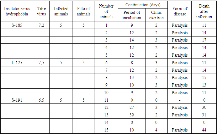
From the data given in the table, it can be seen that when infected with the rabies virus, the duration of the incubation period of the disease in mice and the infectious titer of the virus depend on the method of inoculation.In this experiment, white mice were relatively highly sensitive when they were infected intracerebrally.The results of the studies conducted on mice made it possible to divide the studied isolates mainly into two groups: (a) isolates isolated in particularly rabies-prone farms with short incubation periods (5-10 days) and a high infectious titer (6.2-7.7 log LD50/0.03 ml); b) isolates isolated in rabies-hazardous farms with long incubation periods (11-20 days) and a low infectious titer (4.0-4.1 log LD50/0.03 ml);c) isolates isolated in less rabies-prone farms with long incubation periods (17-25 days) and low infectious titer (4.0-4.1 log LD50/0.03 ml).Based on the studies, it can be said that isolates isolated from carnivores and rodents living in natural foci of rabies, i.e., isolated from especially dangerous rabies farms, are relatively highly pathogenic for guinea pigs and white mice with different methods of infection.Of the 10 field isolates of the rabies virus isolated from dogs, foxes, and gray rats studied, 4 isolates showed great pathogenicity to white mice and 3 isolates to guinea pigs.Thus, a comparative study of the pathogenic activity of the street rabies virus for guinea pigs and white mice isolated from wild animals made it possible to divide the studied isolates into three groups of rabies virus strains:1. high-level isolates isolated on the territory of especially dangerous rabies farms.2. active isolates isolated in the territory of rabies-prone farms.3. weakly active isolates isolated in the territory of less dangerous rabies farms.The scientific work carried out showed that foxes are currently the main natural focal sources of infection, and gray rats are carriers of the rabies virus. The intensity and seasonality of the epizootic process among farm and domestic animals is primarily related to the ecology of wild carnivores. A number of issues related to this disease have not yet been sufficiently deciphered, and of particular importance among them is the improvement of preventive measures due to the spread of rabies among wild carnivores. We had six series of experiments on 36 foxes, 36 dogs, 36 jackals, 60 white mice, 36 rabbits, 48 domestic mice and 48 grey rats.For infecting all experienced animals was used epizootic stamp (S-33) on rabbits of street virus of hydrophobia, singled out from sick with hydrophobia jackals with titre 6,0-6,6 LD50/0,03 ml.According the diagram of experiment we have done preparations from tissue of fox, we came from central nervous system of dead experienced animals. Besides those, we bio tested 6 white mice, infecting their brains with 0,03 ml of 10% virus containing suspension, prepared from tissues of mice of brain. On the results of that we noticed the virus with which infected the hip of the dogs can be discovered in 24 h, on the fox – in 24-48 h, on jackals – 48 h, and then the virus may be discovered in brains in seven, eight, nine days.On the second type of experience on 16 foxes, dogs and jackals which was led 10% virus containing suspension of rabbit brain with the size of 3 ml physiological solution which was led into two points of chewing tissue in one time. Experience on the base of described method as it is given on page 2 on the results of investigation was fixed that the virus on the dogs was determined it was revealed 12 days after infection, in brain – after 6 days. But we couldn’t find out infected tissue after 24 h. and on the foxes virus was discovered till 24 h., in brain – till 7 days. After infecting jackals with virus of hydrophobia was discovered till 24 h., in brain – till 8 days.During the third experience we used 30 white mice and 18 rabbits. We infected those animals with 10% virus containing suspension of rabbit brains on physiological solution, under the skin of tip of the nose in the size: 0,03 ml on white mice, 0,2 ml on rabbits and investigated the presence of virus hydrophobia on the base of described methods. On the results of investigation we singled out that infected virus was kept till 12 h, in brain – till two days in white mice. But in rabbits virus of hydrophobia was discovered in 12 h, in brain – till 3 days. In 24 hours and more time it was not available to reveal virus in infected place of rabbits. As it is seen on the results of the experiment virus on domestic mice was kept in active state till 12 hours after infected, in brain can be revealed for 3 days. In 24 hours and more period of time virus in infected muscle was not revealed. And on grey rats can be discovered after 12 hours, in brain can be revealed of the 5th day.The sixth series of experiences of 30 domestic mice and 30 grey rats which were infected with 10% virus containing suspension of rabbit brain with physiological solution in muscles of thigh was infected with 0,1 ml and 0,5 ml and 3 of animals of every type were dead after infecting in 12, 24, 48, 72, 96, 120, 144, 168, 192 and 216 hours which were investigated on the base of the method in the presence of hydrophobia virus. On the results of this we singled out that after infecting with virus in domestic mice we discovered in 24 hours, in brain – on the 5th day. In the latest period of time it is not available to reveal virus in infected muscles.On the sample of hydrophobia virus of grey rats can be discovered in 12 and 24 hours after infecting, in brain – in 5-6 days. Investigation of virus in infected muscles is not discovered in other periods of time.So, the experiment on white and domestic mice, grey rat, rabbits, foxes, jackals and dogs were shown in different periods of time the revealed hydrophobia virus in brain from infected animals, on the dependence from the place of its inoculation. At any case in 12-48 hours virus was not available to single out of the infected place by different methods. In brain hydrophobia virus was singled out in different periods of time i.e. from 3 till 10 days. The results of investigation which we got have definite clearness pathogenesis of illness in different animals on the dependence of used methods.The hydrophobia virus of three types were investigated on 15 karakul sheep of both sex, the age of 1,5-2 years, dividing into 3 group, each group consisted of 5 sheep. Sheep were bound in normal place for hydrophobia; blood serum wasn’t antibodies to the virus of hydrophobia.After 30days of quarantine sheep was infected 10% in brain containing virus of hydrophobia in different isolation as S-185, L-125 and S-191, on the level of 2-3 passage.Virus containing suspension were passed into chewing muscles of three group sheep in the size of 2,0 ml, by 1,0 ml on each side. Infecting titre of experimented insulations became equal from 6,5 till 7,5 lg LD50/0,03 ml in infected white mice in the size of 6-7 gr. We examined infected animals during 3 months. Results of investigation is given in the table 3.Table 3. Pathogenesis of street virus of hydrophobia of karakul sheep
 |
| |
|
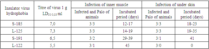
It is seen from the diagram, that the incubated period of sheep, infected S-185 were from till 14 days (average 11,8), in the second group of sheep, infected insulation L-125 during 8-13 days (average 10,4 days) and in the third group of sheep, infected insulation S-191 27-40 days (average 35,3 days). On the results of this ten sheep infected insulation S-185 and L-125 were dead, five sheep of infected insulation S-191 three sheep were dead, which were ill after long incubated period, continuing 10-39 days, two sheep of this group were stable to infection.Experimental infected sheep with hydrophobia virus and their illness was in paralytic form, it was clinically appeared during 2-4 days. The main symptoms were expressed in apathy, tremor, salivation and paralysis.The results of infected sheep with hydrophobia virus allow us to notice that two insulation (S-185, L-125), which were singled out in stationery bad zone, were seemed high pathogenesis for sheep, and insulation S-191, singling out in stationary conditionally good zone – less pathogen. These data allow us to conclude, that in the territory Of Uzbekistan circulate stamps of street virus of hydrophobia, having different degrees of pathogenesis in infecting different types of animals.While investigating the accumulation of virus in brain of infected sheep with insulation S-185, pointed out titre 5,6-6,1 lg LD50/0,03 ml and with insulation L-125, the titre was 5,3-6,0 lg LD50/0,03 ml and with stamps of hydrophobia virus S-191 the titre was fluctuated 2,5-3,9 LD50/0,03 ml.On the 90th day after infecting 2 not diseased sheep killed and were examined with virological investigation. Hydrophobia virus in brain of those sheep by means of bio testing method and MFA was not discovered.For investigation of pathogenesis of stamps of street virus of hydrophobia of donkeys were experimented on 24 donkeys with age of 1,5-2 years old. The donkeys were bound from stationery good into having hydrophobia zone, before infecting they were in quarantine for 45 days. In blood serum slavish imitation of antibody was absent. For infecting donkeys we used 10% tissue brain of white mice, infected by insulation of hydrophobia virus – S-185, L-122, S-191, L-125.Experimented donkeys virus containing suspension, in one group we operated into chewing muscle and in others (12 donkeys) – under the skin of neck is each side in the size of 2,5 ml which consisted of 5,0 ml. The infected donkeys were controlled during three months. The results of infecting are given in table 4.Table 4. Pathogenesis of stamps of street virus of hydrophobia of donkeys in different ways of infecting
 |
| |
|
It is seen on the diagram 2 that the donkeys, infected inner muscle insulation of hydrophobia virus S-185 and L-125 were ill and dead. Incubated period of illness was 12-19 days (average – 15,5 days). The donkeys after infecting under the skin with those stamps they were ill and dead. In this case incubated period in both cases consisted of 18-25 days (average – 21,2).Clinical features of infected donkeys were observed during 2-3 days.Three donkeys were infected inner muscle insulation S-191, from which two were dead. Incubated period lasted 29-39 days (average 34 days). In infecting under the skin with the same stamps one of three was ill, incubated period lasted 41 days. Illness lasted for 2-4 days. From three donkeys which were infected inner muscle stamps L-122, only one was dead. Incubated period was 45 days. Illness lasted for 5 days. To stamps L-122, donkeys which were infected under the skin seemedstable and during 90 days control animals were not ill.Accumulation of virus in brain of donkeys, infected inner muscle insulation S-185 and L-125 was high and its titre was 5,0-5,5 lg LD50/0,03 ml, and in brain of donkeys infected with these insulation under the skin, titre of virus was 3,0-4,1 lg LD50/0,03 ml. Titre of virus in brain of donkeys, infected with inner muscle insulation S-191, was 2,1-3,0 lg LD50/0,03 ml, in infecting with these insulation titre of virus was 0,03 ml, 1,5 lg LD50/0,03 ml and after infecting inner muscle with insulation L-122 titre of virus was equal to 2,0 lg LD50/0,03 ml. On the 90th day after infecting 8 donkeys which were not ill they were killed and had to test to virological investigation of brain with the methods of using bio testing on young muscles and MFA. We were not able to distinguish virus from brain of those animals.
4. Conclusions
So, the investigated 12 insulation of hydrophobia virus, we singled out in the territory of Uzbekistan from fox, jackal, wolf, red gopher, domestic and field mice and bat turned out to be pathogenic for white mice and white rats at infecting in brain, intraplacentically and hypodermically. The investigated 23 insulation of hydrophobia virus there were also singled out from fox, jackal, wolf, domestic and field mice, grey rat, dog and cattle and they turned out to be pathogenic for rabbits at using it hypodermically, into muscle and abdominal cavity. There were studied 4 types of street viruses at experimental infecting dog, sheep and donkey inner muscularly. 2 insulation of hydrophobia virus, singled out from dog and fox turned out to be highly virulent. One of the insulations singled out from foxes, was virulent and weakly virulent, and hypodermically infecting donkeys it turned out to be virulent. Red gophers and eagles didn’t react on infecting, but they were bearers of the virus for a long time.Mentioned above wild and domestic animals, according to the results of investigation, are potential sources of hydrophobia virus in the territory of Uzbekistan. In conclusion above pointed investigation and experiments are given table 5.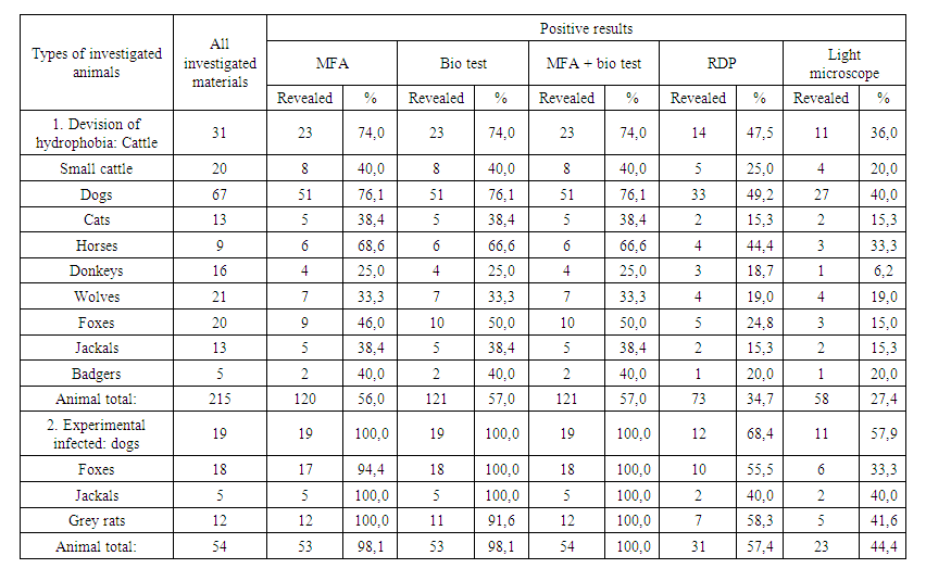 | Table 5. Comparative investigation of sensibleness and specification of different methods of laboratory diagnostics of hydrophobia |
References
| [1] | Борисов А.В., Гусева М.Н. Хранение вируса бешенства с различными стабилизаторами // Мат. межд. научно-практ. конф. ВНИИВВиМ. 30-31 мая 2001. -С. 61-63. |
| [2] | Вагабов Р.М, Баринский И.Ф. Вирусы группы бешенства // Вопросы вирусологии. -М., 1979. -№ 5. -С. 451-458. |
| [3] | Кантарович Р.А. Вирусы группы бешенства // Микробиология, эпидемиология и вирусология. -М.: Медицина, 1972. -№ II. -С. 9-15. |
| [4] | Маматов Н.М. Сравнительное изучение скорости проникновения вируса бешенства в ЦНС и значение места инфицирования в развитии заболевания. Болезни сельскохозяйственных животных. Тр.Уз.НИВИ, 1978, Т.27, -С.71-74. |
| [5] | Маматова М.Н. Эпизоотология бешенства и способы пероральной иммунизации плотоядных животных // Автореферат диссертации на соискание ученой степени кандидата вет. наук. –Ташкент, 1996. -С. 77-97. |
| [6] | Метлин А.Е., Рыбаков С.С. Выделение антигена вируса бешенства в консервированных формалином пробах головного мозга мышей // Мат. межд. научно-практ. конф. ВНИИВВиМ. 30-31 мая 2001. -С. 46-49. |
| [7] | Официальный сайт Всемирной Организации Здравоохранения. 2018. [Эл.ресурс]. Режим доступа: http://www.who.int/mediacentre/factsheets/fs099/ru/ Аvailable at: www.oie.int/wahis; http://www.oie.int/en/animal-health-in-the-world/rabies-portal/. |
| [8] | ШайкуловХ.Ш., Исакулова М.М., Маматова М.Н. Степень бактериоциногенности антибиотикорезистентных штаммов стафилококков, выделенных в Самарканде // Journal. Eurasian jornal of Medical and natural Sciences. www.in-academy.uz. |
| [9] | International Committe on Taxonomy of Viruses [electronic resource]. - URL: http://www.ictvonline.org/virusTaxonomy.asp. - 13.11.2013. -Р. 177. |
| [10] | Mamatova M.N. Biological properties of rabies virus and immunogenicity of oral antirabic vaccines in granules // Journal. American Journal of Medicine and Medical Sciences. 09.12. 2022. http://creativecommons.org/licenses/by/4.0/. |
| [11] | Rabies - World Health Organization (WHO). 2020. https://www.who.int/health-topics/rabies#tab=tab_1. |
| [12] | Terrestrial Animal Health Code Chapter 7.7, 6th Edition. World Organisation for Animal Health (OIE), France, 2017. Available at http://www.oie.int/en/international-standard-setting/terrestrial-code/access-online/, accessed March, 2018. |
| [13] | WHO. 0 by 30 our catalytic response. Available at http://www.who.int/rabies/United_against_Rabies/en/, accessed December 2017. |
| [14] | WHO Expert Consultation on Rabies, third report: WHO Technical Series Report No. 1012, Geneva, 2018 (ISBN 978-92-4-121021-8). |
| [15] | WHO fact sheet on rabies. Available at http://www.who.int/mediacentre/factsheets/fs099/en/, accessed March 2018. 5 Birhane MG. |

 Abstract
 Reference
 Full-Text PDF
 Full-text HTML