Achilova Donohon Nutfilloevna1, Shukurov Bakhtiyor Kodirovich2
1Bukhara State Medical Institute Named after Abu Ali Ibn Sino, Uzbekistan
2Dermatovenerologist, Bukhara City Polyclinic № 6, Uzbekistan
Copyright © 2023 The Author(s). Published by Scientific & Academic Publishing.
This work is licensed under the Creative Commons Attribution International License (CC BY).
http://creativecommons.org/licenses/by/4.0/

Abstract
Vitiligo greatly affects the patient's psychological state and self-esteem, especially when the skin of the face and hands is damaged, such patients are more likely to be mentally ill, and various mental disorders are detected. Vitiligo is a multifactorial disease that develops as a result of a combination of genetic, metabolic and immunological disorders. Disruption of the processes of regeneration and proliferation of melanocytes indicates the presence of defects in these cells. To date, it is not known what processes lead to the onset of vitiligo. According to a number of researchers, genetic factors and defects of the antioxidant system cannot be the cause of this disease by themselves, and the influence of external factors is necessary for the manifestation of vitiligo. In the exacerbation phase of the disease, the latent inflammatory reaction and autoimmune processes play a certain role.
Keywords:
Vitiligo, Neurogenic (neuroendocrine), Autodestruction (self-destruction), Genetic, Autoimmune (immune), Theory of biochemical disorders (oxidative stress)
Cite this paper: Achilova Donohon Nutfilloevna, Shukurov Bakhtiyor Kodirovich, Results of Personal Research in Adolescent Vitiligo Disease Clinical Description of Adolescents with Vitiligo, American Journal of Medicine and Medical Sciences, Vol. 13 No. 6, 2023, pp. 821-824. doi: 10.5923/j.ajmms.20231306.10.
1. Introduction
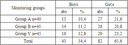
Our study was conducted in adolescent children aged 12 to 15 years with vitiligo, and the results were analyzed, the average age was 14.5±0.3.Table 1 shows the gender distribution of adolescents with vitiligo by treatment method.Table 1. Distribution of patients by gender depending on the treatment plan
 |
| |
|
In the study, the percentage of male patients in group A was 10.4% (13 boys), in group B - 11.2% (14 boys), in group V - 12.8% (16 boys). The number of girls in the research groups was much higher: 27 (21.6%) in group A and 26 (20.8%) in group B, 82 (65.6%) girls prevailed in group V. There was no statistically significant difference between the groups by gender (r>0.05).Age distribution in study groups also did not differ significantly (Figure 1).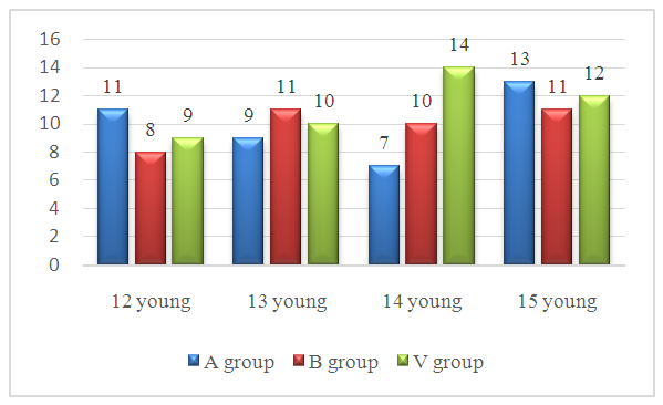 | Figure 1. Age distribution of patients in study groups |
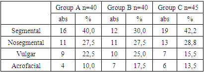
The percentage of 15-year-old patients was the maximum - 13 in group A, 11 in group B, and 12 in group V. this showed 1.5 times more than the total number of patients between groups, respectively. The number of patients aged 14 years was 7 in group A, 10 in group B and 14 in group V. 14-year-old patients were in the 2nd place in terms of contingent participation in research groups. The group of 12-year-old patients was the smallest group: the number of such patients was 11 in group A, 8 in group B, and 9 in group V. 13-year-old patients were 9 in group A, 11 in group B, and 10 in group V, their proportion was 19.5%, respectively; It was equal to 12.5% and 17.9%. There was no significant difference between the groups when they were divided by age.During the study, we divided the patients into groups according to the form of vitiligo. It is detailed in Table 2Table 2. Distribution of vitiligo by form in study groups
 |
| |
|
As can be seen from Table 2, the analysis of the distribution of patients according to the form of vitiligo, the segmental form of the disease in group A - 16 (40.0%) patients, group B - 12 (30.0%) patients, group V - 19 (42.2%) ) showed that it is determined in the case.The non-segmental, in particular, focal form of the disease was observed in 11 (27.5%) patients in group A, in 11 (27.5%) patients in group B, in 13 (28.8%) patients in group V.The vulgar form of vitiligo was found in group A - 9 patients (22.5%), group B - 10 (25.0%) patients, group V - 7 (15.5%) patients.The acrofacial form of vitiligo was initially detected in 4 cases in group A, 10.0%, and in 7 patients in group B, 17.5%. In group V, this form of the disease was recorded in 6 (13.5%) patients.Table 3. Distribution of vitiligo by duration in study groups
 |
| |
|
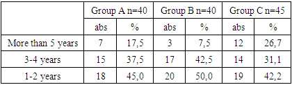
As can be seen from Table 3, patients whose vitiligo lasted more than 5 years included 7 (17.5%) patients in group A, 3 (43.8%) patients in group B, and 12 (26.7%) patients in group V. organized, in which no significant differences between groups were observed. The number of patients whose disease lasted for 3-4 years was 15 (25.0%) patients in group A, 17 (21.9%) patients in group B, and 14 patients (31.1%) in group V.In the study, the incidence of vitiligo at 1-2 years was 18 (45.0%) patients in group A, 20 (50.0%) patients in group B, and 42.2% in 19 patients in group V. The number of patients with vitiligo lasting more than 5 years was minimal.During the study, the body was noted as the most common area of vitiligo - approximately one third of patients in each group - 40.0-45.3% of cases (Table 4).Table 4. Distribution of vitiligo in the study groups according to prevalence in areas
 |
| |
|
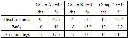
Head and neck was injured in 9 (22.2%) patients in group A, 7 (17.5%) patients in group B, 12 patients (26.7%) in group V. Vitiligo occurred in the body part of the procedure in 16 (40.0%) patients in group A, 18 (45.0%) patients in group B, and 19 patients in group V (42.2%).In the study, arm and leg injuries occurred in 15 (37.5%) patients in group A, 15 (37.5%) patients in group B, and 14 (31.1%) patients in group V.Table 5. The presence of concomitant diseases in the study patients
 |
| |
|
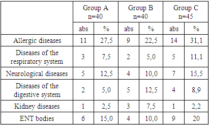
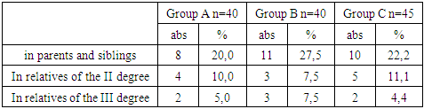
As can be seen from Table 5, information on the presence of additional diseases in the patients included in the study is presented.Allergic diseases were recorded in 11 (27.5%) patients in group A, 9 (22.5%) patients in group B, and 14 (31.1%) patients in group V.Diseases of the respiratory system were recorded in 3 (7.5%) patients in group A, 2 (5.0%) patients in group B, and 5 (11.1%) patients in group V.Neurological diseases were noted in 5 (12.5%) patients in group A, 4 (10.0%) patients in group B, and 7 (15.5%) patients in group V.Gastrointestinal diseases were detected in the examined patients (chronic gastritis, stomach and duodenal ulcer diseases), they were found in 2 (5.0%) patients in group A, 5 (12.5%) patients in group B, 4 patients in group V (8.9%) was recorded in the patient.Kidney diseases were noted in 1 (2.5%) patient in group A, 3 (7.5%) patients in group B, and 1 (2.2%) patient in group V.Diseases of larynx organs were noted in 6 (15.0%) patients in group A, 4 (10.0%) patients in group B, and 9 (20.0%) patients in group V.In our opinion, another important point during the study was that all vitiligo patients were studied by questioning and examining some relatives of the mother and father of adolescent children (Table 6). Vitiligo was among the diseases of unknown etiology in 48 children (38.4%), including parents and siblings, in 8 patients in group A (20.0%), in 11 patients in group B (27, 5%) and in 10 patients (22.2%) in group V.Table 6. Classification of close relatives in patients with vitiligo in study groups
 |
| |
|
In II-degree relatives, it was detected in 4 patients (10.0%) in group A, 3 patients (7.5%) in group B and 5 patients (11.1%) in group V.In III-degree relatives, it was detected in 2 patients (5.0%) in group A, 3 patients (7.5%) in group B, and 2 patients (4.4%) in group V.During our study, the living conditions and social environment of all patients with vitiligo were studied (Table 7).Table 7. Distribution of patients in study groups by living conditions
 |
| |
|
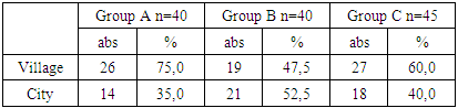
It was found that 26 patients (75.0%) in group A, 19 patients (47.5%) in group B and 27 patients (60.6%) in group V lived in rural areas.It was found that 14 patients in group A (35.0%), 21 patients in group B (52.5%) and 18 patients in group V (40.0%) lived in urban conditions.Characterization of parameters of humoral immunity and cytokine status in patients with vitiligo.After describing the humoral factors of the immune system in patients with vitiligo, their characteristics were determined and evaluated. Diagnostic and prognostic features specific to this nasological unit were determined. Taking this into account, all patients were divided into groups A, B and V, whose brief definition was given at the beginning of this chapter, so we did not dwell on them.
2. Result and Discussion
We presented the results obtained in the study in comparison with each other in these study groups and with the parameters of the control group.The study of the concentration of the main immunoglobulins (IgA, IgM, IgG) in the blood serum, which provide the humoral immunity of the body's immune system, showed that the parameters of both compared groups were reliably higher than those of the control group (healthy individuals).The increase of studied IgA concentration in groups A, B and V to the amounts of 1.66±0.10 ng/ml and 1.50±0.09 ng/ml, respectively, is reliable compared to the parameters of the control group (1.22±0.07 ng/ml). showed an increase in the level, this increase was 1.36 and 1.23 times, respectively (r<0.05), indicating an increase in the synthesis of this immunoglobulin in the body.Due to the long follow-up period, we see such a situation, but, despite the continuous process over time, the increase in the concentration of IgA in group B with a strong corresponding antigen stimulation ensured the strengthening of local immunity and contributed to the extinguishing of the purulent-inflammatory process to a certain extent.A similar result was obtained when determining and analyzing the concentration of IgM in the blood serum of patients. Compared to the control group, it was observed to be increased in groups A, B and V - 1.02±0.07 ng/ml against 1.22±0.09 ng/ml and 1.12±0.08 ng/ml, respectively. if the numbers in group 1 and the control group were significantly different from each other (1.20 times r<0.05).But this did not negate the tendency of this immunoglobulin to increase, considering that its presence in the blood serum for a short time (10 days) is of little importance in the primary immune response, we make sure that all the obtained numbers are close to each other. We would like to acknowledge that the importance of this immunoglobulin in determining the prognosis of vitiligo, the course of the disease and the end is very low.The results of the study and analysis of the amount of IgG also showed that a trend similar to the above was observed. As the parameters of groups A, B and V did not reliably differ from each other (15.73±0.26 ng/ml and 15.56±0.20 ng/ml, р>0.05), they were reliably higher than the parameters of the control group. It is noteworthy that it was found in large quantities (r<0.005) up to .96 and 1.94 times. This condition is associated with the production of IgG, which is the main immunoglobulin in blood serum, in large quantities in accordance with the stimulation of antigenic aggression. If we take into account the fact that IgG mainly provides the secondary immune response, it is produced after some time in the primary immune response, it shows the great role of this immunoglobulin in antigen elimination.Thus, determining the concentration of the main immunoglobulins (IgM, IgG, IgA) in the blood serum of patients with vitiligo showed that their directions of change and increasing trends were practically the same in patients of groups A, B and V. The amount of IgA increased by 1.36 and 1.23 times in both groups compared to the control group, respectively (r<0.05), and the concentration of IgM increased by 1.20 times (r<0.05) and 1.10 times, respectively (r>0.05) increased, these parameters of IgG increased reliably by 1.96 and 1.94 times (r<0.005). Recognizing the importance of IgA for sIgA produced on mucosal surfaces, we note that there were no significant changes in IgM concentrations. Although both immunoglobulins have a pathogenetic value for the nasological entity under study, we recognize that they are of little value in determining the diagnostic and course of the disease and the outlook of the end.However, we believe that a sharp increase in IgG is related to the duration and degree of the disease, therefore, a sharp increase in the amount of IgG from the indicators of the control (healthy) group or from the reference indicators is recommended as a diagnostic and prognostic immunological criterion.In contrast to IgA, IgM and IgG described above, the concentration of IgE in the blood serum has changed to an extent that attracts attention. Unlike other immunoglobulins, their amount was different not only from the control groups, but also between the compared groups.But the parameters of group A reliably increased by 6.33 times compared to the parameters of control groups (up to 154.80±8.14 ng/ml, r<0.005), in groups B and V, this indicator increased even more - 167.58±6, 67 ng/ml (6.85 times r<0.005). A large (6.33 and 6.85 times) increase of IgE in the blood serum of patients with vitiligo indicated that an allergic background had appeared in the body.
3. Conclusions
Thus, it was found that the concentration of IgE in the blood serum of the patients in the observation group reliably increased by 6.33 times (group A) and 6.85 times (in groups B, V) compared to the parameters of the control group. It is noteworthy that the amount of this immunoglobulin was significantly higher in group B and V than group A (by 1.08 times), but the result was not significantly different (r>0.05). Their long-term persistence in the body is explained by the continuous elimination of pathogens. Taking into account the above, a comprehensive approach to vitiligo treatment tactics is necessary. It is also recommended to use the level of IgE detection as an additional diagnostic and prognostic immunological criterion that determines the course and outcome of the disease.
References
| [1] | Achilova D.N. Clinical analysis of morphophysiological changes in immune organs in children after past illnesses // A new day in medicine. - Bukhara, 2021. - No. 5 (37). - P. 111 - 114. |
| [2] | Achilova.D.N. Specific course of allergic reactions in children // Web of scientist international scientific research journal. ISSN: 2776-0979 Vol. 2 No. 09 (2021), Indonesia. – P. 10-17. |
| [3] | Achilova.D.N. Modernassays for the treatment of allergic skin diseases in children // International journal of formal edication. Polsha, 2021. Volume: 01 Issue: 04. – P. 6-17. |
| [4] | Achilova D.N., Amonov R. A., Sharipova L. Kh., Yomgurova O. R., Rustamov B. B. Clinical, Immunological and Medico-Social Aspects of Allergic Diseases in Children // Annals of the Romanian Society for Cell Biology. Romania, 2021: Vol 25: Issue 3. – P. 6736-6740. |
| [5] | Achilova D.N., Yomgurova O.R. Clinical-immunological and medico-Social aspects of allergic diseases in children, development of criteria for early diagnosis and prognosis of the course of the disease (literature review) // British medical journal. London, 2022. Vol-2, No 2. – P. 46-53. |
| [6] | Achilova D.N. Current issues of food allergy diagnostics in pediatric practice // International Journal of Early Childhood Special Education. Issue 02 2022. (INT-JECSE) DOI: 10.9756/INT-JECSE/V14I2.677 ISSN: 1308-5581 Vol 14, – P. 5991- 5995. |
| [7] | Achilova D.N., Khushvaktova M.F. Clinical-Immunological Methods of Clinical-Immunological Research of Atopic Dermatitis in Children Permanently Residing in the Area of Oil Refining Enterprises // American Journal of Medicine and Medical Sciences. USA, 2022, 12(10): DOI: 10.5923/j.ajmms. 20221210.05. – P. 1034-1038. |
| [8] | Achilova D.N., Nuraliev N.A. The study of clinical and diagnostic features of various manifestations of allergic diseases in children // Medicine and Innovations. - Tashkent, 2022. - No. 4 (8). - P. 297-303. |
| [9] | Achilova D.N., Nuraliev N.A. Studying the effectiveness of long-term observation, treatment and prevention of bronchial asthma and allergic diseases in children // Medicine and Innovations. - Tashkent, 2022. - No. 4 (8). - P. 261-267. |
| [10] | Sharipova Gulnihol Idiyevna. The effectiveness of the use of magnetic-infrared-laser therapy in traumatic injuries of oral tissues in preschool children//Academic leadership. ISSN 1533-7812 Vol:21Issue 1 |
| [11] | Sharipova Gulnihol Idiyevna. Discussion of results of personal studies in the use ofmil therapy in the treatment of trauma to the oral mucosa // European Journal of Molecular medicine, Volume 2, No.2, March 2022 Published by ejournals PVT LTDDOI prefix: 10.52325Issued Bimonthly Requirements for the authors. |

 Abstract
 Reference
 Full-Text PDF
 Full-text HTML