Karshiyeva Dilovar Rustamovna
Bukhara State Medical Institute named after Abu Ali Ibn Sino, Uzbekistan
Correspondence to: Karshiyeva Dilovar Rustamovna, Bukhara State Medical Institute named after Abu Ali Ibn Sino, Uzbekistan.
Copyright © 2023 The Author(s). Published by Scientific & Academic Publishing.
This work is licensed under the Creative Commons Attribution International License (CC BY).
http://creativecommons.org/licenses/by/4.0/

Abstract
Much of the literature provides information comparing the long-term effects of smoking with the economic benefits of tobacco farming and tobacco production. Such studies show that tobacco-related diseases, losses due to premature deaths of smokers, and economic losses due to smoking-related diseases are among them. All this indicates that tobacco smoking is an important socio-economic problem facing the government of any country. The effect of the components of tobacco smoke on the human body during smoking can directly cause various diseases in the mucous membrane of the oral cavity, nose and bronchi, which is a number of diseases in the body of smokers. transformations, blood flow enters the salivary gland and is removed from the saliva to the mouth. Changes in oral fluid, oral mucosa, and minor salivary glands can be the first symptoms to diagnose diseases caused by smoking.
Keywords:
Tobacco, Oral cavity, Smoking, Gingivitis, Toothpastes
Cite this paper: Karshiyeva Dilovar Rustamovna, Results of a Clinical Study on the Dental Status of Tobacco Smokers, Clinical and Dental Examination Results, American Journal of Medicine and Medical Sciences, Vol. 13 No. 6, 2023, pp. 801-805. doi: 10.5923/j.ajmms.20231306.06.
1. Introduction
During the research period, we divided tobacco smokers into 2 large groups, depending on the duration of smoking: group A (those who have smoked for at least 5 years up to 10 years) and group B (those who have smoked for more than 10 years).
2. Materials and Methods
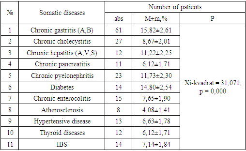
In patients with chronic catarrhal stomatitis, chronic catarrhal goitre, leukoplakia, and small salivary gland lesions, diabetes during examination was 14.80±2.54%, thyroid diseases 6.12±1.71%; cardiovascular pathology (atherosclerosis in 4.08±1.41% cases, hypertensive disease - 6.63±1.78%), diseases of the gastrointestinal tract (chronic gastritis (A, B) 15.82±2.61; There were concomitant diseases such as chronic cholecystitis 8.67±2.01, chronic hepatitis (A, B, C) 11.22±2.25, chronic pancreatitis 6.12±1.71. Diseases of the urinary system 11.73± It was found in 2.30% cases (Chi-square = 31.071; r = 0.000) (Table 1).Table 1. Comorbidities in smokers
 |
| |
|
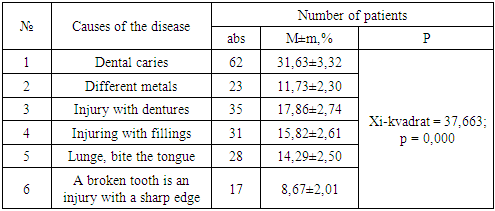
The most common local injurious factor in the development of chronic catarrhal stomatitis, chronic catarrhal gout, leukoplakia and minor salivary gland damage in OShQ was damage to the mucous membrane of the oral cavity in 31.63±3.32% of cases, in 11.73±2.30% of cases - mouth the simultaneous presence of different metals in the cavity. Injuring with dentures was 17.86±2.74%, injury with fillings was 15.82±2.61%, injury with a broken tooth was 8.67±2.01%. Harmful habits such as biting the tongue and mucous membrane of the tongue were recorded in 14.29±2.50% of cases (Chi-square = 37.663; r = 0.000) (see Table 2).Table 2. The state of the oral cavity of smokers before remediation
 |
| |
|
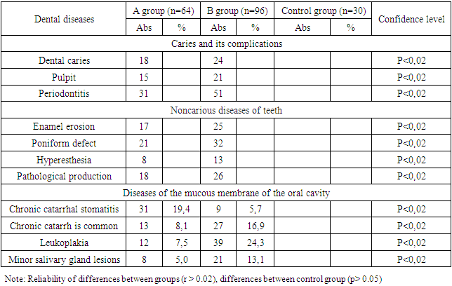
Based on the results of a retrospective analysis, the analysis of the effectiveness of the traditional local treatment scheme of patients with red flat iron showed improvement in 48.7% of cases. Prednisolone in the form of tablets: administered orally every other day after breakfast in a dose of 20 mg in the 1st week, in a dose of 15 mg in the 2nd week, in a dose of 10 mg in the 3rd week, and in a dose of 5 mg in the 4th week; nicotinic acid in the form of tablets in a dose of 0.05 g 2 times a day; vitamin A (oil) 10 drops 3 times a day.In 9% of cases, chronic catarrhal stomatitis, chronic catarrhal goitre, leukoplakia, and minor salivary gland lesions were transformed into a simple form of erosive-ulcerative form. Resistance to conventional therapy was observed in 42.3% of patients, which was manifested by frequent exacerbations and unstable remission from 2 to 5 times a year.Thus, the results of the retrospective analysis show that the frequency of chronic catarrhal stomatitis, chronic catarrhal goitre, leukoplakia, and minor salivary gland damage among other diseases of the mucous membrane of the oral cavity is 44.3% (out of 442 patients). Associated diseases of chronic catarrhal stomatitis, chronic catarrhal gout, leukoplakia, and minor salivary gland lesions are diabetes in 14.80±2.54% of cases, thyroid diseases in 6.12±1.71% of cases; cardiovascular pathology (atherosclerosis in 4.08±1.41% cases, hypertensive disease in 6.63±1.78% cases), diseases of the gastrointestinal tract (chronic gastritis in 15.82±2.61 cases (A, B) ; chronic cholecystitis in 8.67±2.01 cases; chronic hepatitis (A, B, C) in 11.22±2.25 cases; chronic pancreatitis in 6.12±1.71 cases). Diseases of the urinary system were found in 11.73±2.30% of cases. At the same time, among all forms of chronic catarrhal stomatitis, chronic catarrhal goitre, leukoplakia and minor salivary gland damage, the erosive-ulcer form was found in 33.67±3.38% (66) patients, and 68.8% in people aged 51-60 years and older. % (135) increases. The most common location of morphological elements, in 95 (48.5%) patients is the lung mucosa, in the retromolar area - 30.1% (59). Low efficiency of traditional local treatment, high tolerance to treatment of patients with chronic catarrhal stomatitis, chronic catarrhal goitre, leukoplakia and minor salivary gland lesions prompts to search for new methods and means of pathogenetic treatment.Examination of patients began with their general condition, skin color, palpation of local lymph nodes. An examination of the oral cavity was performed to assess the dental condition.We studied the condition of the lips, tongue, mucous membrane of the tongue, the presence of tooth marks or bite marks, the condition of the tongue and labial folds, the depth of the oral cavity, determined the condition of the gingival margin (color, shape, presence of swelling, canals), mineralized and non-mineralized, determined the presence of supragingival and subgingival stones, the presence and depth of periodontal pockets, the presence of exudation. The prevalence of dental diseases in smokers in the study group during the dental examination was studied (Table 3).Table 3. Prevalence of dental diseases in smokers
 |
| |
|
During the results of the examination, hyperesthesia, pathological tooth decay, and sarcoid defect were leading among dental diseases in both groups. We saw that in smokers with more than 10 years of smoking experience, these indicators increased by 1.5 times compared to smokers with 10 years of smoking experience. Pathological swelling was observed in 26 patients, follicular defect in 32 patients, and hyperesthesia in 13 patients. Diseases of the mucous membrane of the oral cavity are characteristically important in smokers. Chronic catarrhal stomatitis, chronic catarrhal goitre, leukoplakia, minor salivary gland lesions were the main disease background of smokers. The severity of problem in each smoker in the study was calculated and the main point of view of our study was calculated.
3. Result and Discussion
Examination of dental diseases in smokers in the control group showed that caries and pulp disease showed a greater amount compared to the study groups. However, it was found that enamel erosion, plaque defect, pathological swelling, and hyperesthesia from non-carious diseases of teeth were 2.5 times less common in the control group compared to all research groups. Oral mucosal diseases were hardly seen in control group participants.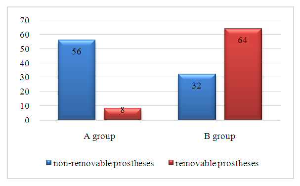 | Figure 1. Proportion of orthopedic devices in smokers in the study group |
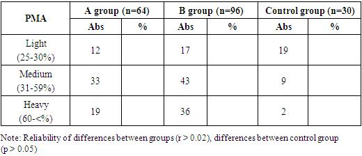
During the research, a relatively large number of orthopedic devices were seen and studied in our patients. In smokers with more than 10 years of smoking experience, we saw that orthopedic devices increased 2 times compared to smokers with 10 years of smoking experience. It can be seen that year after year the use of tobacco products has been proven to increase the need for orthopedic devices.According to the results of the investigation carried out during the study, periodontal diseases were very common among smokers in all research groups, and we used the classification of periodontal diseases of WHO in 2018 (Jebsen, Caton et al., 2018; Albandar et al., 2018) (Figure 2).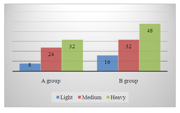 | Figure 2. Distribution of periodontal tissue damage in smokers depending on severity |
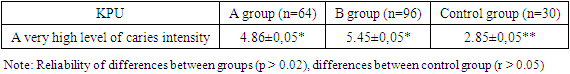
During the study, complaints of smokers were heard and an objective examination was conducted. Pain in the gums, bleeding from the gums, and bad breath were more common in smokers. Bleeding from milk (25.7%) and dry mouth (17.1%) complain 2.7 times less often (60%; 40%, respectively). 57.1% of patients on the main background in the study complained of bad breath, 10 times more than in the participants of the control group (4.7%). Smokers were 3 times more likely to have white spots on the tongue (28.1%, 4.3%, respectively). A feeling of bitterness in the oral cavity was noted in 15.7% of patients against the background of the main disease and in 1.5% of patients in the control group. Seminars were organized under the motto "We are against smoking", and after the research patients were admitted to the club, their lifestyle and questionnaires were taken from them. During the questionnaire, various complaints were presented and summarized.As a result of the oral examination of the smoker, non-carious lesions of the tooth such as erosion were noted, which were found in 82.8% of patients in group A. Pupillary defects were observed in 62.8% of patients in group B. Pathological tooth decay was found in 74.2% of patients in group A and 27.1% in group B, but no significant difference between groups was observed (p< 0.02).When the intensity of damage to teeth with caries was studied, the average value of KPU index in group A (very high level of caries intensity), KPU index in group A - 4.86±0.05 (very high level of caries intensity), KPU index in group B - 5.45±0.05 (high level of caries intensity) was found. A statistically significant difference was found between groups A and B (p<0.02). KPU index in patients of the control group was 2.85±0.05 (p<0.05). Table 4 shows the comparison data of KPU, the index of caries intensity in teeth between both groups, obtained during the examination of smoking patients.Table 4. Results of the KPU index study among smokers
 |
| |
|
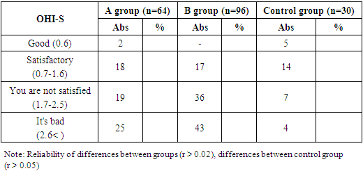
In the process of studying the dental status of smokers, the level of hygiene of the oral cavity is important, therefore, when this indicator is studied, the OHI-S hygiene index, which takes into account the amount of dental caries and tartar, was 3.01±1.1 in group A and 2.94±1 in group B. was 4.A statistically significant difference was found between groups A and B (p<0.02). OHI-S hygiene index in control group patients was 1.25±1.5 (p<0.05). The comparison data of the OHI-S hygiene index between the two groups, obtained during the examination of the patients in the study, is presented in Table 5. Before professional hygiene was carried out in the oral cavity of the patients who were diagnosed with bad periodontitis during the study, they were explained about this stage.Table 5. Results of the OHI-S index study among smokers
 |
| |
|
The PI periodontal index was used to determine the severity of periodontal disease in smokers, which showed that the value of this index was 5.51±2.29 in group A patients, which corresponded to the severe level of periodontal pathology in group B patients, and it was 2 times that of group A patients, control more than 10 times than the group. The data of periodontal index PI between the three groups, obtained during the examination of patients, are presented in Figure 3.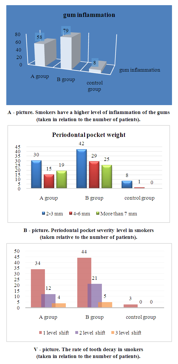 | Figure 3. Results of the PI periodontal index study in smokers |
Papilla-marginal alveolar index (RMA) scores were high in both study groups (98%). The presence of severe gingivitis in 34 (77.3%) and 24 (58.5%) patients in group A and group B, respectively, indicates that the disease has already developed severe gingivitis and periodontitis (Table 6).Table 6. Analysis of RMA index results in smokers
 |
| |
|
During the study M. in smokers. M. Sialometry according to the Pojaritskaya method allows to determine the amount of unstimulated mixed saliva. Sialometry was performed in the morning for 10 minutes, on an empty stomach, by spitting (usually 4.1 ml of mixed saliva is released in 10 minutes, on average). The greatest hyposalivation was found in the oral cavity of group B smokers, and the average value for this group was 3.0 ± 0.28 ml. A statistically significant difference was found between groups A and B (p<0.02). In the patients of the control group, the indicators approached the norm (p<0.05). The sialometry data between the two groups, obtained during the examination of the patients, is presented in Figure 4.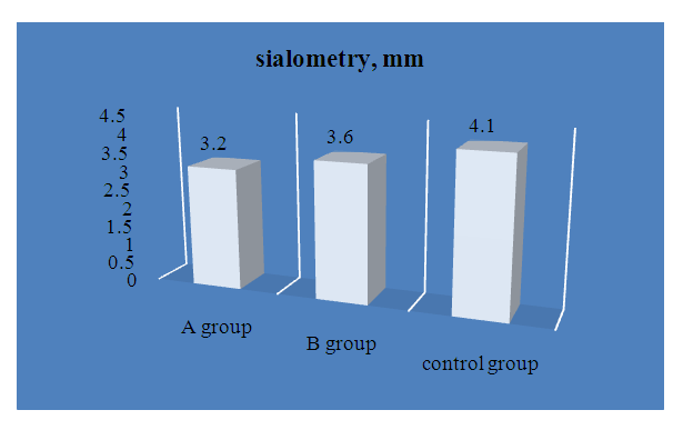 | Figure 4. M. in smokers. M. The results of the study of sialometry by the Pojaritskaya method |
References
| [1] | Almas K., Maroof F., Mcallister C, Freeman R. Smoking behaviour and knowledge in high school students in Riyadh and Belfast. II Odontostomat. Trop. - 2002. - Vol. 25. - № 98. - P. 40 - 44. |
| [2] | Bendick C, Scheifele C, Reichart P.A. Oral manifestations in 101 Cambodians wit HIV and AIDS. II J. Oral. Pathol. Med. - 2002. - Vol. 31. - № 1. - P. 1 -4. |
| [3] | Casiglia J., Woo S.B. A comprehensive review of oral cancer. II Gen. Dent. -2001. - Vol. 49. - № 1. - P. 72 - 82. |
| [4] | Faddy M.J., Gullinan M.P., Palmer J.E. et al. Ante-dependence modeling in a longitudinal study of periodontal disease: The effect of age, gender, and smoking status. II J. Periodontol. - 2000. - Vol. 71. - № 3. - P. 454 - 459. |
| [5] | Sharipova Gulnihol Idiyevna. DISCUSSION OF RESULTS OF PERSONAL STUDIES IN THE USE OFMIL THERAPY IN THE TREATMENT OF TRAUMA TO THE ORAL MUCOSA // European Journal of Molecular medicinevolume 2, No.2, March 2022 Published by ejournals PVT LTDDOI prefix: 10.52325 Issued Bimonthly Requirements for the authors. |
| [6] | Sharipova Gulnihol Idiyevna. THE EFFECTIVENESS OF THE USE OF MAGNETIC-INFRARED-LASER THERAPY IN TRAUMATIC INJURIES OF ORAL TISSUES IN PRESCHOOL CHILDREN // Academic leadership. ISSN 1533-7812 Vol: 21 Issue 1. |
| [7] | Данилевский Н.Ф., Леонтьев В.К., Несин А.Ф., Рахний Ж.И. Заболевания слизистой оболочки полости рта. - М.: Издательство ОАО "Стоматология", 2001.-272 с. |
| [8] | Кулик И.В., Миргородская Л.В. ВИЧ-инфекция. Проявления в полости рта. Журнал: Институт стоматологии. - 2001. - № 2. - С. 36 - 40. |
| [9] | Курение как фактор заболеваний пародонта, кариеса и потери зубов. // Стоматологическое обозрение. - 2003. - № 2. - С. 3 - 4. |
| [10] | Трубачева И.А., Перминова О.А., Шатров СВ. Популяционные аспекты табакокурения у взрослого населения Томска // III Международная конференция по восстановительной медицине (реабилиталогия). — М.: Злато-граф, 2000. - С. 426 – 427. |

 Abstract
 Reference
 Full-Text PDF
 Full-text HTML