American Journal of Medicine and Medical Sciences

p-ISSN: 2165-901X    e-ISSN: 2165-9036

2023;  13(5): 633-636

doi:10.5923/j.ajmms.20231305.18

Received: Apr. 22, 2023; Accepted: May 12, 2023; Published: May 17, 2023


Shifts as Markers of Perinatal Damage of the Central Nervous System

Fayzullaeva Khilola Bahronovna

Assistant of the Department of Biological Chemistry, Samarkand State Medical University, Samarkand, Uzbekistan

Correspondence to: Fayzullaeva Khilola Bahronovna, Assistant of the Department of Biological Chemistry, Samarkand State Medical University, Samarkand, Uzbekistan.

Email:

Copyright © 2023 The Author(s). Published by Scientific & Academic Publishing.

This work is licensed under the Creative Commons Attribution International License (CC BY).
http://creativecommons.org/licenses/by/4.0/

Abstract

Perinatal lesions of the Central nervous system occupy a leading place in the structure of morbidity in newborns. In this regard, we set ourselves the task of evaluating the activity of the S100B neurotest as a marker of perinatal CNS damage in posthypoxic syndrome, depending on the severity of asphyxia; compare metabolic shifts with neurotest indicators in dynamics; highlight the correlation between S100B and Ca, Mg serum levels as components of the formation of perinatal lesions of the central nervous system. According to the results obtained from the isolated signs of multiple organ failure, a high level of S100 neurotest in combination with hypoglycemia can be considered a certain clinical and laboratory predictor of the formation of neurological symptoms and long-term consequences against the background of obvious clinical signs. Correlation analysis of the relationship between Ca and Mg showed a high positive statistically significant relationship with neonatal asphyxia, especially with body weight shifts, regardless of the severity of asphyxia, and a moderately and weakly positive and in some cases negative correlation with S100/Ca, S100/Mg, which confirms the predominance of S100 as the leading laboratory neurotest in neonatal asphyxia.

Keywords: Newborns, S100 neurotest, Hypocalcemia, Hypomagnesemia

Cite this paper: Fayzullaeva Khilola Bahronovna, Shifts as Markers of Perinatal Damage of the Central Nervous System, American Journal of Medicine and Medical Sciences, Vol. 13 No. 5, 2023, pp. 633-636. doi: 10.5923/j.ajmms.20231305.18.

1. Introduction

Hypoxic - ischemic encephalopathy resulting from hypoxia, underlies the delay in psychomotor development in the first year of life, the formation of attention deficit hyperactivity disorder, cerebral palsy, motor and cognitive disorders [6,7].
In this regard, early diagnosis of the severity of hypoxia and the associated CNS damage is of particular importance for the choice of adequate therapy and in the preparation of health procedures and medical correction during further medical examination, taking into account the risk of developing long-term neurological symptoms [1,3,10].
A great role is given to the assessment of the marker of perinatal damage to the central nervous system according to the S100B neurotest. Also, the role of hypoglycemia, hypocalcemia, hypomagnesemia and a decrease in the level of phosphorus in the blood of a newborn is not excluded, as components of the formation of metabolic disorders indirectly creating a background for neurological symptoms [2,4,9].

2. Literature and Methodology

To accomplish this task, 60 full-term newborns born with a history of asphyxia were examined. Diagnosis according to WHO criteria (2008). Depending on body weight and severity of asphyxia, groups of patients were
I- body weight less than 2500.0 g
II- Normal body weight at birth
III- body weight over 4000g
According to the severity of asphyxia, each group is divided into subgroups: a-moderate asphyxia, b-severe asphyxia.
Research methodology-marker neurotest S100B by ELISA, the level of glucose, calcium, magnesium, phosphorus was determined on the Merilyzer Clini Quant apparatus.

3. Results and Discussion

Of the observed newborns, up to 60% were dominated by boys. Analysis of anamnestic data on maternal health showed the predominance of:
Leading factors of antenatal hypoxia: acute infections of the upper respiratory tract - 80%; anemia - 63%; placental insufficiency - 43%; caesarean section - 40%; thyroid disease - 40%; pathology of the urinary system - 36%; hypertension disorder - 34%; inflammatory diseases of the pelvic organs in women - 28%.
Leading intranatal factors of acute asphyxia: childbirth and delivery, complicated by fetal stress - 80%; cord entanglement - 40%; premature detachment of the placenta - 36%; violations of labor activity - 35%; severe preeclampsia - 30%; the onset of labor after a 24-hour waterless period - 28%; premature rupture of membranes - 22%. In more than half of the cases, a combination of 2-3 factors was identified. Thus, there is such a picture of a combination of leading factors: deterioration of maternal blood oxygenation 76%; interruption of blood flow through the umbilical cord 45%; violation of gas exchange through the placenta 40%; insufficiency of respiratory efforts of the newborn 28%; inadequate hemoperfusion of the maternal part of the placenta 25%;.
Patients are monitored in accordance with the rule of clinical, instrumental and laboratory monitoring according to indications and to the extent necessary. The neurotest marker S100B was tested in observed newborns in the first hours and on the fifth day of life. (tab. 1)
Table 1. Dynamics of protein S-100 concentration in children in comparison groups
     
As can be seen from Table 1, in the first hours of life in low-weight newborns and those with large body weight, S100B increases sharply by almost 5 times compared to healthy newborns; in patients with normal body weight, neurotest markers increase moderately up to 2.5 times. On the 5th day of life in heavy newborns, although the S100B values gradually decrease, they remain 1.5 times high compared to healthy newborns; in other groups, the neurotest values are reduced to normal or closer to normal. Against this background, hypoglycemia is observed from 52% in group IIb, to 60-62% in groups IIIb and Ib.
Given the role of the S100B neurotest as a marker of perinatal CNS damage in newborns, and neurological symptoms are pronounced in our observations, the neurotest with the indicated S100B marker was performed in the first hours of life and in dynamics on the 5th day. In the group of small children and large children, S100B increases sharply above the norm 5 times, in children with normal body weight, the indicators increase moderately by 2.5 times. On the 5th day of life in overweight patients, although the S100B values gradually decrease, they remain 1.5 times high compared to healthy newborns; in other groups, the neurotest values are almost reduced to normal or already within the range of indicators in healthy newborns. Against this background, we observe hypoglycemia from 52% in group IIb, to 60-62% in groups IIIb and Ib. In parallel with these changes, which aggravate the neurological status in asphyxia of newborns, we observe a sharp decrease in the level of phosphorus in the blood in all groups of newborns with asphyxia, a decrease in the level of magnesium and ionized Ca in all groups, a sharp decrease in plasma Ca in groups Ib and IIIb, namely in newborns with changes in body weight (table 2).
Table 2
     
It is well known that phosphorus is involved in neuron myelination, hypocalcemia and hypomagnesemia indirectly aggravate the state of vascular wall permeability and muscle tone. It is believed that neonatal hypoglycemia can lead to immediate and long-term neuropsychiatric disorders, the later it is detected, the more likely these complications are [5,8].
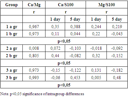
Comparing these features, it is clear that the S100B marker is the leading neurotest in assessing perinatal CNS damage, but the presence of hypocalcemia, hypomagnesemia, hypoglycemia, and a decrease in phosphorus levels. Given the activity of these metabolites in the structure of the cell membrane and neuron myelination, it serves as an aggravating background in the formation of perinatal CNS damage in newborns.To confirm this relationship between the neurotest marker S100B, Ca and Mg, the correlation dependence of these indicators was assessed. A high positive relationship was noted statistically significant S100 Ca / Mg in all groups of patients, regardless of the severity of asphyxia, except for group IIa, where the relationship is weakly positive.
Indicators S100/Ca in underweight patients and patients with normal weight on the first day of life from a weakly positive to moderately positive correlation, and in patients with a large body weight, the correlation is negative and weak. On the 5th day of life in underweight patients, the correlation remains weak in severe asphyxia and moderate direct statistically significant. In patients with normal body weight, the correlative relationship is negative, weak, statistically significant, in large children with moderate asphyxia, the relationship is inversely weak, and in severe asphyxia, the relationship is direct moderate. The relationship S100/Mg is mostly inverse, weak, statistically significant.

4. Conclusions

Of the identified signs of multiple organ failure, a high level of S100 neurotest in combination with hypoglycemia can be considered a certain clinical and laboratory predictor of the formation of neurological symptoms and long-term consequences against the background of obvious clinical signs.
Correlation analysis of the relationship between Ca and Mg showed a high positive statistically significant relationship with neonatal asphyxia, especially with body weight shifts, regardless of the severity of asphyxia, and a moderately and weakly positive and in some cases negative correlation with S100/Ca, S100/Mg, which confirms the predominance of S100 as the leading laboratory neurotest in neonatal asphyxia.

References

[1]  Butolin E. G. et al. ROLE OF BIOMARKERS OF ORGANIC MATRIX OF BONE TISSUE IN CHRONIC HEMATOGENOUS OSTEOMYELITIS IN CHILDREN // European journal of molecular medicine. – 2022. – Т. 2. – №. 5.
[2]  G’ayratovna S. X. et al. PRINCIPLES OF THE USE OF ANTIGENS IN THE IMMUNITY DIAGNOSIS OF ECHINOCOCCOIS DISEASE // Thematics Journal of Social Sciences. – 2022. – Т. 8. – №. 2.
[3]  Kenjayevich B. A. et al. STUDIES OF REPARATIVE REGENERATION OF CHITOSAN DERIVATIVES IN EXPERIMENTAL THERMAL BURNS // ResearchJet Journal of Analysis and Inventions. – 2022. – Т. 3. – №. 4.
[4]  Н.К.Мurtazayeva Diagnosis of Diabetes Measurements witha Glucometer Volume 6| March, 2022 ISSN: 2795-7624. http://www.geniusjournals.org/.
[5]  Mamadalieva Z. R. et al. DETERMINATION OF ALBUMIN IN BLOOD ON A BIOCHEMICAL ANALYZER BY VIRTUAL LABORATORY METHOD // Thematics Journal of Chemistry. – 2022. – Т. 6. – №. 1.
[6]  Mamadaliyeva Z. R. et al. DETERMINATION OF ALANINE AMINOTRANSFERASE IN BLOOD BY VIRTUAL LABORATORY METHOD ON A BIOCHEMICAL ANALYZER // Thematics Journal of Chemistry. – 2022. – Т. 6. – №. 1.
[7]  Mamadaliyeva Z. R., Nazarova M. Kediyorova Sh. X, & KM Xalikov. (2022). Determination of alanine aminotransferase in blood by virtual laboratory method on a biochemical analyzer // Thematics Journal of Chemistry ISSN. – С. 20-22.. – С. 1-6.
[8]  Nabieva F. S., Fayzullayeva K. B., Rayimova F. S. The importance of enzyme immunoassay in the diagnosis of infectious diseases // Central Asian Research Journal for Interdisciplinary Studies (CARJIS). – 2022. – Т. 2. – №. 10. – С. 46-49.
[9]  Saidmurodova Z. A. Toshmurodov DA Nuklein kislotalar kimyosi, ularning tuzilishi va ahamiyati //Вестник магистратуры. –2021. – 2021. – С. 2-1.
[10]  Saidmurodova Z. A., Halimova S. A. HUJAYRALARNING BOLINIB KOPAYISHI VA ULAR HAQIDA BA'ZI TUSHUNCHALAR // Eurasian Journal of Medical and Natural Sciences. – 2022. – Т. 2. – №. 5. – С. 93-95.
[11]  Saidmurodova Z. A., Nazarova M. E., Keldiyorova S. E. DNK TUZILISHI GENETIK TRANSFORMATSIYA JARAYONI, TADQIQOTLAR TAXLILI // Eurasian Journal of Academic Research. – 2022. – Т. 2. – №. 4. – С. 121-124.
[12]  Sh N. G. et al. The specifics of neurosanographic changes in the diagnosis of posthypoxic complications in children born in asphyxia // Colloquium-journal. – Голопристанський міськрайонний центр зайнятості, 2020. – №. 19 (71). – С. 6-7.
[13]  Xusanovich U. G. et al. The fauna of mosquites (diptera: Phlebotomina) and its epidemiological importance in the skin leishmaniosis of uzbekistan // Web of Scientist: International Scientific Research Journal. – 2022. – Т. 3. – №. 4. – С. 1123-1128.
[14]  Азим Б. и др. Состояние свободнорадикального окисления липидов при экспериментальном инфаркте миокарда у крыс // Европейский журнал молекулярной и клинической медицины. – 2021. – Т. 8. – №. 3. – С. 816-820.
[15]  Алимова О. Б., Карабаев А. Г., Ким О. В. Клинико-Иммунологические Особенности Острой Диареи У Детей С Синдромом Гемоколита // Central Asian Journal of Medical and Natural Science. – 2022. – Т. 3. – №. 5. – С. 617-623.
[16]  Бакроновна Ф.Х., Негматовна А.М., Саломат Х. Анализ специфики антенатальных и интранатальных факторов риска у новорожденных с внутриутробной гипоксией.
[17]  Бердиярова Ш. Ш., Юсупов Ш. А., Назарова Г. Ш. КЛИНИКО-ЛАБОРАТОРНЫЕ ОСОБЕННОСТИ ХРОНИЧЕСКОГО ГЕМАТОГЕННОГО ОСТЕОМИЕЛИТА // Central Asian Research Journal for Interdisciplinary Studies (CARJIS). – 2022. – Т. 2. – №. 5. – С. 116-125.
[18]  Ибрагимова Н. С., Келдиѐрова Ш. Х. К., Назарова Г. Ш. ЗНАЧЕНИЕ ФОЛИЕВОЙ КИСЛОТЫ, ГОМОЦИСТЕИНА И ЭНДОТЕЛИНА-1 ПРИ РАЗВИТИИ СИНДРОМА ПОЛИКИСТОЗНЫХ ЯИЧНИКОВ У ЖЕНЩИН РЕПРОДУКТИВНОГО ВОЗРАСТА // Central Asian Research Journal for Interdisciplinary Studies (CARJIS). – 2022. – Т. 2. – №. 10. – С. 41-45.
[19]  Ким Д. В., Ким О. В. ФИЗИОЛОГИЯ ПИЩЕВАРЕНИЯ У ДЕТЕЙ // Oriental renaissance: Innovative, educational, natural and social sciences. – 2022. – Т. 2. – №. 6. – С. 308-312.
[20]  Саткыналиев Т. Т. и др. Формирование правовых аспектов регулирования предпринимательства в Кыргызской Республике // Вестник Кыргызского государственного университета строительства, транспорта и архитектуры им. Н. Исанова. – 2018. – №. 2. – С. 179-183.
[21]  Саттарова Х. Г. и др. ПРИМЕНЕНИЕ «МЕСТНЫХ АНТИГЕНОВ» В ИММУНОЛОГИЧЕСКОЙ ДИАГНОСТИКЕ ЭХИНОКОККОЗА // VOLGAMEDSCIENCE. – 2021. – С. 592-593.
[22]  Сувонкулов У. Т. и др. Распространенность кожных лейшманиозов среди населения в эндемичных районах Узбекистана // Общество и инновации. – 2020. – Т. 1. – №. 1/S. – С. 592-597.
[23]  Файзуллаева Х. Б. и др. КОРРЕКЦИЯ КОМПЛЕКСНОГО ЛЕЧЕНИЯ ПРИ МЕТАБОЛИЧЕСКОМ АЦИДОЗЕ У НОВОРОЖДЕННЫХ С ТЯЖЕЛОЙ АСФИКСИЕЙ // ЖУРНАЛ ГЕПАТО-ГАСТРОЭНТЕРОЛОГИЧЕСКИХ ИССЛЕДОВАНИЙ. – 2022. – №. SI-3.
[24]  Файзуллаева Х. Б. и др. ОСОБЕННОСТИ ФЕРМЕНТАТИВНЫХ ПОКАЗАТЕЛЕЙ ПРИ ДИАГНОСТИКЕ ПОСТГИПОКСИЧЕСКИХ ОСЛОЖНЕНИЙ СО СТОРОНЫ СЕРДЕЧНО-СОСУДИСТОЙ СИСТЕМЫ В ПЕРИОД НОВОРОЖДЕННОСТИ // АКТУАЛЬНЫЕ ПРОБЛЕМЫ БИОМЕДИЦИНЫ-2020. – 2020. – С. 339-340.
[25]  Файзуллаева Х. Б., Абдуллаева М. Н. ПРОГНОСТИЧЕСКАЯ ИНФОРМАТИВНОСТЬ КЛИНИКО-ЛАБОРАТОРНЫХ ПОКАЗАТЕЛЕЙ ПОСТГИПОКСИЧЕСКОГО СИНДРОМА У НОВОРОЖДЕННЫХ // ЖУРНАЛ РЕПРОДУКТИВНОГО ЗДОРОВЬЯ И УРО-НЕФРОЛОГИЧЕСКИХ ИССЛЕДОВАНИЙ. – 2022. – Т. 3. – №. 4.
[26]  Файзуллаева Х. и соавт. Метаболический статус как показатель постгипоксических осложнений у новорожденных, родившихся в условиях асфиксии // European Journal of Molecular & Clinical Medicine. ISSN. – 2020. – С. 2515-8260.
[27]  Халимова С. А. Самостоятельные занятия физической культурой в вузе // ИЗБРАННЫЕ ДОКЛАДЫ 64-Й УНИВЕРСИТЕТСКОЙ НАУЧНО-ТЕХНИЧЕСКОЙ КОНФЕРЕНЦИИ СТУДЕНТОВ И МОЛОДЫХ УЧЕНЫХ. – 2018. – С. 931-933.
[28]  Хамраев Х. Т., Хамраева Д. Х., Ким О. В. Особенности функции щитовидной железы у пациентов с метаболическим синдромом // Здоровье, демография, экология финно-угорских народов. – 2017. – №. 3. – С. 52-54.
[29]  Халиков Кахор Мирзаевич, Мамадалиева Зарина Рахмат Кизи. (2022). Интернет тармоғида виртуальная лаборатория ишларини бажариш имкониятини берувчи конструкторлари. Публикации Novateur (1), 1–76. Получено с http://novateurpublication.org/index.php/np/article/view/17.
[30]  Ш БС и соавт. Клинико-лабораторная характеристика хронического остеомиелита у детей. – 2022.
[31]  Ш БС и соавт. Клинико-лабораторные особенности хронического гематогенного остеомиелита // Тематический журнал микробиологии. – 2022. – Т. 6. – №. 1.
[32]  Шукуруллаевна Б.С. и соавт. СОВРЕМЕННЫЕ АСПЕКТЫ ЛАБОРАТОРНОЙ ДИАГНОСТИКИ ХРОНИЧЕСКОГО ОСТЕОМИЕЛИТА У ДЕТЕЙ //Web of Scientist: International Scientific Research Journal. – 2022. – Т. 3. – №. 10. – С. 19-23.
[33]  Шукуруллаевна Б.С. и соавт. СОВРЕМЕННЫЕ АСПЕКТЫ ЛАБОРАТОРНОЙ ДИАГНОСТИКИ ХРОНИЧЕСКОГО ОСТЕОМИЕЛИТА У ДЕТЕЙ //Web of Scientist: International Scientific Research Journal. – 2022. – Т. 3. – №. 10. – С. 19-23.
[34]  Эгамова М.Т. и соавт. Игровой метод реабилитации детей с детским церебральным параличом // Индийский журнал судебной медицины и токсикологии. – 2020. – Т. 14. – №. 4. – С. 7979-7983.