-
Paper Information
- Next Paper
- Previous Paper
- Paper Submission
-
Journal Information
- About This Journal
- Editorial Board
- Current Issue
- Archive
- Author Guidelines
- Contact Us
American Journal of Medicine and Medical Sciences
p-ISSN: 2165-901X e-ISSN: 2165-9036
2023; 13(5): 612-615
doi:10.5923/j.ajmms.20231305.14
Received: Apr. 22, 2023; Accepted: May 10, 2023; Published: May 13, 2023

Clinical and Neurophysiological Features of Children Born Prematurely
 Abstract
 Reference
 Full-Text PDF
 Full-text HTML
Ergashev Sukhrob Saidovich1, Niyazov Shukhrat Toshtemirovich2, Jurabekova Aziza Takhirovna3
1Basic Doctoral Student, Department of Neurology, Samarkand State Medical University, Uzbekistan
2Doctor of Medical Sciences, Associate Professor, Department of Neurology, Samarkand State Medical University, Uzbekistan
3Doctor of Medical Sciences, Professor, Head of the Department of Neurology, Samarkand State Medical University, Uzbekistan
Copyright © 2023 The Author(s). Published by Scientific & Academic Publishing.
This work is licensed under the Creative Commons Attribution International License (CC BY).
http://creativecommons.org/licenses/by/4.0/

One of the risk factors for failure to develop the central nervous system in children is prematurity - a child born at less than 37 full weeks of gestation. Extremely important for determining the nature of prematurity, it is in preterm birth division of premature babies by body weight. But, the modern approach to the problem, dividing children only by gestational age or only by birth weight may be insufficient. Thus, a complicating factor (cytomegalovirus infection, toxoplasmosis in the mother) or current viral diseases (influenza, covid, infection of the genitourinary system) play a role in not fully mature children. Chronic background (diabetes mellitus; hypothyroidism) comorbid condition of a pregnant woman, is equally important to maintain a healthy gestational age and correspondingly full development of the child's body.
Keywords: Neurophysiological features of children, Premature infants, Brain
Cite this paper: Ergashev Sukhrob Saidovich, Niyazov Shukhrat Toshtemirovich, Jurabekova Aziza Takhirovna, Clinical and Neurophysiological Features of Children Born Prematurely, American Journal of Medicine and Medical Sciences, Vol. 13 No. 5, 2023, pp. 612-615. doi: 10.5923/j.ajmms.20231305.14.
1. Introduction
- In the pathogenesis of insufficient brain formation, blood circulation problems during pregnancy (severe anemia, hepatitis) are considered. According to the scientific hypothesis, decay products of extravasated red blood cells cause narrowing of blood vessels in the arterial channel, causing vasospasm, which results in depletion of blood supply, further leading to neuronal death, causing brain tissue damage and persistent neurological disorders. Thus, the multifactorial nature of adverse neurological outcomes in premature babies [3,8,11], is beyond doubt, but a thorough analysis of the leading etiological factors in premature babies requires.Purpose of the study: To study risk factors leading to premature birth, on the basis of indicators of clinical, neurological, neurophysiological data at birth and in 6 (7) months, to determine unfavorable prognosis, severe neurological symptoms.
2. Material and Methods
- The use was carried out on the basis of MKSAMS, Department of Neonatology from 2021 to 2023, 69 premature infants were examined, children were divided into two groups depending on their birth weight: 1) children weighing from 2 to 2.5 kg (40 children), 2) children weighing from 1.5 to 2 kg (29 children). All children had gestational ages of 34-36 weeks. The main methods of investigation in the work were standard examinations by a neonatologist and neurologist, at the birth of the child and observation in dynamics; the rules of diagnosis using scales to identify developmental limitations were followed. Additional research methods, as the gold standard, are neurosonography. Amplitude-integrated EEG (AEG) was performed selectively on the patients. All research methods were performed with the parents' (or mother's) written permission. Need for proof of this study, a group of children (healthy) who were born at term and had no problems during delivery, a group of children was selected according to the same pattern of standard examination and scales to detect complete well-being of the infant 2- (26). The outcome of children born prematurely, various neurological complications are possible, to clarify the effect of low birth weight and underdevelopment of the brain and the body as a whole, the objectives of this study included follow-up of the same children (6-7 months later), according to the same indicators (clinical and neurological examination, NSG, aEEG). Standard deviation set was used for statistical analysis, t-test (Student's t-criterion) was used to compare the groups. Differences were considered statistically significant at p<0.05; all parameters were calculated on an individual computer.
3. Results
- The initial stage of the study was to find out which factors led to premature pregnancy and preterm birth. For this purpose, a thorough interview was conducted to collect the pregnancy history (the course of this pregnancy and labor). The anamnesis (survey) revealed a significant difference between the groups in the presence of factors that aggravated the period of normal pregnancy. Group 1 included threatened termination of pregnancy in 93%, feto-placental insufficiency in 27%, ARI in 26.6%, water shortage in 10.8%, and arterial hypertension in about 9%. The nature of delivery in the greater percentage through natural childbirth, it is -89%, the rest were born by cesarean section (placenta adherence, not the correct position of the head) - 11%, where p equals 0.05. Body length at birth was on average 4.5 cm lower relative to the healthy child group (p<0.001). Apgar score at the first minute of life was 5. Apgar score at the fifth minute of life was 6.
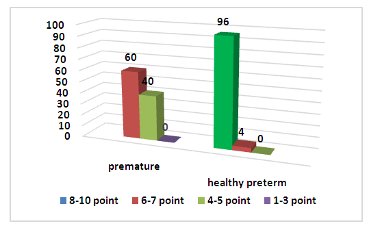 | Figure 1. Apgar scores in the study groups |
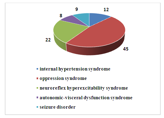 | Figure 2. Clinical characteristics of premature infants |
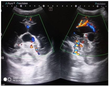
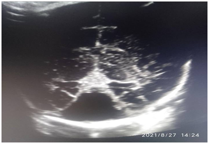 Evaluating the results of the analysis of structural abnormalities, according to the NSG data, noted different cerebral abnormalities, compared with healthy children. Thus, antenatal and postnatal VVCs in group 1 were found in 29.5% of cases. Subarachnoid hemorrhage was diagnosed in only one child. Porencephalic cyst was diagnosed in one case. Periventricular leukomalacia was detected in 4 children. Evaluating the results of the analysis of structural abnormalities, according to NSG data, different abnormalities of the cerebral structure were noted in comparison with healthy children. Thus, antenatal and postnatal VVF in group 1 was found in 29.5% of cases. Subarachnoid hemorrhage was diagnosed in only one child. Porencephalic cyst was diagnosed in one case. Periventricular leukomalacia was detected in 4 children.
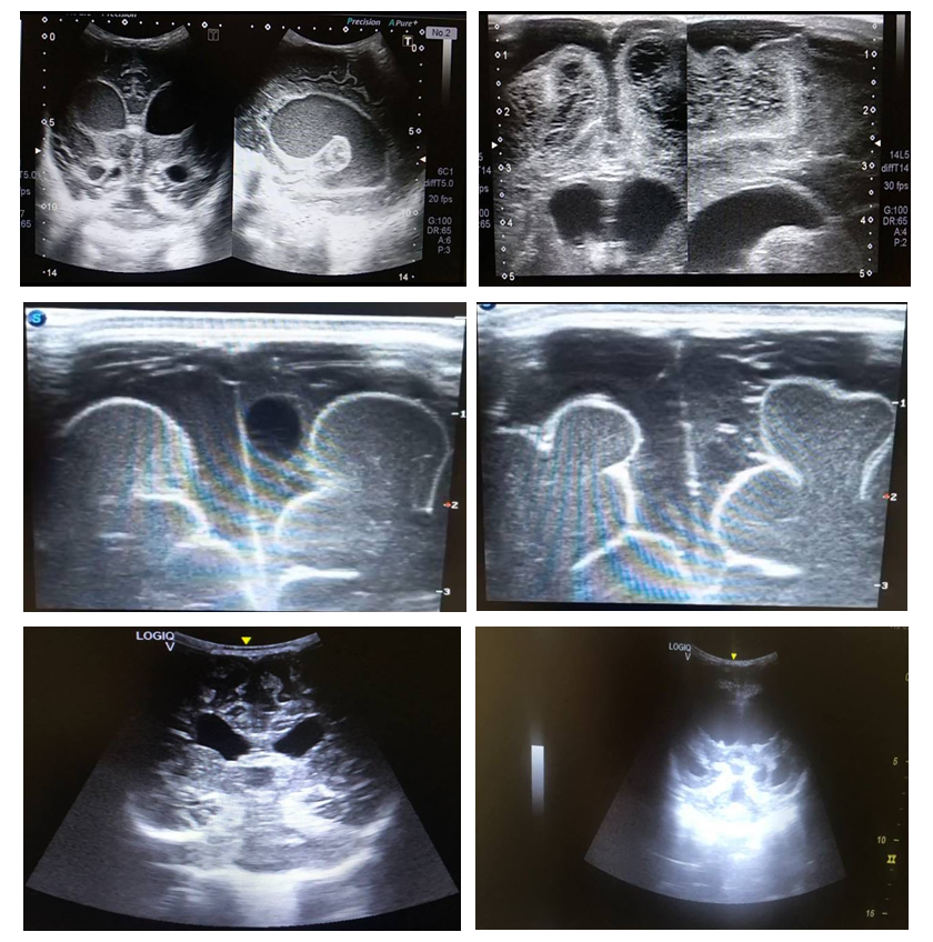 | Figures 3-5. Premature infants (main group), 32 -34 weeks. According to the neurosonogram, dilated lateral ventricles, periventricular leukomalacia, verified dilation of external cerebral spaces (interhemispheric cleft, subarachnoid space) |
4. Conclusions
- Thus, children born prematurely, with low birth weight, with a low Apgar score, with the consequence of intrauterine asphyxia, constitute a high risk group for perinatal CNS lesions and the formation of adverse neurological outcome in the remote period, due to which, the birth of such children leads to an increase in neurological deficit. Premature infants, are a multifactorial disease, in the development of which the degree of maturity of the whole organism and the degree of prematurity, anatomical and physiological features of the brain, the level of hypoxia suffered, the combination of all the stressful aspects for the child, determines the features of the clinical picture, the severity of the course and the prognosis of the disease are important. The leading factors of neurological disorders in children are the threat of termination of pregnancy, low gestational age, severe asphyxia at birth, low Apgar score in the first minutes of birth. Children born with lower birth weight were significantly more likely to show delayed motor and psycho-speech development, as confirmed by the study after 6 (7) months. In addition, diagnosis according to the NSG, reveals abnormalities of cerebral structures, in most cases of premature birth, and is one of the important indicators of adverse neurological outcome. Currently, neuroimaging parameters of the brain of children born with low birth weight and prematurely, which requires further research in this direction, remain incompletely studied (because of the difficulty of conducting).