-
Paper Information
- Next Paper
- Previous Paper
- Paper Submission
-
Journal Information
- About This Journal
- Editorial Board
- Current Issue
- Archive
- Author Guidelines
- Contact Us
American Journal of Medicine and Medical Sciences
p-ISSN: 2165-901X e-ISSN: 2165-9036
2023; 13(4): 521-524
doi:10.5923/j.ajmms.20231304.36
Received: Apr. 13, 2023; Accepted: Apr. 25, 2023; Published: Apr. 26, 2023

Kidney Microcirculation Condition in Extrahepatic Cholestasis Dynamics
K. X. Akhmedov, D. A. Xurmatova, S. T. Kurbonova, J. Sh. Surabova
Termiz Branch of Tashkent Medical Academy, Uzbekistan
Copyright © 2023 The Author(s). Published by Scientific & Academic Publishing.
This work is licensed under the Creative Commons Attribution International License (CC BY).
http://creativecommons.org/licenses/by/4.0/

In the next 15 years, an increase in the global incidence of the biliary system by 30-50% is predicted, which is explained by lifestyle and nutrition, hereditary factors [2]. Over the past 10 years, there has been a steady increase in diseases accompanied by the development of extrahepatic cholestasis.
Keywords: Experimental cholestasis, «LUMAM-IZ», Kidneys, Laparotomy, Endotoxicosis, Bile duct, Biomicroscopy
Cite this paper: K. X. Akhmedov, D. A. Xurmatova, S. T. Kurbonova, J. Sh. Surabova, Kidney Microcirculation Condition in Extrahepatic Cholestasis Dynamics, American Journal of Medicine and Medical Sciences, Vol. 13 No. 4, 2023, pp. 521-524. doi: 10.5923/j.ajmms.20231304.36.
Article Outline
1. Introduction
- The mechanism of kidney damage in cholestasis is largely due to the toxic effect of bilirubin secreted through the glomerular membrane. Unconjugated bilirubin accumulates in large quantities in the interstitium, disrupting the function of the tubules and interstitium, which causes destruction of the papillary zone [5]. Mortality in the group of patients with acute renal failure on the background of cholestasis is 57%. The frequency of deaths correlates with the level of hyperbilirubinemia. Thus, with a serum bilirubin content of more than 342 µmol/l, lethality was 85%, and below 171 µmol/l - 33% [5].Thus, with hyperbilirubinemia, there is severe endotoxicosis, hemodynamic disorders, as well as a direct effect of bilirubin on the renal tissue, causing severe local toxic damage. Conducted studies indicate the effect of the duration of jaundice on the severity of impaired renal function. The effectiveness of preoperative preparation directly depends on the duration of cholestasis, contributing to the improvement of the course of the postoperative period with the restoration of liver and kidney functions in patients with 10-day cholestasis and a significant improvement in these indicators with prolonged obstructive jaundice.
2. Purpose of the Study
- To examine the microhemodynamics of the kidneys in the dynamics of the extrahepatic cholestasis.
3. Materials and Methods of the Study
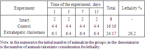
- The experiments were carried out on 48 outbred male rats of a mixed population with an initial weight of 180-200 g, kept in a laboratory diet in a vivarium. Extrahepatic cholestasis was reproduced in rats by ligation of the common bile duct [1]. The total lethality in this group was 29.%. The control was provided by false collated animals (16 rats), which was carried out only laparatomy in aseptic conditions. In these groups lethality was not observed. The intact group was 8 rats. The studies were carried out 1, 3, 7 and 15 days after the models were reproduced. The choice of terms of the study is related to the development of significant morpho-functional changes in the liver at experimental cholestasis [2]. An outline of the experience is presented in table 1.
|
4. The Results of the Research
- It is well known that the liver and kidneys have a close functional relationship, which is mainly expressed in the biotransformation of the endo- and xenobiotics, as well as eliminates of metabolites and and biological oxidation products. Issues related to changes in the peripheral blood circulation system of the kidneys in the event of various types of bile outflow are virtually unexplored. Studies of such a plan, in our view, would be of great practical importance from the point of view of preventing possible complications on the excretory activity of the kidneys.Renal circulation is unique in its structure, which allows each segment of the renal capillary bed to play its special functional role: filtration takes place in glomerulus; cortical peritoubular capillary network is specialized for reabsorption, medullary circulation provides the needs of the renal concentration mechanism [3]. When entering the kidney, the renal artery is divided into numerous branches, which initially go perpendicular to the kidney surface, then bending at the level of the cortico-mullar boundary and further follow the parallel surface. These arc arteries give rise to branches that pass through the cortical layer to the surface of the kidneys. The interlobular arteries are connected to the vascular loops of the glomerulus through arterial vessels, so-called afferent arterioles, and with peritubular capillaries through efferent vessels, usually having an arteriolar structure. This structure is typical of the kidneys of all mammals.Available for vital microscopy are the peritubular capillaries of the cortical layer of the kidneys. The only area in which an efferent vessel of the ball perfuses the canal of the same ball is located in the proximal duct of the surface cortical layer of the kidneys. Thus, the condition of the microcirculatory channel of the cortical part of the kidneys to some extent reflects the dynamics of changes in the glomeral capillary. In intact rats with luminescent biomicroscopy, the kidney tissue is presented greenish brown, parenchyma consists of proximal winding tubules and peritubular capillaries, which have a dark gray shade against the background of a whitish color of the epithelium tubules. There are normally single efferent arterioles and peritubular capillaries. The blood flow in the efferent arteries is rapid, jet, continuous flow. Peritubular capillaries departing from the arteriol, widely anastomosing among themselves form a dense network with polygonal cells, stretched along the canals (pic.1). The boundaries of the duct apparatus and capillaries are clearly delineated due to the whitish luminescent strips that appear when light is refracted by the tubule epithelium.
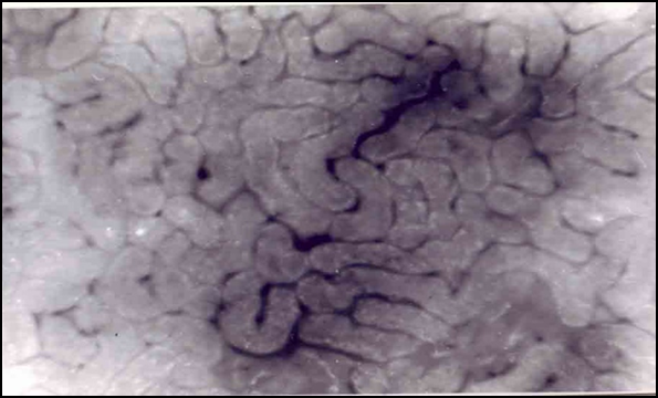 | Picture 1. Autofluorescence in the capillaries of the proximal convoluted tubules of the cortical layer of the kidney of intact animals magnification x 75 |
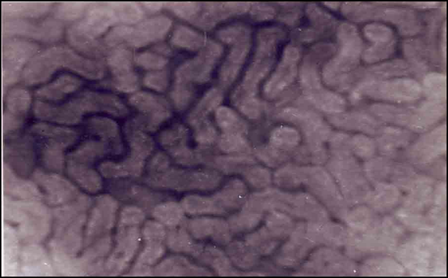 | Picture 2. Autofluorescence in the capillaries of the proximal convoluted tubules of the cortical layer of the kidney of control animals magnification x 75 |
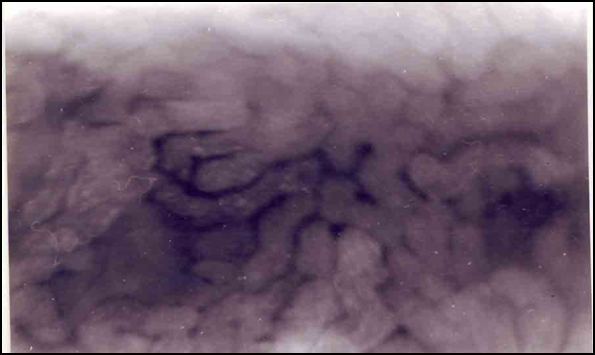 | Picture 3. Autofluorescence of the capillaries of the proximal convoluted tubules of the cortical layer of the kidney 1 day after ligation of the common bile duct magnification x 75 |
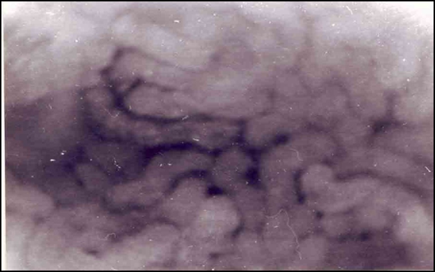 | Picture 4. Autofluorescence of the capillaries of the proximal convoluted tubules of the cortical layer of the kidney 3 days after ligation of the common bile duct magnification x 75 |
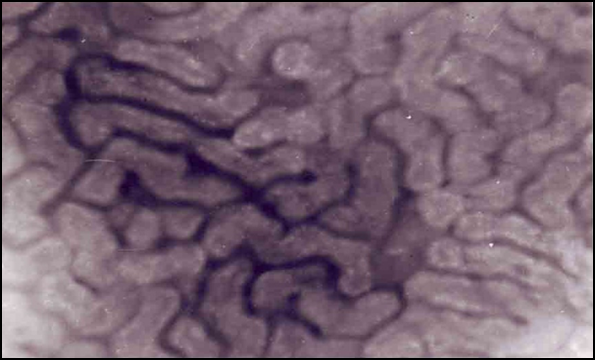 | Picture 5. Autofluorescence of the capillaries of the proximal convoluted tubules of the cortical layer of the kidney 7 days after ligation of the common bile duct magnification x 75 |
5. Сonclusions
- 1. Thus, studies have shown that microcirculation, and, therefore, kidney activity is involved in the circle of pathological changes in the development of cholestasis syndrome.2. The development of extrahepatic cholestasis disrupts systemic kidney microcirculation at almost all levels of the organization. Violations increase with the duration of cholestasis.
 Abstract
 Reference
 Full-Text PDF
 Full-text HTML