-
Paper Information
- Next Paper
- Previous Paper
- Paper Submission
-
Journal Information
- About This Journal
- Editorial Board
- Current Issue
- Archive
- Author Guidelines
- Contact Us
American Journal of Medicine and Medical Sciences
p-ISSN: 2165-901X e-ISSN: 2165-9036
2023; 13(4): 515-520
doi:10.5923/j.ajmms.20231304.35
Received: Apr. 8, 2023; Accepted: Apr. 23, 2023; Published: Apr. 26, 2023

Morphometric Assessment of the Reparative Regeneration Process in the Place of the Bone Tissue Defect of the Diaphyseal Part of the Greater Tibia of Animals under the Conditions of Hypo- and Hypercalcemia in the Experiment
M. Kh. Rakhmatova1, A. Makhmurov2
1Researcher, Tashkent International University of Chemistry, Uzbekistan
2Researcher, Tashkent State Dental Institute, Uzbekistan
Copyright © 2023 The Author(s). Published by Scientific & Academic Publishing.
This work is licensed under the Creative Commons Attribution International License (CC BY).
http://creativecommons.org/licenses/by/4.0/

Today, ideas about reparative regeneration are inextricably linked with advances in regenerative medicine. In the development of solutions to the problems of bone tissue regeneration, the study of the mechanisms of action of factors that optimize osteogenesis, stimulate differentiation and proliferation of bone cells continues. Development of mechanisms for targeted acceleration of bone tissue reparative regeneration, identification of bone tissue-forming cell sources, increase of osteoblast proliferative activity, morphological analysis of endocrine control mechanisms of bone tissue reparative regeneration is considered one of the most important medical and biological issues today [1,2].
Keywords: Vh, Vl-h, Vf-n, Vb-n, Va-n, Practical traumatology, Morphological characteristics, Post-treatment complications, Blood calcium concentration, Hypocalcemia
Cite this paper: M. Kh. Rakhmatova, A. Makhmurov, Morphometric Assessment of the Reparative Regeneration Process in the Place of the Bone Tissue Defect of the Diaphyseal Part of the Greater Tibia of Animals under the Conditions of Hypo- and Hypercalcemia in the Experiment, American Journal of Medicine and Medical Sciences, Vol. 13 No. 4, 2023, pp. 515-520. doi: 10.5923/j.ajmms.20231304.35.
1. Introduction
- In recent decades, fractures of human long bones account for 80% of all bone fractures. Despite the use of modern medical technologies in the treatment of bone fractures, the percentage of post-treatment complications remains high. Applying the results of experimental research to practical traumatology and orthopedics, allows clinicians to manage the process of reparative regeneration of bone tissue after injury, analyze it and choose the right rational method of treatment, as well as predict the patient’s condition after treatment [5]. Accordingly, the morphological characteristics of the types of reparative regeneration of bone tissue of the diaphyseal part of the tibia after the creation of an artificial defect as a result of the hypofunction of the thyroid and parathyroid glands in the conditions of hypo- and hypercalcemia have not been determined. In scientific sources, a lot of information is given about the endocrine management of bone tissue, the importance of calcium, but it turned out that they are mainly biochemical in nature [3,6,7].Therefore, the purpose of the study is to determine the characteristics of the relationship between the structural changes in the reparative regeneration of the bone tissue of the thyroid gland, parathyroid glands and tubular bone diaphysis under conditions of hypo- and hypercalcemia.
2. Materials and Methods of Research
- The experiments were conducted on 42 adult Chinchilla rabbits with a body weight of 2.0-2.5 kg. All rabbits were divided into 3 groups: 1st control group with normal blood calcium concentration (n= 6), and 2nd experimental group with low (hypocalcemia, n= 18) and high (hypercalcemia, n= 18) calcium concentration. In all groups, the diaphyseal part of the tibia formed a defect in the bone tissue, and the morphological characteristics of the reparative regeneration process were studied. Animals were euthanized 3, 7, 14, 21 and 30 days after the creation of the defect in accordance with the European Convention for the Protection of Animals Used in Scientific Research [4]. In all groups, pieces of the thyroid gland and parathyroid glands, as well as the greater tibia, were taken for morphological studies. Serum hypocalcemia was induced by intraperitoneal injection of a 2.5% aqueous solution of ethylenetetradiamine-tetra-acetic acid sodium salt (EDTA; 1.0 ml per 100 g animal weight). Hypercalcemia, on the other hand, was achieved by intraperitoneal injection of 10% calcium gluconate (1.0 ml per 100 g of body weight). Serum calcium concentration was determined using an atomic absorption spectrophotometer (Beckman, Belgium). The defect of bone tissue in the area of the tibia was formed by the method of A.I. Volotovsky [3]. After all animals were examined by a veterinary specialist and found to be disease-free, local anesthesia was performed with 0.5% novocaine solution, and an incision was made in the skin and subcutaneous tissue in the middle third of the greater tibia. With the help of a sharp scalpel, two long 1.0 cm and two transverse incisions connecting them were made at a distance of 0.5 cm from each other. After removing the periosteum, an endosteal bone tissue defect was created in the bone with a bur machine measuring 0.5 cm in width and 1.0 cm in length. The sutures were removed 8-9 days after the creation of the bone defect. No complications were observed after surgery. Bone fragments taken from the defect area of the diaphyseal part of the tibia were first fixed in alcohol, then decalcified in an alcoholic solution of nitric acid. For preparation of histological preparations, decalcified bone fragments were embedded in paraffin. Sections taken with a microtome were stained with hematoxylin-eosin. Micropreparations were studied under a light microscope “OPTIKA” (Italy).Morphological changes in the process of reparative regeneration developed at the site of a bone defect were calculated using the morphometric method of G. G. Avtondilov’s “multi-point test” (160 points), where the points corresponding to each structural unit were counted in a x10 objective and the percentage of the total number of points was calculated. All results were processed using conventional variance statistical methods. For this purpose, the average arithmetic magnitude (M), average arithmetic error (μ), and relative indicators (%) were calculated. Significance of differences was determined by the Fisher-Student test (P). Statistical processing was performed on a Pentium IV processor-based personal computer using a software package for medical and biological research.
3. Research Results
- The following results were obtained in the morphometric study of reparative regeneration of bone tissue under different conditions at the site of an artificially formed defect in the diaphysis of the tibia. In the bone defect of control group animals, on the 3rd day of the experiment, lymphohistiocytic infiltration in the inflammatory focus was 8.7±1.4%, on the 7th day it increased to 17.5±1.9%. From the 14th day, this indicator decreased by 10.0±1.5%, by the 30th day by 7.5±1.3%. The area occupied by fibro-reticular tissue occupied 13.1±1.6% of the area on the 3rd day of the experiment, and by the 7th day, this indicator increased by 4 times, i.e. 46.2±2.4%, on the 14th day of the experiment, the proportion of osteoid tissue due to the increase, it was observed that the area occupied by fibro-reticular tissue decreased by 35.0±2.2% and by 31.2±2.3% on the 30th day. Around the bone defect, new bone columns were detected from the 7th day of the experiment, the area occupied by them was 15.6±1.8%. By the 14th day, it was observed that the area of the new bone columns increased sharply, i.e. it was 51.3±2.4%, and by the 30th day it was 61.2±2.4% (table 1, diagram 1).
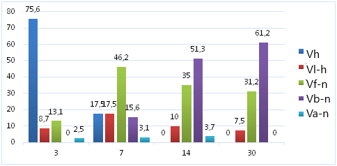 | Diagram 1. The relative area occupied by the tissue structural units in the focus of reparative regeneration developed in the bone defect of the control group |
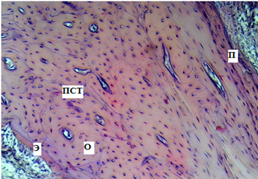 | Figure 1. Control rabbit tibia. 30th day. P- periosteum; E- endosteum; PST- plate-like bone tissue; O- osteons. The paint is GE. Size x100 |
|
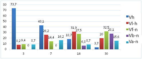 | Diagram 2. Relative area occupied by tissue structural units in the center of reparative regeneration developed in the hypocalcemic group bone defect |
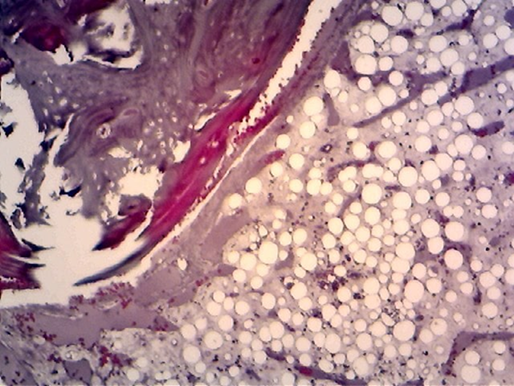 | Figure 2. Experimental hypocalcemia. 7 days. Adipose tissue accumulated around the bony defect. The paint is GE. Size x100 |
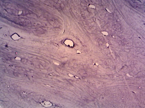 | Figure 3. Experimental hypocalcemia. 14th day. Bone defect area. Formation of fibrotic tissue. The paint is GE. Size x100 |
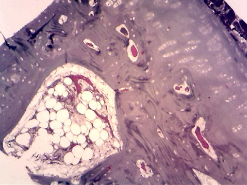 | Figure 4. Experimental hypercalcemia. 14th day. Bone defect area. Formation of fibrotic tissue. The paint is GE. Size x100 |
|
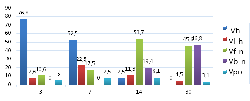 | Diagram 3. Relative area occupied by tissue structural units in the focus of reparative regeneration developed in the bone defect of the experimental group under conditions of hypercalcemia |
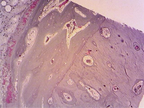 | Figure 5. Experimental hypercalcemia. Day 30. Bone defect area. Formation of osteoid tissue. The paint is GE. Size x100 |
4. Conclusions
- 1. In the experiment, after an artificial defect was induced in the bone tissue of the diaphyseal area of the tibia in the animals, the reparative regeneration in the animals of the control group had an adaptive character, and the morphological signs of complete reparative regeneration were revealed step by step in the dynamics of the experiment. In this case, the structure of the bone tissue around the defect approached its primary structure. 2. In the experiment, it was found that the artificial defect in the bone tissue of the diaphyseal area of the tibia in hypocalcemic animals had an incomplete type of reparative regeneration, that is, it had a secondary character. In this case, the development of fibro-reticular and then bone tissue was observed instead of adipose tissue in the defect area. It was found that the bone tissue was formed late and less than the fibro-reticular tissue. 3. In the experiment, it was found that the artificial defect in the bone tissue of the diaphyseal area of the tibia in hypercalcemia was incomplete type of reparative regeneration, that is, it had a secondary character. However, in this condition, the development of fibro-reticular tissue, then fibro-reticular tissue was observed at the site of the defect. In this case, it was also found that the ossification period was delayed and the osteoid tissue occupied half the area of the bone pack.
References
| [1] | Avrunin A.S., Parshin L.K. A hierarchically organized model of the relationship between cellular and tissue mechanisms of calcium exchange between bone and blood // Morphology. -2013. -T. 143, No. 1. -WITH. 76-84. |
| [2] | Brusko A.T., Gaiko G.V. Modern ideas about the stages of reparative regeneration of bone tissue in fractures // Visnik of orthopedics, traumatology and prosthetics. -2014. -№2.- P.5-8. |
| [3] | Volotovsky A.I., Makarevich E.R., Chirak V.E. Regulation of bone tissue in health and disease (Methodological recommendations) Minsk BSMU, 2010. - C. 45-54. |
| [4] | European Convention for the Protection of Vertebrate Animals Used for Experimental and Other Scientific Purposes. // Vop. Reconstructive and Plastic Surgery. -2003. -№4. -S.34-36. |
| [5] | Shilin V.A., Safronov A.A., Kozhanova T.G. Stimulation of reparative osteogenesis in an experiment on a false joint model in rats // Bulletin of the Orenburg State University. -2015. -№3/178. - P.218-222. |
| [6] | Yadykina T.K., Mikhailova N.I., Bugaeva M.S. Experimental studies of bone tissue metabolism and mechanisms of regulation of mineral homeostasis in the dynamics of the development of toxic fluoride osteopathy // Bulletin of the East Siberian Scientific Center of the Siberian Branch of the Russian Academy of Medical Sciences. -2018. -T.17, No. 1. - P.45-51. |
| [7] | Bellido T. Osteocyte-driven bone remodeling. // Calcif Tissue Int. 2014 – Vol. 94(1) –Doi: 10.1007/s00223-013-9774-y. pp. 25-34. |
 Abstract
 Reference
 Full-Text PDF
 Full-text HTML

