-
Paper Information
- Next Paper
- Previous Paper
- Paper Submission
-
Journal Information
- About This Journal
- Editorial Board
- Current Issue
- Archive
- Author Guidelines
- Contact Us
American Journal of Medicine and Medical Sciences
p-ISSN: 2165-901X e-ISSN: 2165-9036
2023; 13(4): 511-514
doi:10.5923/j.ajmms.20231304.34
Received: Apr. 12, 2023; Accepted: Apr. 24, 2023; Published: Apr. 26, 2023

Analysis of Pathomorphological and Morphometric Changes in Indices of Uterine Myoma
Alibekov O. O.1, Israilov R. I.2, Mamataliev A. R.3
1Senior Lecturer, Andijan State Medical Institute, Uzbekistan
2Doctor of Medical Sciences, Director of the Republican Pathoanatomical Center of the Ministry of Health of the Republic of Uzbekistan
3Candidate of Medical Sciences, Associate Professor, Head of the Department, Andijan State Medical Institute, Uzbekistan
Copyright © 2023 The Author(s). Published by Scientific & Academic Publishing.
This work is licensed under the Creative Commons Attribution International License (CC BY).
http://creativecommons.org/licenses/by/4.0/

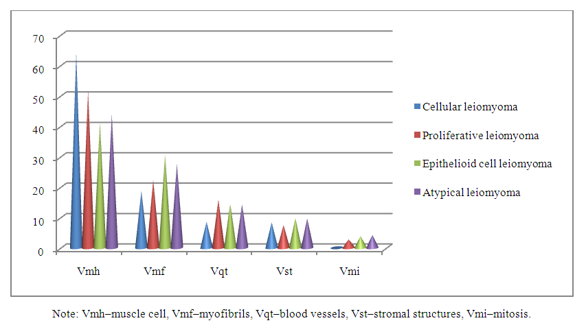
With the help of a dotted grid G.G. Avtandilov made morphometry of various histological forms of uterine fibroids. At the same time, it was noted that the highest indicators of smooth muscle cells in cellular myoma, myofibrils in the epitheloid form, mitoses in the atypical form.
Keywords: Cellular myoma, Proliferative myoma, Epithelioid cell myoma, Atypical myoma, Morphometry, Smooth muscle cells, Vessels, Mitoses
Cite this paper: Alibekov O. O., Israilov R. I., Mamataliev A. R., Analysis of Pathomorphological and Morphometric Changes in Indices of Uterine Myoma, American Journal of Medicine and Medical Sciences, Vol. 13 No. 4, 2023, pp. 511-514. doi: 10.5923/j.ajmms.20231304.34.
1. Relevance
- Uterine fibroids are benign tumors of the uterus, consisting of muscle (phenotypically altered smooth muscle cells of the myometrium) and connective tissue, a hormone–dependent disease and a type of hyperplastic processes of the uterus. In recent years, there has been a trend of “rejuvenation” of uterine fibroids, so the number of young women with the disease at the age of 20–25 years is increasing [1,2,3,5].Research goal: proliferation leiomyocytes based on morphometric studies of various histological forms of uterine fibroids, study the relationship between parenchyma and stroma.
2. Materials and Methods
- In this study, 50 hysterectomies performed during 2019–2022 were analyzed by general morphological and morphometric analysis, and 5 uterine fibroids amputated for other reasons.When conducting morphometric studies, we modified the point study method by transferring it to a computer screen, that is, we took 10 pictures of histological preparations prepared for each group of the material under study from different areas and applied a linear grid of 200 cells on a computer monitor corresponding to these pictures, and we the points of intersection of the lines were counted depending on what tissue structure they correspond to. The fact that the grid nodes G.G. Avtandilova are distributed unevenly over all areas of the surface of the painting of the fabric, which corresponds to the law of relativity. The area of all existing structural units in the picture is taken as Vv, i.e. for 100%, the area of each calculated structural unit is specified by putting the name of this structure, for example: Vmh (muscle cell), Vmf (myofibrils), Vqt (blood vessels), Vst (stromal structures and Vmi (mitosis). In this regard, in As a result of counting the points, the relative area of the studied structural units in the tissue was calculated. The area occupied by all structural units in the studied tissue is equal to Vv, i.e. if it is equal to 100%, then the points evenly distributed in it are denoted by z, and if the ratio of each point to the structural unit is taken as R, then its formula is expressed as follows: P=Vv/100.Correspondence of points to other structural subdivisions is determined by the following formula: Q=100–Vv/100.If we take the points corresponding to the structural units under study as x, then its error coefficient is calculated by the formula: x / z – P, the percentage of absolute error is calculated by the following formula: e \u003d (x / z – R ). 100 \u003d 100 x / z – Vv.The calculation error according to the theory of relativity – x / z – R, is calculated using a different formula: \u003d t. √Rq/z.In this formula: x is the number of points corresponding to the studied structural units; z is the total number of all points in the test system; R is the unit of relativity of the points falling on the structures under study; q is the unit of relativity of points that fall into other structural units; t is the normalized difference between the indicators.Based on the foregoing, the absolute error of quantitative indicators is calculated by the following formula: e= t √Vv (100 – Vv)/z.Using the morphometric method, G.G. Avtandilova “counting points – a test system” counted uterine leiomyoma in the following histogenetic forms by dividing the composition of cells and tissue structures into the following groups: 1) cellular leiomyoma; 2) proliferative leiomyoma; 3) epithelioid cell leiomyoma; 4) atypical leiomyoma (BSST, 2003) [4]. These histogenetic forms of uterine myometrial leiomyoma were isolated and isolated by microscopic examination of preparations stained with hematoxylin and eosin. There were an average of 10 points from each group.The area occupied by the following tissue and cellular structures corresponding to all histogenetic forms of leiomyoma was calculated in %: smooth muscle cells –Rmh; myofibrils – Rmf; blood vessels – Rqt; stroma structures – Rst; mitosis – Rmi.Each structural unit was added 10 points, numbered in the figure, the average value was calculated and the area occupied by the structural unit (V) was calculated using the following formula, for example: in cellular leiomyoma, the area occupied by the cell muscle was calculated using the following formula – Vmh = Rmh / R x. In this regard 1) cellular leiomyoma; 2) proliferative leiomyoma; 3) epithelioid cell leiomyoma; 4) in histogenic forms of atypical leiomyoma, the area occupied by all cellular and tissue structures was calculated: Vmh, Vmf, Vqt, Vst, Vmi. The obtained digital data were subjected to statistical processing.
3. Results and Discussions
- The most common histogenetic form of leiomyoma, the area occupied by smooth muscle cells in the tissue of simple fibroids, had the highest rate and averaged 64.3%. It was confirmed that it makes up 3/2 of the entire area of leiomyoma tissue, and it was found that the main place in the tissue of this histogenetic form of leiomyoma is occupied by smooth muscle cells (Table 1). It was determined that the area occupied by myofibrils of smooth muscle cells is 3.4 times less than the area of cells. So, with this leiomyoma, smooth muscle cells proliferated and the reproduction process was confirmed. It has been established that blood vessels supplying smooth muscle cells consist of small capillaries and their area is much smaller; equals 8.6%. In the simple form of this leiomyoma, there were no structural changes indicative of mitosis, which are characteristic of the mitotic process of cells.
|
 | Diagram 1. Morphometric Indicators of Histogenetic Forms of Uterine Leiomyoma (%) |
4. Conclusions
- It has been observed that the area occupied by smooth muscle cells in leiomyoma tissue is highest in cellular form, followed by proliferative form, and relatively less in epithelioid cell form. It has been established that the area occupied by muscle tissue myofibrils is the smallest in cellular leiomyoma and relatively large in the epithelioid–cellular form. It has been confirmed that the vessels occupy a smaller area in cellular leiomyoma and the same extent in other forms. It was found that mitoses account for 2.8% in proliferative leiomyoma, 3.8% in epithelioid cell form and 4.2% in atypical form.
 Abstract
 Reference
 Full-Text PDF
 Full-text HTML