-
Paper Information
- Next Paper
- Previous Paper
- Paper Submission
-
Journal Information
- About This Journal
- Editorial Board
- Current Issue
- Archive
- Author Guidelines
- Contact Us
American Journal of Medicine and Medical Sciences
p-ISSN: 2165-901X e-ISSN: 2165-9036
2023; 13(4): 479-482
doi:10.5923/j.ajmms.20231304.26
Received: Apr. 8, 2023; Accepted: Apr. 20, 2023; Published: Apr. 22, 2023

Expression Levels of the Antiapoptosis Protein Bcl-2 in Women's Urethral Polyps
Boboev R. A., Qosimhojiev M. I.
Andijan State Medical Institute, Andijan, Uzbekistan
Copyright © 2023 The Author(s). Published by Scientific & Academic Publishing.
This work is licensed under the Creative Commons Attribution International License (CC BY).
http://creativecommons.org/licenses/by/4.0/

Levels of expression of the antiapoptosis protein Bcl-2 have been found in women's urethral polyps. The results showed that in the urethral covering epithelium in the control group, this protein was found to be expressed only at a low level in the basal floor. During the onset of the metaplastic process in the variable epithelium during the initial period of the polyp, it was observed that Bcl-2 protein expression was elevated in the cells of the basal floor of the epithelium where acanthosis developed. During the formation of the polyp, it was observed that all basement cells of the epithelium metaplasia and were located vertically, with relatively greater levels of expression of Bcl-2 protein in their basal and intermediate basement cells.
Keywords: Women, Urethra, Polyp, Leukoplakia, Immunogystochemistry, Bcl-2 protein
Cite this paper: Boboev R. A., Qosimhojiev M. I., Expression Levels of the Antiapoptosis Protein Bcl-2 in Women's Urethral Polyps, American Journal of Medicine and Medical Sciences, Vol. 13 No. 4, 2023, pp. 479-482. doi: 10.5923/j.ajmms.20231304.26.
Article Outline
1. Introduction
- Bcl-2, located on human chromosome 18, which has antiapoptosis from proteins 16, is a homologous protein that slows down the process of domain 6 apoptosis. This protein, with a molecular weight of 22 KDA, is located in the cell and nuclear membranes, sarcoplasmic and mitochondrial membranes [2,4,6,8]. Hyperexpression of this protein prevents the release of calcium ions and slows lipoperexidation by inhibiting antioxidant activity as well as slowing down non-centetase activity. The primary function of Bcl-2 prevents cytochrome s, AIF, ATP, which are antiapoptose molecules in mitochondria, from exiting through bribery holes. Why Bcl-2 closes bribe holes by adhering to the membrane of the mitochondria, proapoptosis disconnects signals and apoptosis does not develop [1,3,5,7]. Urethral polyps are a relatively common safe tumor of the women's peshob release tube and affects the zinc status of pasints, altering the quality of the style of the ghost. Polyps are in most cases localized in the area of the external excretory opening of the urethra, clinically causing aching, thirst when the forehead comes, pollacuria, stranguria, urethrorrhagia and urine dimming. The causes of urethral polyps have been little studied and remain unclear. According to most scientific studies, polyps of the urethra are a hyperplastic process, which, in most cases, is determined by proliferation of the urethral wall with mesenchymal tissues and epithelium due to a violation of their interaction for unknown reasons, without being considered a safe tumor. As the cause of this process, dysgormonal changes in the epithelium of the urogenital area in women and inflammatory processes of chronic infection confirm as significant. The results of immunogystochemical examination of the antiapoptosis protein in its enveloping epithelium in the women's urethral polyp provide assistance that has determined the morphogenesis and developmental cycles of the disease. The purpose of the study. Determination of the degree of expression of the antiapoptosis protein BCL-2 in the polyps of women's urethra.
2. Material and Methods
- As material, 32 biopsy material from patients who underwent treatment in 2018-2021 was histologically studied in the Urology Department of the multidisciplinary Hospital of the Andijan region. The biopsy flakes were dehydrated in alcohols with increased concentration and paraffin was poured and the bricks were prepared after they had dried in a 10% solution of formalin for 48 hours. Histological cuts of 4-5 µm were made from paraffin bricks, painted in hematocilin-eosin, van-Gison Paints, and an immunogystochemical examination of the BCL-2 marker was carried out, the light was seen under a microscope, the kelaky areas were photographed.
3. Research Results and Their Discussion
- To determine the pathomorphological and immunogistochemical changes that occur in the lining epithelium of women's urethra, human-derived biopsy material with no pathology in the urethra was initially studied as a control group. Then, patomorphological and immunogistochemical changes in the epithelium covering the surface of the urethral polyp were studied in comparison with each other in terms of the clinical-morphological forms and developmental periods of the urethral polyp.Control group women it is determined that the lining epithelium of the urethral mucosa consists of the usual multilayered variable epithelium, and that its epithelial cells located on the basal floor are located relatively large, hyperchromic, lined up on the basal membrane, most of their nuclei are Aval and oblong in shape. In the surface folds of the multilayered epithelium, it is observed that the cells are relatively thinned, the nuclei are both reduced in size and the staining is opened, the location is flattened. The results of an immunogystochemical examination of the epithelial cell's antiapoptosis protein show that in the control group, expression of this protein is observed at very low levels in the cytoplasm of certain cells of relatively young and cambial levels located only on the basal membrane, and in cells of other intermediate and surface layers (Figure 1-2).
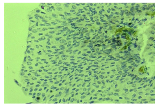 | Figure 1. Urethral, Meyor, Bcl-2 protein is low in some cells of the basal floor. Paint: immunogystochemistry. Size: 10x40 |
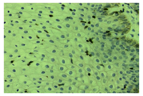 | Figure 2. Polyp, initial period, Bcl-2 protein is eskpressed in some cells of the basal floor and intermediate floor. Paint: immunogystochemistry. Қшяу: 10x40 |
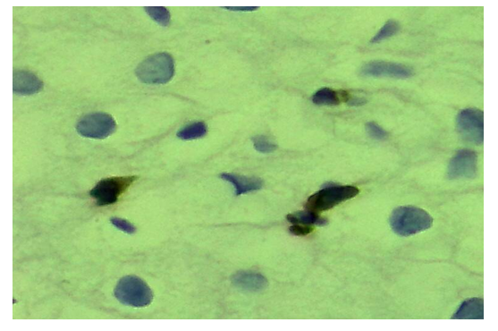 | Figure 3. Urethral polyps, Bcl-2 protein intermediate floor epithelial cells are expressed close to the nucleus. Paint: immunogystochemistry. Size: 10x100 |
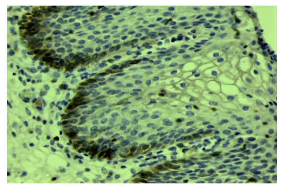 | Figure 4. Urethral polyps, BCL-2 basal floor expressed in 2-3 rows. Paint: immunogystochemistry. Size: 10х40 |
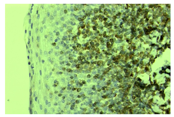 | Figure 5. Urethral polyps, Bcl-2 are expressed in most cells of the basal and intermediate floors. Paint: immunogystochemistry. Size: 10х40 |
4. Conclusions
- Immunogystochemical study of urethral polyps, which determine in which layers of the enveloping multilayer variable epithelium the Antiapoptosis Bcl-2 protein is expressed, is an important factor in the diagnosis of this disease. In a control group with no urethral disease, a low level of expression of Bcl-2 protein only in the basal floor indicates that apoptosis activity is maintained. Early urethral polyps, during the onset of the metaplastic process in the variable epithelium, expression of the Bcl-2 protein in the cells of the basal floor of the epithelium where acanthosis has developed suggests that the antiapoptosis gene is activated. During the secondary period of polyp development, it was found that all floor cells of the epithelium were metaplased and located vertically, with the Bcl-2 protein expressed to a relatively greater extent in their basal and intermediate floor cells.During the differentiated period of urethral polyps, it is observed that the cells of all layers of the epithelium develop proliferative activity and metaplasia, the presence of inflammation in their private plate, high expression of Bcl-2 protein in all epithelial cells.
 Abstract
 Reference
 Full-Text PDF
 Full-text HTML