Shomirov D. A., Nadirxonova N. S.
Republican Specialized Scientific and Practical Medical Center for Obstetrics and Gynecology, Uzbekistan
Copyright © 2023 The Author(s). Published by Scientific & Academic Publishing.
This work is licensed under the Creative Commons Attribution International License (CC BY).
http://creativecommons.org/licenses/by/4.0/

Abstract
The article studied the state of the content of autoantibodies to native class G DNA in the blood serum of women with genital prolapse after hysterectomy. 89 women aged 26 to 64 were examined. Among 89 patients, the main group consisted of 52 women with GPPGE, and the control group - 37 healthy women without GPPGE. All patients underwent clinical and instrumental, functional, cytological and immunological studies.
Keywords:
Genital prolapse, Autoantibodies, Immunoglobulin G, DNA-DS
Cite this paper: Shomirov D. A., Nadirxonova N. S., Autoimmune Aspects of Genital Prolapse after Hysterectomy, American Journal of Medicine and Medical Sciences, Vol. 13 No. 4, 2023, pp. 456-458. doi: 10.5923/j.ajmms.20231304.20.
1. Introduction
According to scientific research, in the pathogenesis of the development of genital prolapse after hysterectomy (GPHE), it is more likely that the connective tissue of the body will change under the influence of endo- and exogenous factors in the presence of genetic features of the immune system [1,4,5,8].In the mechanism of development of pathological processes in many diseases, autoimmune processes are of great importance, which are the main component of the development of a number of systemic diseases, including collagenases, structural formations of connective tissue, metabolic syndrome, etc [3-10].At the same time, the autoimmune mechanism for the development of GPHE becomes important, in which the immune system attacks normal, healthy tissues of its own body, that is, the body loses tolerance to its own tissue antigens. A common feature of most autoimmune diseases is B-cell activation, leading to hyperglobulinemia and excessive production of autoantibodies (AAB) with different specificity [3,4,7,11].With the increase in the number of hysterectomies around the world, including in Uzbekistan, the number of patients with GPHE tends to increase, which requires close attention and study from all sides.The purpose of the research. To study the state of the content of autoantibodies to native DNA of class G in the blood serum of women with GPHE.
2. Material and Research Methods
89 women aged 26 to 64 were examined. Among 89 patients, the main group consisted of 52 women with GPHE, and the control group included 37 healthy women without GPHE. All patients underwent clinical and instrumental, functional, cytological and immunological studies. The level of autoantibodies (AAB) of class G (IgG) to native single-stranded (DNA-SS) and double-stranded (DNA-DS) DNA in blood serum was determined by the method of solid-phase ELISA - research (company "Vector-Best").
3. Results
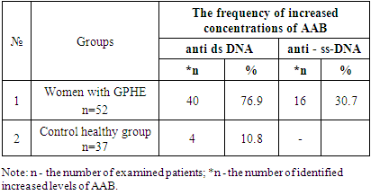
The results of an ELISA research on the detection of AAB in patients with GPHE showed that among 52 women of the main group, the blood serum of 40 patients showed an increase in the level of AAB to native double-stranded DNA, which accounted for 76.9% of cases. While single-stranded DNA was detected in 16 patients, which accounted for 30.7% of cases. In the group of healthy individuals, among 37 individuals, only 4 had an increased level of AAB of class G to DNA-DS, which accounted for 10.8% of cases. (Table 1).Table 1. The frequency of detection of IgG antibodies to double- (anti ds DNA)- and single-stranded DNA (anti - ss-DNA) in the blood serum of patients with GPHE (abs.%)
 |
| |
|
The results obtained indicate the presence of an autoimmune process in patients with GPHE. Analysis of the quantitative characteristics of autoantibodies (AAB) to native DNA in the blood serum of women with GPHE revealed an increase in the concentration of autoimmune antibodies of class G autoimmune antibodies to double-stranded AAT - DNK -DS by 3.9 times compared with the control group and averaged 38.3± 4.1 IU/ml and was statistically significant. (P<0.05). Whereas the concentration of AAB of class G to single-stranded DNK - SS averaged 9.1 ± 2.4 IU / ml, which was 1.6 times higher than in control healthy individuals and tended to increase, however, the indicators were not statistically significant. (P>0.05).Table 2. Indicators of denatured antibodies DNK-DS et DNK-SS in women with GPHE (M+m)
 |
| |
|
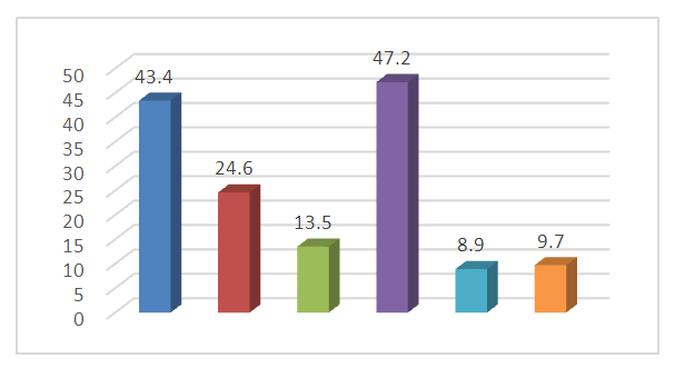
Interesting data showed the state of AAT depending on the age of the patients. With an increase in age from 51 to 55 years in 11 patients, the concentration of AAB of class G to native double-stranded DNA-DS averaged 51.7 + 7.2 IU / ml, while at the age of 40-50 years in 7 women it was on average - 21+2.1 IU/ml, which is 5.3 and 2.2 times higher than in control healthy individuals. The data obtained were statistically significant (P<0.05).A high concentration of AAB to DNA -DS, in our opinion, is associated with background diseases in the body of women. (Fig.1) | Figure 1. Indicators of concomitant diseases in patients with AAT to DNA-DS (%) |
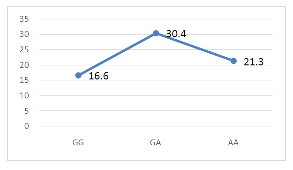
As follows from the figure, women with an increased concentration of DNA - DS were most often diagnosed with varicose veins, in 47.2% of cases, urinary tract infections in 43.4%, hypertension in 24.6%, diabetes mellitus 13.5% and liver disease in 9.7% of cases, respectively.The results of the ELISA study were analyzed taking into account the association of polymorphism of the genotypes of the IL-23 gene (rs11209026) in the examined women with GPHE. (Fig.2.) | Figure 2. DNA-DS indicators in women with GPHE with associations of IL-23 gene genotype polymorphisms (IU/mL) |
As can be seen from the figure, in women with associations of polymorphism of the IL-23 gene genotypes with favorable GG genotypes, the level of DNA-DS averaged 16.6 + 6.7 IU / ml, with heterozygous GA genotypes - 30.4 + 3.1 IU /ml, while in mutant homozygous genotypes of the IL-23 AA gene, the level of DNA - DS averaged -21.3 + 6.2 IU / ml, respectively. The level of AAB to DNA-DS was 1.7, 3.1 and 2.2 times higher than in healthy women, which was statistically significant. (P<0.05)The analysis of the data obtained indicates that the concentration of DNA -DS increased most in women with a heterozygous variant of the association of polymorphism of the IL-23 gene compared with favorable indicators - by 1.8 and mutant homozygous - by 1.4 times of variants of the IL-23 gene genotypes respectively.Thus, the analysis of the obtained results shows that in women with GPHE, there is a high detection of AAB of class G to native double-stranded DNA in 76.9% of cases, whereas to single-stranded DNA -SS in 30.7% of cases. Analysis of the quantitative characteristics of autoantibodies (AAB) to native DNA in the blood serum of women with GPHE revealed an increase in the concentration of autoimmune antibodies of class G to double-stranded AAT - DNK -DS by 3.9 times compared with the control group and averaged 38.3± 4.1 IU/ml and was statistically significant. (P<0.05).Taking into account the association of IL23 gene polymorphism among 40 women with heterozygous G/A variant, 18 patients showed an increase in the concentration of DNA - DS, which accounted for 45% of cases, and the level of DNA-DS averaged 30.4 + 3.1 IU / ml (P<0.05).
4. Conclusions
1. In women with GPHE, in 76.9% of cases, the detection of AAB to native double-stranded DNA-DS with a high concentration by 3.9 times compared with the control group and averaged 38.3 ± 4.1 IU / ml and was statistically significant. (P<0.05), which indicated the development of an autoimmune process in the body.2. Women with AAB to DNA-DS were most often diagnosed with varicose veins (varicocele) in 47.2% of cases, urinary tract infections in 43.4%, hypertension in 24.6%, diabetes mellitus 13.5% respectively.3. An increase in the concentration of autoantibodies to native double-stranded DNA-DS was most often observed in women with a heterozygous G/A variant of the IL-23 gene, which accounted for 45% of cases.4. In women with associations of polymorphism of IL-23 gene genotypes with favorable G/G genotypes, the level of DNA-DS averaged 16.6+6.7 IU/ml, with heterozygous G/A genotypes - 30.4+3.1 IU/ml, while in mutant homozygous genotypes of the IL-23 A/A gene, the level of DNA - DS averaged -21.3+6.2 IU/ml, respectively. The level of AAB to DNA-DS was 1.7, 3.1 and 2.2 times higher than in healthy women, which was statistically significant. (P<0.05)5. In our opinion, the data obtained are of diagnostic and prognostic value in the clinical course of GPHE, and the determination of the AAB titre of class G will contribute to the further choice of individual treatment.
References
| [1] | Rapp, D.E. Comprehensive evaluation of anterior elevate system for the treatment of anterior and apical pelvic floor descent: 2-year followup / D.E. Rapp, A.B. King, B. Rowe, J.P. Wolters // J Urol. – 2014. – Vol. 191(2). – pp. 389-394. |
| [2] | Reynolds, S. et al. Immediate effects of the initial FDA notification on the use of surgical mesh for pelvic organ prolapse surgery in medicare beneficiaries / W.S. Reynolds, K.P. Gold, S Ni, M.R. Kaufman, R.R. Dmochowski, D.F. Penson // Neurourol Urodyn. - Apr 2013. – Vol. 32(4). – pp. 330-335. |
| [3] | Rusina, E.I. Lower urinary tract dysfunction in women with pelvic organ prolapse: diagnostic problems // Journal of Obstetrics and Women’s Diseases. – 2018. – Vol. 67 (4). – pp. 4-12 (in Russian). |
| [4] | Shkarupa D.D. Combined pelvic floor repair on levels I and II support defects: Posterior intravaginal sling and subfascial colporrhaphy/ D.D. Shkarupa, N.D. Kubin, E.A. Shapovalova, A.O. Zaytseva, A.V. Pisarev // Akusherstvo i Ginekologiya. – 2016. – Vol. 8. pp. 99-105 (in Russian). |
| [5] | Shkarupa D.D. Patent 2661042 RU. S1 MPK A61B 17/00, A61B 17/42, A61F 2/02. The method of surgical reconstruction of the pelvic floor / D.D. Shkarupa, N.D. Kubin, E.A. Shapovalova, A.O. Zaytseva. - № 2017119821; request 06.06.17; published 11.07.18. – Bulletin № 20 (in Russian). |
| [6] | Su, T.H. Single- incision mesh repair versus traditional native tissue repair for pelvic organ prolapse: results of a cohort study / T.H. Su, H.H. Lau, W.C. Huang, C.H. Hsieh, R.C. Chang, C.H. Su // Int Urogynecol J. – 2014. – Vol. 25(7). – pp. 901–908. |
| [7] | Thomas V., Shek C., Guzman Rojas R.A. et al. The latency between pelvic floor trauma and presentation for prolapse surgery. Ultrasound Obstet.Gynecol. – 2013; 42 (S1): 39. |
| [8] | Trutnovsky, G. Pelvic floor dysfunction - does menopause duration matter? / G. Trutnovsky, R. Guzman-Rojas, A. Martin, H.P. Dietz // Maturitas. – 2013. – Vol. 76(2). pp. 134-138. |
| [9] | Utomo, E. Validation of the Pelvic Floor Distress Inventory (PFDI-20) and Pelvic Floor Impact Questionnaire (PFIQ-7) in a Dutch population / E. Utomo, B.F. Blok, A.B. Steensma, I.J. Korfage. // Int Urogynecol J. – 2014. – Vol. 25(4). – pp. 531-44. |
| [10] | Vitale, S.G. Transvaginal bilateral sacrospinous fixation after second recurrence of vaginal vault prolapse: efficacy and impact on quality of life and sexuality / S.G. Vitale, A.S. Lagana, M. Noventa // Biomed Res Int. – 2018. – P. 6. |
| [11] | Volloyhaug I., Wong V., Shek K.L. et al. Does levator avulsion cause distension of the genital hiatus and perineal body? Int. Urogynecol. J. – 2013; 24: 1161-65. |
| [12] | Wilkins, M. F. Epidemiology of Pelvic Organ Prolapse / M. F. Wilkins, J.M. Wu // Current Obstetrics and Gynecology Reports. – 2016. – Vol. 5(2). – pp. 119–123. |
| [13] | Wright, J.D. Nationwide trends in the performance of inpatient hysterectomy in the United States / J.D. Wright, T.J. Herzog, J. Tsui // Obstet Gynecol. – 2013. –Vol. 122 (2 Pt 1). – pp. 233-241. |
| [14] | Zebede, S. Reattachment of the endopelvic fascia to the apex during anterior colporrhaphy: does the type of suture matter? / S. Zebede, A.L. Smith, R. Lefever, V.C. Aguilar, G.W. Davilla // Int Urogynecol J. – 2013. – Vol. 24 (1). – pp. 141-145. |


 Abstract
 Reference
 Full-Text PDF
 Full-text HTML
