-
Paper Information
- Next Paper
- Previous Paper
- Paper Submission
-
Journal Information
- About This Journal
- Editorial Board
- Current Issue
- Archive
- Author Guidelines
- Contact Us
American Journal of Medicine and Medical Sciences
p-ISSN: 2165-901X e-ISSN: 2165-9036
2023; 13(3): 286-289
doi:10.5923/j.ajmms.20231303.19
Received: Feb. 20, 2023; Accepted: Mar. 7, 2023; Published: Mar. 15, 2023

Undifferentiated Connective Tissue Dysplasia in the Development of Genital Prolapse in Women of Reproductive Age
Rozagul Urinova, Dilnoza Saijalilova
Tashkent Medical Academy, Uzbekistan
Copyright © 2023 The Author(s). Published by Scientific & Academic Publishing.
This work is licensed under the Creative Commons Attribution International License (CC BY).
http://creativecommons.org/licenses/by/4.0/

Actuality: The article studies and evaluates the level of hydroxyproline in the daily urine of women with genital prolapse against the background of undifferentiated connective tissue dysplasia, which makes it possible to judge the state of collagen metabolism in diseases accompanied by destructive processes in the connective tissue. In women with genital prolapse, 57.1% had undifferentiated connective tissue dysplasia. Purpose of the study: to study the level of magnesium in the blood and hydroxyproline in the urine in women with genital prolapse and to determine their relationship with the severity of the disease. Material and methods of research. 83 women of reproductive age were examined, of which 63 women had genital prolapse (main group). The remaining 20 women without genital prolapse formed the comparison group. Results of the study. We conducted a clinical and anamnestic analysis, including somatic, gynecological and reproductive pathology of women, as well as the current condition and complaints. The average age of women in the main group was 26.4 ± 2.2 years, in the comparison group - 24.5 ± 0.6 years, which was in an unreliably significant range. Conclusions: We found that UCTD determines the features of the development of pelvic floor pathology and affects the formation of genital prolapse.Understanding the features of connective tissue metabolism, namely, an increase in the level of hydroxyproline in the urine, and early detection of its disorders can form the basis for preventing the formation and progression of genital prolapse in reproductive age.
Keywords: Genital prolapse, Connective tissue dysplasia, Hydroxyproline
Cite this paper: Rozagul Urinova, Dilnoza Saijalilova, Undifferentiated Connective Tissue Dysplasia in the Development of Genital Prolapse in Women of Reproductive Age, American Journal of Medicine and Medical Sciences, Vol. 13 No. 3, 2023, pp. 286-289. doi: 10.5923/j.ajmms.20231303.19.
Article Outline
1. Relevance
- Genital prolapse is the most common pathology of the pelvic floor in women [2,3,6,7], the share of which reaches about 25-38% among all gynecological diseases [7]. In recent years, there has been a tendency to increase the number of patients of reproductive age worldwide who have a clinical picture of pelvic floor insufficiency, which brings this problem to another level - social. Thus, genital prolapse in women under the age of 45 years is 30-38%, of which women under 30 years old make up 10.1%. At the same time, 2-12% of young women have severe prolapse [5]. It has now been proven that the cause of the development of genital prolapse in young women in most cases is hereditary diseases of the connective tissue [4,6]. Magnesium ions play an important role in maintaining the integrity of the connective tissue structure, which are necessary for the normal course of many physiological processes in the body. In this regard, it is of interest to determine the characteristics of the level of magnesium ions in the blood with varying degrees of severity of genital prolapse, since the literature describes the adverse effect of deficiency of this element in the peripheral blood on the development of obstetric and gynecological pathology and the structure of connective tissue. At present, the theory of systemic connective tissue dysplasia as the leading cause of prolapses has become widespread. UCTD is an anomaly of the tissue structure and is a systemic pathology, it would be logical to assume that the pelvic floor muscles cannot but respond to it with their characteristics. T.Yu. Smolnova et al. [4,5] believe that the prolapse and complete prolapse of the internal genital organs in women is one of the manifestations of nCTD at the level of the reproductive organs. Oxyproline is one of the main amino acids in collagen. The need for a biochemical study of the metabolism of the structural components of the connective tissue as an assessment of the state of the pelvic floor muscles is obvious. All this became the subject of our research.
2. Purpose of the Study
- To study the level of magnesium in the blood and hydroxyproline in the urine in women with genital prolapse and to determine their relationship with the severity of the disease.
3. Material and Methods of Research
- 83 women of reproductive age were examined, of which 63 women had genital prolapse (main group). The remaining 20 women without genital prolapse formed the comparison group. According to the severity of genital prolapse (according to the POP-Q classification), the main group was divided into 3 subgroups: 1 A subgroup consisted of 29 women with I degree of prolapse; 1 The subgroup consisted of 23 women with II degree of genital prolapse and 1 C subgroup consisted of 11 women with severe degree. The inclusion criteria for the group were: POP-Q 1-3 degree prolapse of the genitals, preserved menstrual function, the absence of diseases that increase intra-abdominal pressure and are accompanied by chronic cough, and the absence of surgical intervention on the genitals.The exclusion criteria from the group were: the presence of chronic pathologies that increase intra-abdominal pressure, a history of operations on the genital organs, including hysterectomy, hysterectomy, Manchester operation, etc. Recruitment into groups was carried out by "case - control". In the groups, an anamnesis was taken, a physical examination was performed, and the leading clinical syndromes of uCTD were identified. Methods for diagnosing UCTD included registration of phenotypic stigmas, determination of the magnesium level in the blood serum and the level of hydroxyproline in the daily urine. The significance of the difference in quantitative data with a normal distribution was carried out using Student's t-test for independent samples, the arithmetic mean and standard deviation M (SD) were calculated. To assess differences, the critical level of significance was p<0.05.
4. Results of the Study
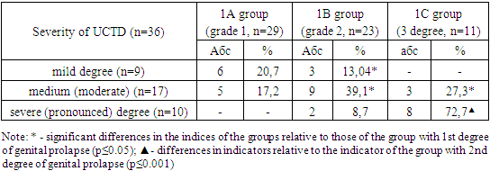
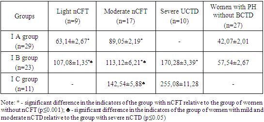
- We conducted a clinical and anamnestic analysis, including somatic, gynecological and reproductive pathology of women, as well as the current condition and complaints. The average age of women in the main group was 26.4 ± 2.2 years, in the comparison group - 24.5 ± 0.6 years, which was in an unreliably significant range.The presence of UCTD in the studied women was determined by identifying 8 or more signs of undifferentiated connective tissue dysplasia out of 16 highly informative markers [1]. These include: joint hypermobility, thin skin, dentine defects, asthenic syndrome, mitral valve prolapse, lower extremity varicose veins, arachnodactyly, skin hyperextensibility, gothic palate, striae, scoliosis, neurocirculatory dystonia, deviated septum, systolic murmur on auscultation of the heart due to small anomalies in the development of the heart, congenital dislocation of the hip, keloid scars. The severity of connective tissue dysplasia was assessed according to the scale of clinical criteria for the severity of UCTD. Thus, in the main group of women with genital prolapse, uCTD was detected in 36 women, which amounted to 57.1%. Whereas, in the group of women without genital prolapse, this indicator was 8.7%, which is 6.6 times less than in the group with genital prolapse. UCTD in 2 women without genital prolapse was observed in mild mild severity.When studying the severity of UCTD in groups of women with genital prolapse, interesting data were obtained (Table 1).
|
|
5. Discussion
- Magnesium ions play an important role in maintaining the integrity of the connective tissue structure, which are necessary for the normal course of many physiological processes in the body. In this regard, it is of interest to determine the characteristics of the level of magnesium ions in the blood with varying degrees of severity of genital prolapse, since the literature describes the adverse effect of deficiency of this element in the peripheral blood on the development of obstetric and gynecological pathology and the structure of connective tissue.We believe that the prolapse and complete prolapse of the internal genital organs in women is one of the manifestations of nCTD at the level of the reproductive organs. Oxyproline is one of the main amino acids in collagen. The need for a biochemical study of the metabolism of the structural components of the connective tissue as an assessment of the state of the pelvic floor muscles is obvious. All this became the subject of our research.Thus, we have revealed the relationship between the severity of genital prolapse in women and the presence and severity of UCTD: the more severe the severity of UCTD, the more severe the degree of genital prolapse. This is confirmed by increased excretion of OP in daily urine in the women under study.
6. Conclusions
- 1. We found that UCTD determines the features of the development of pelvic floor pathology and affects the formation of genital prolapse.2. Understanding the features of connective tissue metabolism, namely, an increase in the level of hydroxyproline in the urine, and early detection of its disorders can form the basis for preventing the formation and progression of genital prolapse in reproductive age.
Conflicts of Interest
- The authors declare that they have no conflict of interest.
Financial Support
- This research did not receive any specific grant from funding agencies in the public, commercial, or not-for-profit sectors.
ACKNOWLEDGMENTS
- All authors participated in the research process and data collection.
 Abstract
 Reference
 Full-Text PDF
 Full-text HTML
