-
Paper Information
- Next Paper
- Previous Paper
- Paper Submission
-
Journal Information
- About This Journal
- Editorial Board
- Current Issue
- Archive
- Author Guidelines
- Contact Us
American Journal of Medicine and Medical Sciences
p-ISSN: 2165-901X e-ISSN: 2165-9036
2023; 13(2): 176-180
doi:10.5923/j.ajmms.20231302.31
Received: Feb. 10, 2023; Accepted: Feb. 26, 2023; Published: Feb. 28, 2023

The Effect of Endotoxin on the Body of a Teenager Athlete During Adaptation to Physical Activity
Z. I. Valizhanova1, D. R. Abdulkhayeva1, G. R. Yuldasheva2, A. A. Khadzhimetov3
1Republican Scientific and Practical Center of Sports Medicine, Tashkent, Uzbekistan
2Center for the Development of Professional Qualifications of Medical Workers, Tashkent, Uzbekistan
3Tashkent State Dental Institute, Tashkent, Uzbekistan
Copyright © 2023 The Author(s). Published by Scientific & Academic Publishing.
This work is licensed under the Creative Commons Attribution International License (CC BY).
http://creativecommons.org/licenses/by/4.0/

This study aimed to study the relationship between stress response and indicators of endogenous aggression, HDL, and leukocyte levels in adolescent athletes before and after physical activity. 32 young athletes aged 13-15 years engaged in various sports were examined. It was revealed that intestinal endotoxin in dysbiosis is one of the components affecting the parameters of lipogram and phagocytosis. Therefore, it is advisable to use endotoxemia indicators to assess the health status of adolescent athletes.
Keywords: Adolescent athlete, HDL, Polymorphonuclear leukocytes, Endogenous toxins, Stress
Cite this paper: Z. I. Valizhanova, D. R. Abdulkhayeva, G. R. Yuldasheva, A. A. Khadzhimetov, The Effect of Endotoxin on the Body of a Teenager Athlete During Adaptation to Physical Activity, American Journal of Medicine and Medical Sciences, Vol. 13 No. 2, 2023, pp. 176-180. doi: 10.5923/j.ajmms.20231302.31.
1. Introduction
- In the hemodynamic system during exercise, according to [Yakovleva M.Yu.2021], the delivery of oxygen and nutrients to the heart and skeletal muscles is activated with simultaneous depletion of blood flow in other internal organs, in particular, in the intestine. So, after 15 minutes of training on the treadmill, athletes have a decrease in blood flow in the upper mesenteric artery by 43%, and blood flow is restored 40-60 minutes after the end of training. Accordingly, longer and more intense workouts contribute to the development of ischemic status in the intestine. The lack of oxygen and nutrients to which the intestines of athletes are regularly exposed leads to metabolic disorders in the intestinal wall, a decrease in the protective properties of the mucous membrane, and an increased risk of inflammatory processes. Stress, in the process of intensive training, is also one of the factors leading to disruption of the intestine, in particular, there is an increase in the permeability of the intestinal wall, the development of local inflammatory reactions, as well as the state of endotoxemia. Experiencing acute stress reduces the content of lactobacilli and bifidobacteria in the intestine in patients for several days. The effects of stress hormones on the intestinal microflora are associated with the most important feature of bacterial survival — the formation of so-called bacterial films [Bailey M. T., Coe C. L. 1999; Lyte M., Ernst S., 1992; Hall-Stoodley L., Costerton J. W., Stoodley R, 2004]. Thus, stress can lead to intestinal dysbiosis, and dysbiosis, in turn, to central changes that affect the reduction of resistance to stress.The process of endotoxemia is accompanied by the appearance of symptoms in athletes such as bloating, discomfort, problems with stool (constipation, diarrhea, and their alternation), belching, vomiting, diarrhea, and high susceptibility to intestinal infections. According to [Vyshegurov Ya.Kh., Zakirova D.Z., Rascheskov A.Yu., Yakovlev M.Yu., 2006], high cortisol levels, in stressful situations, activate tryptophan-2,3-dioxygenase, which leads to a decrease in serotonin synthesis in the brain. In turn, insufficient serotonin levels are considered a significant causal factor in the development of anxiety, aggression, affective disorders, and stress [Vyshegurov Ya.Kh., Zakirova D.Z., Rascheskov A.Yu., Yakovlev M.Yu., 2006].Endotoxin or otherwise LPS of the intestinal microflora takes an active part in the processes of homeostasis and supports all links of the immune system in various conditions of the body [M. Yu. Yakovlev, 1993]. It should be noted that when endotoxin enters the general bloodstream and anti-endotoxin immunity (AEI) is insufficient, endotoxin aggression (EA) is transformed, characterized by the manifestation of a wide range of pathogenic actions of lipopolysaccharide (LPS). As is known, an intact colon mucosa of a healthy person is, in all likelihood, a fairly reliable barrier that prevents endotoxin from entering the bloodstream in large quantities. On the way of penetration of endotoxin from the intestine into the general bloodstream, the second barrier after the intact intestinal mucosa is liver cells and fixed liver macrophages. Under the conditions of the practical norm, 31% of endotoxin can still enter the blood [M. Yu. Yakovlev 2003]. This is confirmed, in particular, by data that in practically healthy people, polymorphonuclear leukocytes (about 3.5%) are detected in peripheral blood smears, binding endotoxin using the so-called Fe-mediated binding, that is, using antibodies fixed on Fc receptors of leukocytes [Likhoded V.G. et al., 1996b]. Such leukocytes, in particular, polymorphonuclear leukocytes (PML), play an important role in the detoxification of endotoxin, and they can be considered factors of cellular anti-endotoxin immunity. When LPS interacts with PML, many metabolic characteristics of the latter change, including stimulating the formation of secondary mediators by monocyte-macrophage cells. The mechanism of this phenomenon is reduced to the "preparation" of neutrophils for the accelerated production of reactive oxygen species in response to the effects of various stimuli, which has killer activity against a wide range of bacteria, fungi, viruses, and protozoa. At the same time, phagocytic activity decreases in hyperactivated forms of PML, which reduces the effectiveness of the body's anti-endotoxin barriers-a and decreases the surface negative charge of PML membranes by endotoxin. The process of endotoxin aggression increases with damage to the intestinal mucosa and dysbiosis, which are accompanied by the translocation of bacteria and their waste products into the small intestine.There are isolated reports in the scientific literature about the possible role of intestinal LPS in the body's disadaptation to physical exertion (overload). However, the solution to these issues has not been possible until now.Several blood plasma components also can bind endotoxin. These are, first of all, several acute-phase reactants, a C-reactive protein, as well as a factor causing the disaggregation of LPS macromolecules - lipoproteins of high specific density, which, due to the presence of hydrophilic and hydrophobic ends in their structure, usually form spheroidal structures [BogHansen T. et al. 1978; Tobias P. et al. 1982]. Disaggregation of LPS with high specific density lipoproteins (HDL), capable of binding more than half of the endotoxin within a few minutes. [Apollonin A.B. et al. 1990; Freudenberg M. et al. l985]. Binding to HDL leads to a significant decrease in the toxicity of LPS [Mathison J., Ulevitch R. 1979; Mathison J., Ulevitch R. 1981], Endotoxin complexes with HDL can circulate in the bloodstream for 24 hours, are absorbed by tissues, and are gradually excreted. The greatest concentration of these complexes is found in the adrenal glands. The binding of HDL to LPS plays an important role in protecting against endotoxin. It is necessary to emphasize the possibility of LPS penetration into the general bloodstream in intestinal dysbiosis. A sharp increase in the total number of microorganisms in the colon with dysbiosis may be accompanied by a sufficiently intensive multiplication of Gr" bacteria in the small intestine, the mucosa of which is more permeable to endotoxin and more vulnerable than the mucosa of the colon [Lang Ch., Alteveer R. 1986].The above condition was the basis for studying the relationship between the stress response and indicators of endogenous aggression, the level of HDL, and blood leukocyte,ytes in adolescent athletes before and after physical exertion.
2. Materials and Methods
- The study was conducted based on the children's sports school of the Olympic Reserve in Tashkent. 32 young athletes aged 13-15 years engaged in various sports were examined. The control group of 16 people consisted of practically healthy (medically examined) peers who do not play sports. For these studies, athletes were divided into two groups: I-group (boys) – sport (football) – 18 people, II-group (girls) – sport difficult coordination (rhythmic gymnastics) – 14 people. To assess physical performance (aerobic efficiency), the PWC170 sample (modified by L. I. Abrosimova, 1978) with a single physical load was used. The duration of the load was 3 minutes with a frequency of 30 ascents per minute. The height of the step was selected in such a way that the angle of bending the leg when climbing the step was about 70°.To study the concentration of cortisol, 2 saliva samples were collected: the first before physical activity, and the second after, according to the collection protocol. Portions of saliva were placed in a plastic tube of the Eppendorf type (2 ml volume). The collected saliva was stored at - 20°C before the study. Before testing, the samples were thawed and centrifuged. The determination of cortisol was carried out on immunoassay kits for the quantitative determination of cortisol by the company "HUMAN" on the analyzer. The MSM level was determined by ultraviolet spectrophotometry at wavelengths of 254 and 280 nm. The assessment of intoxication of the body by the imbalance between the accumulation and binding of toxins in plasma was expressed in units quantitatively equal to the extinction indices.The selection of venous blood from athletes for biochemical parameters was carried out before and after physical activity. The concentration of subs high-density density lipoproteins (HDL mmol/L) – was determined by enzymatic methods on the analyzer "MINDRAY". The results of the study were processed using the STATISTICA 6.0 standard software package. by the method of variational statistics with the determination of the arithmetic mean (M) and the standard error of the mean (m). Intergroup differences were assessed by the Student's t-criterion and were considered reliable at a significance level of at least 95% (p<0.05), which is recognized as a reliable indicator in biomedical research.
3. Results and Discussion
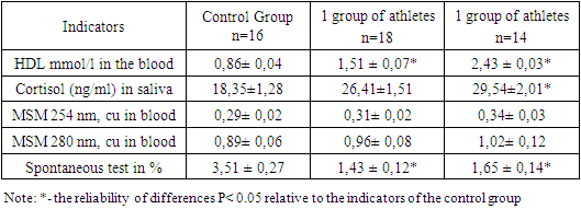
- One of the most informative biochemical indicators from a prognostic point of view was the content of cortisol, as one of the criteria for the effectiveness of training, the development of overtraining, stress, and assessment of the adequacy of the applied training loads. The high level of cortisol in adolescent saliva observed by us (Table-1) is apparently due to the increased neuropsychiatric mobilization of the athlete during physical exertion. Consequently, short-term stress testing with increasing power leads to a significant increase in the concentration of cortisol in saliva.As is known, stress, during intensive training, is one of the factors leading to disruption of the intestine, in particular, increased permeability of the intestinal wall, the development of local inflammatory reactions, as well as the state of endotoxemia, especially with dysbiosis.As can be seen from the results of the studies presented in Table 1, physical activity also leads to a change in the dynamics of HDL in the blood of young athletes. Thus, it was noted that in sports such as football, which train endurance, higher indicators of HDL levels are determined in comparison with the control group in young men. It is not excluded that heavy physical exertion in athletes who train mainly for endurance causes the connection of lipids to the processes of energy supply of muscular activity. It is noteworthy that the level of HDL in the blood of athletes engaged in rhythmic gymnastics is also significantly higher compared to the control group (Table 1). [In the studies of V.A.Kudinov co-author. 2020] it has been shown that HDL is involved in the process of reverse cholesterol transport, in the modulation of the inflammatory process, in blood clotting and vasomotor reactions of the body, also these particles have antioxidant properties and contribute to immune reactions and intercellular signaling. At the same time, the main HDL proteins synthesized in various organs have a high positive charge and can bind free fatty acids, activate lipoprotein lipase, regulate triglyceride metabolism, bind to glycosaminoglycans, bind negatively infected molecules, prevents activation of the blood clotting system, regulates platelet aggregation and binds hydrophobic molecules. Consequently, in adolescent athletes, physical exertion and a psychoemotional state are accompanied by an increase in the level of HDL in the blood, particles of which can bind endotoxins formed in the intestine against the background of dysbiosis.
|
4. Conclusions
- As our studies have shown, a significantly significant decrease in the content of LPS-PN was noted in the peripheral blood of boys and girls athletes, which indicates a decrease in the sorption potential of neutrophils. I.e., the number of neutrophils binding LPS was sharply reduced in the peripheral blood of adolescents. It should be noted that simultaneously with a decrease in the level of LPS-positive neutrophils, the ability of polymorphonuclear leukocytes to absorb an additional amount of LPS on their membrane also decreases. The decrease in the peripheral blood level of LPS-PN, in our opinion, is due to their migration and accumulation in various organs and tissues. According to [V.A. Malova, 1992; Nekhaev, S.G., Grigoriev, Yu.I., 2010] this method eliminates biologically active forms of LPS from the systemic bloodstream, which reduces the direct damaging effects of LPS on the structures of the body. However, granulocytes that have migrated to organs and tissues have a secondary damaging effect on them due to the release of a pool of lysosomal enzymes and mediators. It should also be noted that activated LPS neutrophils also exert their influence on Kupfer cells of the liver, through which the regulation of the protein-synthetic function of the liver is carried out. In addition, an increase in the adhesive and aggregation properties of neutrophils according to [M.E.Wilson, 1985] may lead to their sequestration from the central pool to the marginal one, which reduces their concentration in the bloodstream and may contribute to the migration of these leukocytes into organs and tissues, thereby enhancing their organ pathological effects. The revealed decrease in the sorption capacity of polymorphonuclear leukocytes may be due to a decrease in the sorption capacity of neutrophils, which carries a small number of Fc receptors on the outer membrane of leukocytes. Thus, the analysis of the totality of available data shows that the components of gram-negative and gram-positive bacteria of the intestinal microflora can play an important role in the physiology and pathology of the athletes' bodies. In this situation, intestinal endotoxin in dysbiosis is one of these components, possibly the active one itself. Therefore, it is advisable to use endotoxemia indicators to assess the health status of adolescent athletes.
 Abstract
 Reference
 Full-Text PDF
 Full-text HTML