American Journal of Medicine and Medical Sciences

p-ISSN: 2165-901X    e-ISSN: 2165-9036

2023;  13(2): 43-46

doi:10.5923/j.ajmms.20231302.02

Received: Jan. 8, 2023; Accepted: Jan. 30, 2023; Published: Feb. 13, 2023


Markers of Antitumor Immunity in the Prediction of Uterine Leiomyoma in Women of Reproductive Age

Shakhlo Khamidova1, Shakar Navruzova2

1Basic Doctoral (PhD) Student, Bukhara State Medical Institute, Bukhara, Republic of Uzbekistan

2Department of Pediatrics, Bukhara State Medical Institute, Bukhara, Republic of Uzbekistan

Correspondence to: Shakhlo Khamidova, Basic Doctoral (PhD) Student, Bukhara State Medical Institute, Bukhara, Republic of Uzbekistan.

Email:

Copyright © 2023 The Author(s). Published by Scientific & Academic Publishing.

This work is licensed under the Creative Commons Attribution International License (CC BY).
http://creativecommons.org/licenses/by/4.0/

Abstract

The article contains fragments of a scientific study aimed at developing prognostic criteria for uterine leiomyoma, the authors studied the factors of antitumor immunity, cytokines and growth factors in women of reproductive age. Early diagnosis of uterine leiomyoma contributes to effective conservative and organ-preserving treatment of this disease.

Keywords: Uterine leiomyoma, Immunity, Women of reproductive age, Markers

Cite this paper: Shakhlo Khamidova, Shakar Navruzova, Markers of Antitumor Immunity in the Prediction of Uterine Leiomyoma in Women of Reproductive Age, American Journal of Medicine and Medical Sciences, Vol. 13 No. 2, 2023, pp. 43-46. doi: 10.5923/j.ajmms.20231302.02.

1. Introduction

According to the modern concept, the treatment of uterine leiomyoma should be radical and aimed at preserving reproductive function in women of reproductive age. From the standpoint of evidence-based medicine, uterine leiomyoma is one of the most frequent types of pathology in the modern female population [1].
According to foreign and domestic scientists, uterine fibroids affect 25-30% of women over 35 years old, and in recent years the disease is increasingly detected at a young age (18-35 years) [2].
The study of the role of cytokines and other low–molecular-weight inflammatory mediators in the pathogenesis of tumour and inflammatory diseases in pregnant women with large uterine fibroids is one of the directions of studying the mechanisms of growth of myomatous nodes, possible early prediction of the course and outcome of pregnancy, and the creation of new methods of diagnosis and treatment [3].
A woman's body mass factor, realized in adipose tissue through the conversion of active forms of estrogens from precursors, is closely related to the production of sex steroids. Exceeding the normal values of the body mass index increases the risk of uterine fibroids by 1.2-2.3 times [4], and every 10 kg of excess body weight increases the risk of tumour development by 21% [5,6].
The aim of the study was to evaluate the prognostic significance of immunological indicators in the implementation of antitumor immunity in uterine leiomyoma in women of reproductive age.

2. Materials and Methods

Methods such as observation, experiment, statistic, test system analysis were used in the research. 120 women of reproductive age with uterine leiomyoma hospitalized in the Department of gynaecology were examined on the basis of the Bukhara Regional Perinatal Center. The control group consisted of 30 healthy women of reproductive age. The content of pro-inflammatory, anti-inflammatory cytokines and cancer markers in blood serum was studied by enzyme immunoassay using the "Vector-Best" test systems with a set of reagents A-8768, Russian Federation, Novosibirsk. The test kit is designed to determine the number of cytokines in the blood of patients. The levels of proinflammatory cytokines IL-6, TNF-α, growth and damage factors- transforming growth factor β2 (TGF-β2), insulin-like growth factor-I (IGF-I), fibroblast growth factor FGF), and vascular endothelial growth factor (VEGF), cancer markers in blood serum (СА-125; СА-15-3; СА-19,9; AFP; СЕА) were determined.

3. Results and Discussion

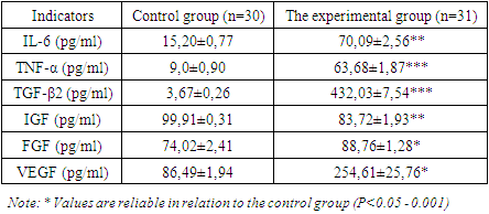
As a result of a study of cytokines in the blood serum of patients of the examined groups, an increase in the level of IL-6 was found to be 4.6 times (up to 70.09±2.56pg/ml) compared to the control (15.20±0.77 pg/ml) (Table 1).
Table 1. Immunological parameters of blood
     
Currently, it is known that the cancer marker CA 125 (Carbohydrate antigen 125) is considered a tumour marker for ovarian cancer. Normally, its concentration is 4.0-8.8 ×10⁹/l (0-30 IU/ml). With an increase in the index above 35 units/ml, ovarian cancer is detected in 90% of cases. An elevated level of CA 125, more than 30 IU /ml may indicate such malignant diseases as cancer of the female genital organs (ovaries – in most cases, less often a cancer of the endometrium (inner layer of the uterus), fallopian tubes, cancer of the respiratory organs (less specifically) and organs of the gastrointestinal tract, pancreas.
In more rare cases, CA 125 is found in non-oncological processes, for example, with endometriosis, excessive growth of the inner layer of the uterus develops; with adenomyosis, the germination of the inner layer of the uterus into muscle tissue is noted; during menstruation and during pregnancy; with inflammation of the female genital organs; inflammatory liver diseases.
Taking into account the above, it is important to determine CA-125 in combination with other cancer markers.
There are scientifically proven facts that the cancer marker CA-15-3 (mucin-like glycoprotein or carbohydrate antigen 15-3 refers to cancer markers of neoplastic (tumour) processes occurring in the mammary gland. Its normal values are - 9.2-38 units / l, and in some laboratories – 0-22 units/ml.
The reason for calling it an oncomarker of breast cancer is the fact that in 80% of cases of breast cancer in women who have metastasized, this oncomarker is enlarged.
There is evidence that the CA 15-3 index can rise with benign neoplasms and inflammatory diseases of the mammary glands; cirrhotic hepatic processes; as a physiological "surge" in the 2nd half of pregnancy and with some autoimmune processes.
Oncomarker CA 19-9 is a carbohydrate antigen 19-9 (CA 19-9), with which early diagnosis of neoplasms of the gastrointestinal tract is carried out. The most informative analysis is for pancreatic tumours. The specificity, in this case, is high and is 82%. In the case of tumour problems of the biliary system and liver, it is specific in 72% of cases. Its normal values are 0-37 units/ml. A concentration of 40 IU/ml or higher is considered dangerous.
Cancer marker CA 19-9 allows you to determine: malignant processes of the gastrointestinal tract (cancer of the stomach, and intestines); cancer of the liver, gallbladder and bile ducts; cancer of the female genital organs and mammary glands; bladder cancer.
Among the processes of non-tumour nature, CA 19-9 increases in the case of inflammatory changes and cirrhotic processes in liver diseases; diseases of the biliary tract and gallbladder (cholecystitis, cholangitis, cholelithiasis); cystic fibrosis (damage to the glands of external secretion and respiratory problems).
Consequently, the obtained results of the study of cancer markers in uterine leiomyoma in women show the presence of other concomitant diseases, in particular gastrointestinal diseases, obesity and disharmony.
Alpha-fetoprotein (AFP) and carcinoembryonic antigens (CEA) were also studied for accurate differentiation in oncopathology. There was a tendency to increase AFP in the blood - up to 13.68±0.90 ng/ml and CEA - up to 6.66±0.64 ng/ml in patients of the examined group. The obtained indicators served as the basis for the diagnosis of uterine leiomyoma.
It is known that the AFP - oncomarker is a glycoprotein in chemical structure and is similar to albumin. Norm: up to 10 ng/ml, (8 IU/ml), content above 10 IU/ml is an indicator of pathology.
Referring to the data from literary sources, the determination of serum cancer markers can be used in the dynamic monitoring of patients. The combined use of CEA and other cancer markers can be useful for determining the prognosis in terms of metastasis, and postoperative complications, and the combined determination of CEA and CA 19-9 can be used for early detection of cancer [7,8,9], AFP should be considered as a potential marker of tumour activity and a predictor of survival [10,11,12].
Carcinoembryonic antigen (CEA) or ANTIGEN CD66E is a non-specific marker. It is produced by the forming of cells in the digestive tract of the fetus. In adults, it is determined in minimal amounts, normally up to 5 ng/ml (according to some data – up to 6.3 ng/ml). According to the literature, there is a slight increase in SEA in smokers. At the level of SEA above 20 ng/ml, a malignant tumour of the gastrointestinal tract (stomach, colon, rectum), a malignant process of the breast, neoplasms of the prostate, the reproductive system of men and women, the thyroid gland, metastatic processes in the liver and bone formations should be suspected.
If the level of SEA is up to 10 ng /ml, then there is a possibility that the patient has pathological processes in the liver (inflammation, cirrhosis), intestinal polyps, Crohn's disease, pancreatic diseases, tuberculosis, lung inflammation (pneumonia), cystic fibrosis and /or postoperative metastatic process.
The study of cytokines in the blood serum of patients of the examined groups revealed a 4.6-fold increase in the level of IL-6, a 7-fold increase in TNFa, an increase in the concentration of TGF-β2 in the blood by 117.7 times, an increase in VEGF by 2.95 times, a decrease in IGF, an increase in FGF.
The obtained result allows us to conclude that, with uterine leiomyoma, there is a high increase in the concentration of growth factors that regulate the processes of angiogenesis and hematopoiesis. At the same time, against the background of a decrease in the protective reparative processes of vascular wall restoration, there is a high risk of stimulating the growth and proliferation of leiomyoma cells. This means that all the data obtained show the state of dysregulation of synthesis, release and transformation of cytokines and protein growth factors in uterine leiomyoma.
The indicators of cancer markers in the blood were increased, with CA-125 increased by 6.26 times, CA-15-3 marker increased by 2.53 times, and CA-19.9 marker increased by 3.75 times. For leiomyoma, the appearance of AFP and CEA markers in the blood was also established, which confirms the diagnosis of leiomyoma in the women of the studied group.
We will continue to work on this research in the future and present these results enriched with new information through more detailed studies in our next studies.

4. Conclusions

Thus, when studying growth factors and pro-inflammatory cytokines in women with leiomyoma, a high increase in IL-6 by 4.6 times, TNFa by 7.0 times, TGF-β2 by 117.7 times and VEGF by 2.95 times found. At the same time, against the background of a decrease in the protective reparative processes of vascular wall restoration, there is a high risk of stimulating the growth and proliferation of leiomyoma cells. This means that all the data obtained show the state of dysregulation of synthesis, release and transformation of cytokines and protein growth factors in uterine leiomyoma.
Uterine leiomyoma is characterized by: relative lymphocytopenia, absolute neutrophilic leukocytosis, and an increase in basophils and ESR against the background of a decrease in the absolute number of eosinophils and monocytes in peripheral blood. And there is also a decrease in AST and an increase in the level of total bilirubin. At the same time, there was also a significant increase in urea levels and a decrease in creatinine and total blood protein in patients of the examination group. The results obtained confirm the violation of the urea cycle, which is clinically manifested by symptoms of renal pathology, which is paraclinical confirmed by hypoproteinemia and uremia. At the same time, hypoproteinemia indicates an increase in the process of catabolism of blood proteins and as an outcome of cow bleeding, characteristic of the tumour process. Coagulogram indicators revealed a significant decrease in PTI with a tendency to hyperfibrinogenemia against the background of discoagulation with the risk of developing DIC syndrome.
Thus, for an accurate diagnosis, it is very important to take into account concomitant diseases and conditions, the pathogenetic mechanism of which allows the causal factor to be disclosed, as well as to predict the recurrence and metastasis of the oncoprocess.

ACKNOWLEDGEMENTS

The authors acknowledge the immense help received from the scholars whose articles are cited and included in references to this manuscript. The authors are also grateful to the authors/ editors/publishers of all those articles, journals and books from where the literature for this article has been reviewed and discussed.

Conflicts of Interest

The authors declare that there is no conflict of interest regarding the publication of this paper.
The Source of funding is nil.

References

[1]  Agaidarov, R.D., 2016. Perinatal outcomes in women with uterine leiomyoma. Problems of Science, (12 (13)), pp.103-105.
[2]  Жукова, Н.П., Дейкало, Н.С., Арестова, И.М., Киселева, Н.И. and Кожар, Е.Д., 2010. Органосохраняющие методы лечения лейомиомы матки. Вестник Витебского государственного медицинского университета, 9(1), pp.76-80.
[3]  Косарева П.В., Конев Р.А., Сивакова Л.В., Самоделкин Е.И., Карипова М.О. 2022. Онкомаркеры в диагностике, прогнозе и выборе метода лечения колоректального рака. Современные проблемы науки и образования. (3). doi: 10.17513/spno.31751.
[4]  Basu, A., Seth, S., Chauhan, A.K., Bansal, N., Arora, K. and Mahaur, A., 2016. Comparative study of tumour markers in patients with colorectal carcinoma before and after chemotherapy. Annals of Translational Medicine, 4(4). p. 71.
[5]  Dolscheid-Pommerich, R.C., Manekeller, S., Walgenbach-Bruenagel, G., Kalff, J.C., Hartmann, G., Wagner, B.S. and Holdenrieder, S., 2017. Clinical performance of CEA, CA19-9, CA15-3, CA125 and AFP in gastrointestinal cancer using LOCI™-based assays. Anticancer Research, 37(1), pp.353-359.
[6]  Feng, Y., Li, Y., Dai, W., Mo, S., Li, Q. and Cai, S., 2018. Clinicopathologic features and prognostic factors in alpha-fetoprotein-producing colorectal cancer: analysis of 78 cases. Cellular Physiology and Biochemistry, 51(5), pp.2052-2064.
[7]  Ganieva Sh.Sh., Radjabova G. B. 2021. Clinical And Laboratory Assessment Of The Health Status Of Patients With Chronic Obstructive Pulmonary Disease Who Have Undergone Coronavirus Infection. Central Asian Journal of Medical and Natural Science, 2021: Special issue on COVID-19: Yesterday, Today, and Tomorrow. pp.76-80. doi: 10.47494/cajmns.vi0.353.
[8]  He, Y., Zeng, Q., Dong, S., Qin, L., Li, G. and Wang, P., 2013. Associations between uterine fibroids and lifestyles including diet, physical activity and stress: a case-control study in China. Asia Pacific journal of clinical nutrition, 22(1), pp.109-117. doi: 10.3316/informit.104887551227284.
[9]  Nguyen, V., Mendelsohn, A. and Larrick, J.W., 2017. Interleukin-7 and immunosenescence. Journal of immunology research, doi: 10.1155/2017/4807853.
[10]  Parker, W.H., 2007. Reproductive characteristics and risk of uterine leiomyomata. Fertility and Sterility, 87(4), pp.725-736.
[11]  Santulli, P., Borghese, B., Lemaréchal, H., Leconte, M., Millischer, A.E., Batteux, F., Chapron, C. and Borderie, D., 2013. Increased serum oxidative stress markers in women with uterine leiomyoma. PloS one, 8(8), p.e72069.
[12]  Liu, Z.Q., Lu, M.Y., Sun, R.L., Yin, Z.N., Liu, B. and Wu, Y.Z., 2019. Characteristics of peripheral immune function in reproductive females with uterine leiomyoma. Journal of oncology, 2019.