American Journal of Medicine and Medical Sciences

p-ISSN: 2165-901X    e-ISSN: 2165-9036

2022;  12(12): 1348-1353

doi:10.5923/j.ajmms.20221212.36

Received: Nov. 26, 2022; Accepted: Dec. 23, 2022; Published: Dec. 30, 2022


Evaluation of the Results of Surgical Correction of Anomal Drainage of the Pulmonary Veins

Abrolov Kh. K., Mamatov M. A., Xamdamov Sh. R., Usmanov X. K.

State Institution “Republican Specialized Scientific and Practical Medical Center for Surgery Named after Academician V. Vakhidov”, Tashkent, Uzbekistan

Copyright © 2022 The Author(s). Published by Scientific & Academic Publishing.

This work is licensed under the Creative Commons Attribution International License (CC BY).
http://creativecommons.org/licenses/by/4.0/

Abstract

The article presents the materials of operated patients with abnormal pulmonary drainage for the period from 2001 to 2021. in the department surgery for congenital heart defects of the RSSPMCH named after academician V.Vakhidov, the age of the patients was 1.4-45 years. In addition to general clinical research methods, all patients underwent echocardiography and multispiral computed tomography for the diagnosis of the defect. A comparative analysis of changes in heart parameters using echocardiography and radiography depending on the type of defect and the degree of pulmonary hypertension. The dynamics and anatomical and functional parameters of operated patients with abnormal pulmonary vein drainage were studied.

Keywords: Abnormal pulmonary venous drainage, Surgical treatment, Echocardiography, Radiography, Pulmonary hypertension, Long-term results

Cite this paper: Abrolov Kh. K., Mamatov M. A., Xamdamov Sh. R., Usmanov X. K., Evaluation of the Results of Surgical Correction of Anomal Drainage of the Pulmonary Veins, American Journal of Medicine and Medical Sciences, Vol. 12 No. 12, 2022, pp. 1348-1353. doi: 10.5923/j.ajmms.20221212.36.

1. Introduction

Congenital heart defects occupy the first place in frequency among all congenital developmental defects, accounting for at least 30% of their total number, about 1% of children are born with congenital heart defects (CHD) [2,4,6,13,16,20]. Despite the fact that at present, the wide availability of surgical care, about 75% of children born with critical congenital heart disease survive to the age of 1 year, about 69% survive to 18 years. Anomalous pulmonary venous drainage (APVD) is a congenital heart disease characterized by a lack of connection between the pulmonary veins and the left atrium. The frequency of APVD is from 0.5% to 9% among all CHD [1,5,7,12,14,19,23]. Among the numerous variants of this defect, the classification proposed by R has become the most widespread. Darling et al. [15]. This classification is based on an anatomical principle depending on the level of entry of the pulmonary veins (PV): Type I - supracardial or supracardiac: PV drains directly into the superior vena cava (SVC) or its tributaries; Type II - cardiac or cardiac: LV drains into the right atrium (RA) or into the coronary sinus; Type III - infracardial or subcardiac: PV drains into the vena cava inferior (VCI) or its tributaries below the level of the diaphragm and IY type - mixed: blood from the PV enters the right parts of the heart along two or three paths described above. Hemodynamic disorders, clinical course, tasks and methods of surgical treatment depend on the type and type of defect, which is divided according to the above principles [1,5,7,12,14,19,23]. However, children who underwent APVD surgery, despite a favorable prognosis for survival, may face a number of problems in the early and late postoperative period, which can significantly affect their quality of life and functional status. Literature analysis proves that studies of long-term results of surgical treatment of APVD are covered in single works [1,5,8,9,10,11,12,14,16,18,21,23]. In conclusion, it should be noted that the analysis of the literature indicates that the study of the results of correction of APVD is an urgent problem and is of great practical and scientific importance.
Purpose: to study the analysis and features of long-term results of surgical treatment of various forms of APVD using echocardiography and radiography.
Objectives:
1. To study the dynamics of the anatomical and functional parameters of operated patients with abnormal pulmonary vein drainage;
2. Conduct a comparative analysis of the results of echocardiographic and radiography examination of patients in the pre- and early postoperative periods;
3. To study echocardiographic and radiography parameters depending on the type of APVD and the degree of pulmonary hypertension defect.

2. Material and Methods

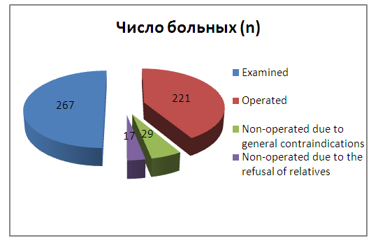
In the Department of Surgery for Congenital Heart Diseases of the RSSPMCH named after A.I. acad. V.Vakhidov State University and in the Department of Cardiac Surgery of ASMI from 2001 to 2021. 267 patients were observed for APVD. For various reasons, 46 (17.2%) patients were not operated on, 29 (10.8%) of them due to general contraindications to surgery and refusal of the patient and/or relatives - 17 (6.3%). 221 (82.7%) patients were operated on; most of them were operated on at the RSNPMCH named after I.I. Acad. V. Vakhidov (Fig. 1).
Figure 1. Scheme of distribution of patients with APVD
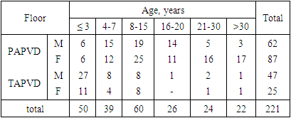
Among the operated patients with APVD - 118 (53.3%0 were men, 103 (46.7%) were women. The age of patients ranged from 1 month to 45 years, averaging 16.3 ± 0.26 years (Table 1).
Table 1. Distribution of patients with APVD depending on gender and age
     
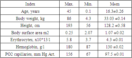
The main parameters of patients with APVD before surgery are presented in Table 2.
Table 2. Characteristics of patients with APVD before surgery
     

3. Results and Discussion

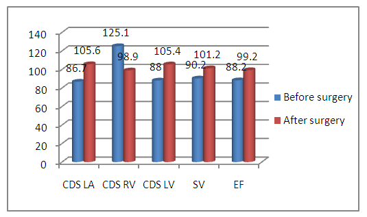
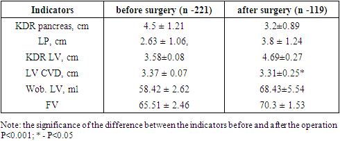
In the late postoperative period, out of 221 operated patients, 119 (53.8%) patients with various anatomical variants of APVD were examined. To study the long -term results of surgery in patients with a defect before and after surgical treatment, a large role belongs to functional research methods, including echocardiography [3,17]. Comparative results of echocardiography with preoperative data obtained in 119 patients in terms of 1 year to 10 (on average, 6.7±0.16) years are shown in Table 3. So, from Table 5 and Fig. 2 it can be seen that the dimensions of the left atrium before the operation were 2.63 ± 1.06 cm, i.e. 96% of the norm. In the remote period, this indicator was 3.2 ± 0.89 cm, i.e. 108%. from the norm. At the same time, we observed normalization or a slight increase in LA, because after correction of the defect, the load on the LA increases. The diastolic size of the right ventricle was 4.5±1.21 cm before surgery or 124% of the norm.
Table 3. Comparative characteristics of the results of echocardiographic studies of patients in the pre- and postoperative periods
     
In the long-term period after the operation, we noted a significant decrease in the value of RV EDR to the limits of the age norm. Thus, the EDR of the pancreas in the long term after the operation was 3.2±0.89 or 98% of the norm. Normalization of the sizes of the right parts of the heart begins immediately after the operation and reaches the norm within 2-3 years after the correction of the defect; the wall thickness of the pancreas still remains increased. The overload of the right parts of the heart, which took place before the operation, was noted in 40.9% of patients, and completely disappeared after adequate correction of APVD.
The left parts of the heart in patients with APVD are relatively small, since these parts, due to the left-right shunt, work in an underload mode (72,165,217). After correction, the load on these departments increases and, in connection with this, an increase in the linear dimensions of the LA and LV occurs. Thus, the initial LA values were 2.83 ± 1.06 cm or 90.05 ± 2.3% of the norm (Fig. 2).
Figure 2. Changes in EchoCG signs in the long term after surgery in patients with APVD (in % - from the norm)
In the postoperative period, the value of the LA CDR was already equal to 3.8±1.24 cm or 108±3.21% of the norm. LV EDR before surgery was 3.58 ± 0.08 cm or 92.5 ± 1.81% of the norm. In remote periods, this indicator reached 4.69 ± 0.27 cm or 106 ± 2.12% of the norm.
An analysis of echocardiographic parameters reflecting the functional state of the left ventricular myocardium showed that the operated patients had an increase in the ejection fraction, and the greatest increase in functional parameters was observed in patients with TALV and in patients with PALV, in whom several pulmonary veins were abnormally drained (Table 3, Fig. 2). So, if the ejection fraction before surgery was only 65.51±2.46% or 98±1.06% of the norm, then in the long term there was a clear increase in this indicator, which was already 70.3±1.53% or 108 ± 1.02% of normal.
Stroke volume before surgery was 52.4±2.62 ml or 90.1±1.01% of the norm (Table 3, Fig. 2). Depending on this, the cardiac index was calculated, which was 3.78±0.19 l/min/m2. In the long-term period, the VO increased and amounted to 68.43±5.54 ml, respectively, the SI increased, up to 3.97±0.25 l/min/m2. The positive dynamics of these parameters indicates the elimination of the left-right shunt and an increase in cardiac output. With the Doppler method of research, inferiority of the interatrial septum in the preoperative period was detected in 217 (98.1%) patients. In the long-term period after surgery, in almost all (99.55%) patients who underwent suturing or plastic surgery of an ASD, the interatrial septum was completely traced; only 1 patient had recanalization of the MPS.
The data obtained after echocardiography indicate that a good result occurred in 106 (89.07%) patients, satisfactory - in 9 (7.5%), and poor - in 3 (2.5%) patients. The latter had indirect signs of overload of the right and left sections, as well as signs of insufficiency of atrioventricular valves in the long-term period. In the group of patients with satisfactory results, who had II- III degree pulmonary hypertension before surgery, signs of pancreatic hypertrophy remained in the long-term period.
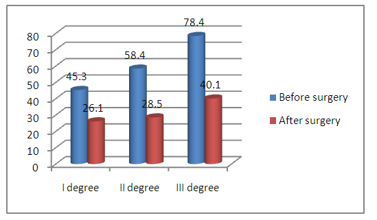
With the help of echocardiography, we tried to study the state of central hemodynamics. At the same time, regression of pressure in the right parts of the heart was studied. Thus, before surgery, according to intraoperative tensiometry, PH degree I was established in 51.1% (113) patients, degree II - in 34.8% (77) and degree III in 10.8% (24) cases with an average value of the ratio systolic pressure RV.LV - 42.6±7.4%; 57.5±8.3% and 80.6±6.5, respectively; only 7 (3.1%) patients had no PH. To analyze the regression of pulmonary hypertension in the long-term after surgery, we measured pressure in the heart cavities using echocardiography (Fig. 3).
Figure 3. Changes in pressure in the pancreas in the long term after surgery in patients with APVD
The figure shows that in addition to 11 (9.2%) of the examined patients, there was a decrease in pressure in the right parts of the heart. Thus, 1st degree PH was observed only in 11 (9.2%) patients with an average value of pressure in the pancreas 40.1±3.5 mm Hg. In other patients, the pressure in the pancreas and LA was within the normal range. Normalization of pressure in the pancreas and LA once again indicates that in patients with APVD, the development of PH and changes in the blood vessels of the ICC are hypervolemic in nature. Residual pulmonary hypertension was noted in patients whose long-term outcome was studied in the first year after surgery and who were operated on at the age of over 18 years. Thus, complex echocardiography made it possible to assess the effectiveness of APVD correction in the long-term period after surgery, taking into account changes in the size of the heart cavities and intracardiac hemodynamics. EchoCG as a simple non-invasive research method provides objective information about the state of intracardiac and central hemodynamics in patients with APVD after surgical correction of the defect [3,22]. The inclusion of echocardiography in the program of examination of operated patients helps to determine the individual system of rehabilitation measures. The use of methods of mathematical processing and analysis of echocardiograms makes it possible to correct the functional parameters of the left and right parts of the heart in dynamics before and after surgery. Simplicity and safety, the absence of contraindications allows us to recommend the use of H echocardiography as one of the main research methods in patients with APVD.
As a result of the study of long-term results, the following long-term results of the operation after APVD correction were noted: good in 105 (88.2%) patients, satisfactory in 11 (9.2%) patients, and unsatisfactory in 3 (2.5%) patients. The measurements of pressure in the cavities of the heart using echocardiography showed that after the operation in the long term, the pressure in the right parts of the heart decreases to normal values; this proves that pulmonary hypertension in APVD is often hypervolemic in nature. The data of postoperative echocardiography prove that in the late postoperative period, the size of the right sections decreases and, conversely, the sizes of the left sections of the heart increase; indicators of the functional ability of the myocardium increase. It should be emphasized that good and satisfactory results were obtained precisely in those patients who had I and II degrees of pulmonary hypertension before the operation and in those who were operated on before the age of 18.
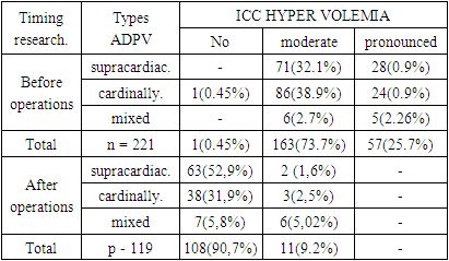
Comparative analysis of X-ray data before surgery was performed in 221 (100%) patients, and in the long-term period it was performed in 119 (53.8%) patients. One of the main indicators of successful ADPV correction is the normalization of the lung pattern. Common to all forms of malformation before surgery is an increase in the lung pattern, which is more pronounced in patients with TADPV. So, if before the operation in 57 (25.7%) patients a pronounced hypervolemia of the pulmonary circulation was noted, then after the operation it disappeared in all patients (Table 4).
Table 4. X-ray signs of ICC hypervolemia in patients with ADPV
     
Moderate hypervolemia was noted before surgery in 163 (73.7%) patients, after surgery it remained only in 11 (7.2%) patients - these were patients of the older age group. The pathognomonic "figure eight" or "snow woman" symptom, characteristic of the supracardiac type of TADPV, was found before surgery in 14 (6.3%) patients; in the long term it disappeared completely.
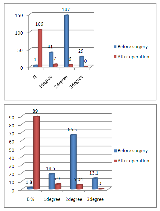
The cardiothoracic index was increased before surgery in 216 (95.6%) patients with ADPV. At the same time, normal CTI was in 4 (1,8%) patients, an increase in grade I was noted in 41 (18,5%), grade II in 147 (66,5%) and grade III in 29 (13.1%) patients (Fig. 4). In the remote period after surgery occupancy, there was a significant positive regression of CTI. So, I degree of increase in CTI was noted in 7 (5,9%) patients, II degree - in 6 (5,04%), in the rest of the examined patients, CTI was within the normal range (Fig. 4).
Figure 4. Changes in CTI in the long-term after surgery in patients with ADPV
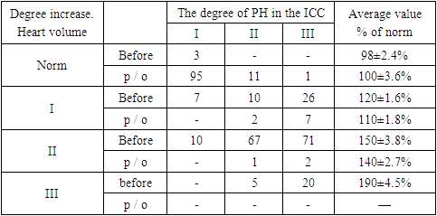
The volume of the heart before correction was increased in 217 (98.2%) patients (Table 5) and only in 3 (1,3%) cases the volume of the heart was within the normal range. It is known that there is a direct relationship between the volume of blood discharge and the degree of increase in the volume of the heart. The volume of the heart in ADPV depends on the size of the right heart. X-ray revealed a significant decrease in the volume of the heart, in some cases to normal values after correction of ADPV. As shown in Table 5, a change in the volume of the heart was noted in all groups with varying degrees of pulmonary hypertension. A noticeable decrease in the volume of the heart is observed during the first 1-3 years after the operation. In the future, as a rule, the dynamics of the reverse development of the volume of the heart was insignificant and this occurs due to a decrease in its right sections. Yes, normal before surgery. heart volume was noted in 3 (1.3%) patients, I degree of increase in 43 (19.4%), II degree in 148 (66.9%) and III degree of increase in 25 (11.3%) patients. In the long term, the volume of the heart returned to normal in 107 (89.9%) patients, the I degree of increase in the volume of the heart remained in 9 (7,5%) and the II degree of increase in 3 (2,5%) patients (Table 5).
Table 5. Dynamics of changes in heart volume in operated patients with ADPV
     
A slight decrease in the volume of the heart was noted mainly in those patients who had II and III degrees of pulmonary hypertension before surgery. Changes in the volume of the heart before and after surgery were studied by us in each age group. The data obtained indicate that its greatest decrease after surgery was found in patients operated on at the age of up to 18 years.
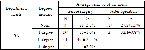
In connection with the arteriovenous discharge at the level of the atria, expansion of the right parts of the heart is observed. These signs are more pronounced in patients with TADPV. An increase in PP before surgery was noted in all 218 patients (Table 6). The greatest increase is determined in patients with II degree of pulmonary hypertension. The cessation of arteriovenous shunting of blood after surgery leads to a decrease in blood supply to the right sections of the heart. In this regard, in the late period after the operation, an increase in PP remained only in 11 (4.9%) patients, and to a lesser extent than before the operation. After adequate correction of the defect, the arteriovenous discharge of blood stops, the minute volume of the systemic circulation increases; in connection with this, the load on the left departments also increases. At the same time, the sizes of the latter increase; in our observations, an increase in the left atrium occurred in 165 (74.6%) patients.
Table 6. Changes in radiological signs in the long-term after surgery in patients with ADPV
     
With ADPV, the entire load of excess blood flow falls on the right heart. The left ventricle functions under “facilitated” hemodynamic conditions, because a certain amount of bypassed blood, bypassing this cavity, enters from the pulmonary veins into the right sections of the heart [24,25,26]. Despite this, out of 221 patients examined by us, only 44 (19.9%) had left ventricular enlargement.
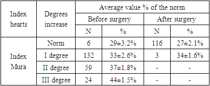
An important radiographic sign of normalization of hemodynamics after the elimination of ADPV is the determination of the degree of pulmonary hypertension, which is expressed on radiographs as a bulging of the pulmonary artery arch [25,26]. Moore's coefficient most accurately reflects the degree of LA bulging. When examining patients after surgery, a significant change in the Moore's index in the positive direction was noted. Thus, before the operation, only in 6 (2,7%) patients the Moore's index was normal, after the operation it became such in 97,4% of patients. Before the operation, the value of the Moore's index was: at I degree 33±2.6%, at II degree 37± 1.8% and at III degree 44±1.5%, and after the operation - at I degree 34±1.6% , and we did not observe II and III degrees of increase (Table 7).
Table 7. Changes in radiological signs in the long-term after surgery in patients with ADPV
     
Analyzing radiological data in patients with ADPV, in the long -term period after surgery, 106 (89.07%) patients had a good result, they had a significant positive dynamics of cardiometric parameters reaching normal values - this is a decrease in signs of pulmonary hypertension, hypervolemia, normalization of the sizes of the right departments of heart. A satisfactory result was obtained in 11 (9.24%) patients - this is a slight decrease in hypervolemia and the size of the right sections, CTI, heart volume and Moore's index. An unsatisfactory result was noted in 2 (1.6%) patients - there was practically no positive dynamics after the operation, there was a significant deterioration in radiographic parameters associated with the development of mitral and three cuspid heart valve insufficiency due to bacterial endocarditis.

References

[1]  Anchabadze, G. O. The results of radical correction of the supracardial form of total anomalous pulmonary venous drainage according to the Tucker method in newborns and children of the first year of life: abstract of the thesis of Candidate of Medical Sciences: 14.01.26 - M., 2011–36.
[2]  Bockeria L.A., Gudkova R.G. Cardiovascular surgery. Moscow. 2016.
[3]  Bondarev, Yu. I., Mitina, I. N. Non-invasive ultrasound diagnosis of congenital heart defects. / Bondarev Yu. I. - M., 2004 - S. 206–300.
[4]  Burakovsky V.I., Bokeria L.A. Cardiovascular surgery. -M.: Medicine, 1989. pp. 100-104.
[5]  Degtereva, E. V. Results of radical correction of total anomalous pulmonary vein drainage // New tasks of modern medicine: materials of the III Intern. scientific conf. - St. Petersburg: Zanevskaya Square, 2014. - S. 42-44.
[6]  Zinkovsky M.F. Congenital heart defects. K .: Book plus. 2008.
[7]  Kupryashov A.A. Atrial septal defect. Partial anomalous pulmonary venous drainage. In book. Bockeria L.A., Shatalov K.V. Children's cardiac surgery. Guide for doctors. FGBU "NMITsSSH named after A.N. Bakulev" of the Ministry of Health of the Russian Federation. 2016. S. 294-312.
[8]  Podzolkov V.P., Kassirsky G.I. (ed.). Rehabilitation of patients after surgical treatment of congenital heart defects. M.: NTSSSH im. A.N. Bakulev. 2015.
[9]  Ruzmetov M.M. Long-term results of surgical treatment of abnormal pulmonary venous drainage: Abstract of the thesis. dis. …cand. honey. nauk. -M., 1993. -S. 4-15.
[10]  Svyazov E.A. Comparative analysis of long-term results of correction of partial anomalous drainage of the pulmonary veins into the superior vena cava // Siberian Medical Journal (Tomsk). - 2017. - T.32. -#1.
[11]  Svyazov EA, Krivoshchekov EV, Podoksenov A. Yu. Comparative analysis of complications after surgical correction of partial anomalous drainage of the right pulmonary veins into the superior vena cava. // CSF. 2016. №2.
[12]  Sobolev Yu.A. Tactical and technical features of surgical correction of anomalous confluence of the right pulmonary veins. Candidate of medical sciences diss. - N. Novgorod. 2008.
[13]  Falkovsky G.E., Krupyanko S.M. Heart of a child: a book for parents about congenital heart defects. - M.: Nikea, 2011.
[14]  Alsoufi B, Cai S, Van Arsdell GS, Williams WG, Caldarone CA, Coles JG. Outcomes after surgical treatment of children with partial anomalous pulmonary venous connection. Ann Thorac Surg. 2007; 84:2020 - 6.
[15]  Darling RS Rothney MB Greig JM Total anomalous pulmonary venous return into the right side of the heart. Lab.Invest. 1957 Vol. N1. P 44-64.
[16]  Davia G Nichols, Ross M Ungtrleider, Philipp J. Spevak,.. “Critical hetart disease in infants and Children” – Elsevier, 2010y. - 1024p.
[17]  Hoffman JIE, Kaplan S. The incidence of congenital heart disease. J Am Call cardiol. 2002; 39: 1890 - 900.
[18]  Friesen CLH, Zurakowski D., Thiagarajan RR, Forbess JM, del Nido PJ, Mayer JE, Jonas RA Total anomalous pulmonary venous connection: an analysis of current management strategies in a single institution. Ann. Thorac. Surg. 2005. 79 (Issue 2): 596–606. DOI: 10.1016/j.athoracsur. 2004.07.005.
[19]  Kim C. at.al. Surgery for partial anomalous pulmonary venous connections: Modifaction of the warden proctdure with a right atrial appendage flap // Korean Jurnal of Thoracic and Cardiovascular Surgery. 2014. N2(47). C.94-99.
[20]  Kouchoukos NT, Blackstone EH, Hanley FL, Kirklin JK Kirklin/Barratt-Boyes cardiac surgery: morphology, diagnostic criteria, natural history, techniques, results, and indications. - 4th ed. Philadelphia: Elsevier. 2013.
[21]  Kumar T.et.al. Pulmonary hypertension due to the presence of isolated partial anomalous puimonary venous connection: A case report // Journal of Cardiovascular Disease Research. 2013. N4(4). C.239-241.
[22]  Notomi Y., Srinath G., Shiota T., Martin-Miklovic MG, Beachler L., Howell K. et al. Maturational and adaptive modulation of left ventricular torsional biomechanics: Doppler tissue imaging observation from infancy to adulthood. circulation. 2006. 113: 2534-41.
[23]  Richard A. Jonas “Comprehensive surgical management of cjngenital heart disease” – second tducation, CRC Press, 2014 y. -704p.
[24]  Baseek I.V., Benken A.A., Grebinnik V.K., et al. Partial anomalous drainage of the pulmonary veins into the inferior vena cava (“Yatagan” syndrome): The role of radiological methods in the primary diagnosis and control of surgical treatment. translational medicine. Volume 7. No. 3. 2020. St. Petersburg.
[25]  Rabkin I.Kh. Grigoryan E.A., Azheganova G.S. X-ray cardiometry. Tashkent. The medicine. -1975. -FROM. 184.
[26]  Meza R., Araujo J., Escobar A. et al. Successful surgical repair of scimitar syndrome in a 38-year-old adult // Int. J. Cardiovasc. Thorac. Surg. 2019. Vol. 5, N 6. P. 80-83.