Abdirazakov I. A.
Tashkent Medical Academy, Uzbekistan
Correspondence to: Abdirazakov I. A., Tashkent Medical Academy, Uzbekistan.
Copyright © 2022 The Author(s). Published by Scientific & Academic Publishing.
This work is licensed under the Creative Commons Attribution International License (CC BY).
http://creativecommons.org/licenses/by/4.0/

Abstract
Morphometric indicators of benign tumors of the thyroid gland depend on quantitative and qualitative changes in pathological changes prevailing in the process and are expressed by different indicators in different histotopographical types of benign tumors. Precisely, the morphometric changes through these indicators are the basis for developing a software system for pre-prognostication and the development of a software system for predicting the risk level of tumors through specific numbers and a mathematical algorithm. At the same time, it serves to apply the parametric precision points required for modern inspections through software indicators. In PET (positron emission tomography), which is one of the modern examination methods, the values indicating the pre-risk level of the tumor are shown through the morphometric points of the organ when determining the level of saturation with glucose isotopes of the tissue and are necessary for further improvement of diagnosis.
Keywords:
Pathological changes, Benign tumors, Atypical, Papillary, Functional, Oxyphilic
Cite this paper: Abdirazakov I. A., Morphometric Parameters of Benign Tumors of the Thyroid Gland, American Journal of Medicine and Medical Sciences, Vol. 12 No. 12, 2022, pp. 1338-1340. doi: 10.5923/j.ajmms.20221212.33.
1. Introduction
In 2017, the 4th edition of the World Health Organization (WHO) Classification of Endocrine Tumors was published. Information about thyroid tumors in this nomenclature has changed significantly as a result of clinical-morphological and molecular-genetic studies. Benign thyroid tumors can develop anywhere on the gland. Benign tumors are divided into two groups: adenomas and cystic (10%) tumors. Adenomas: follicular (15-20%), hyalinized trabecular adenoma, macrofollicular (colloid), atypical, papillary, functional, oxyphilic, and others.Benign tumors of the thyroid gland are more common in women over 40 years of age. These tumors mainly develop when the mechanism of gland cell division is disturbed, the autonomic nervous system is derailed, and thyroid hormones are overproduced. Tumor development is clinically observed with weight loss, hypermetabolism, mood disorders, profuse sweating, intolerance to heat, rapid fatigue, and voice changes.Purpose: to further improve the application of specific aspects of morphometric indicators of thyroid tumors and the methods of modern examination of the determined indicators.
2. Materials and Methods
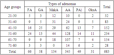
423 adenomas of the тhyroid gland were surgically removed during 5 years in the practice of the Republican Specialized Oncology and Radiology Scientific and Practical Medical Center and branch of the Tashkent region of the Ministry of Health of the Republic of Uzbekistan.Research results and their discussion: For morphometric examination, thyroid gland was removed from 683 patients who were surgically removed. It was determined that the age of the patients was from 21 to 76 years. The frequency of adenomas according to the age of patients is presented in the table below (Table 1).
3. Research Results and Their Discussion
For morphometric examination, thyroid gland was taken from 683 patients who were surgically removed. It was determined that the age of the patients was from 21 to 76 years. The frequency of adenomas according to the age of patients is presented in the table below (Table 1).Table 1. Distribution of the examined material by age groups and types of adenoma, numbers
 |
| |
|
As a result of histological examination of the biopsy material, 6 types of thyroid adenomas were identified: 1) follicular adenoma (FA), 2) hyalinized tubular adenoma (GA), 3) macrofollicular adenoma (MakA), 4) atypical adenoma (AA), 5) papillary (PA) and 6) oxyphilic adenoma (OksA).From the total number of adenomas, 88 were distinguished according to histological staining according to histological examination: 1) follicular adenoma (19), 2) hyalinized tubular adenoma (13), 3) macrofollicular adenoma (20), 4) atypical adenoma (16), 5) papillary (12) and 6) oxyphilic adenoma (8).Hematoxylin-eosin-stained histological sections were scanned and morphometric examination was performed based on the program. The length and width of the epithelial cells, the length and width of the nuclei, the length and width diameter of the follicles and the colloid were measured. Based on the obtained quantitative indicators, the area of colloid and follicles was calculated using the following formula: 𝑆=𝜋𝑎𝑏/4, where S is the area; a – longitudinal diameter; b – transverse diameter.The size of the core was calculated based on the spheroid size using this formula: 𝑉=4𝜋ℎ𝑎2/3, where V is the volume; h – core length; a is the width of the nucleus.The volume of the cell was calculated based on the volume of the cylinder in the following formula: 𝑉=𝜋ℎ𝑑2/4, where V is the volume; h – cell length; d is cell width.The size of the cytoplasm was calculated based on the difference between the size of the cell and the size of the nucleus. As a result of them, "The difference of the nuclear-cytoplasmic ratio was determined.As shown in the table of morphometric indicators, it was found that in safe adenomas of the thyroid gland, the height of the epithelium of the follicle wall was 4-5 μm, and the thickness was 6-7 μm, while it was the smallest in follicular and macrofollicular forms, and relatively large in atypical, papillary, and oncocytic adenomas. Accordingly, the size of the epithelial cell was the lowest (112.5±4.56) in the hyalinized adenoma, and the highest (171.5±4.8) in the oncocytic adenoma. It can be concluded that due to the expansion of follicles in normal forms of adenoma, the epithelium covering its wall atrophies and shrinks, and in atypical and oxyfid forms, epithelial cells become morpho-functionally activated, and their size and volume naturally increase.The results of morphometric calculations of follicular epithelial nuclei were also observed to change in accordance with the above changes. It was observed that in normal forms of adenoma, the nuclei of epithelial cells are relatively small (3.23±0.08), and in atypical forms they increase in size (4.54±0.06). It was found that the volume of the nucleus in follicular adenoma was only 58.12±3.56 μm3, while in oncocytic adenoma it increased almost 3 times, that is, it was 142.7±4.9 μm3 (Table 2). Studies have shown that if the size of the nucleus is small, the area of the cytoplasm is large, and as the size of the nucleus increases, the area of the cytoplasm decreases.The nuclear-cytoplasmic index, which shows the proliferative level of the cell, as determined in any cell. According to the determination of different forms of thyroid adenomas, it was found that this index has a low index compared to normal forms of adenoma, and high indices in atypical forms (Table 2).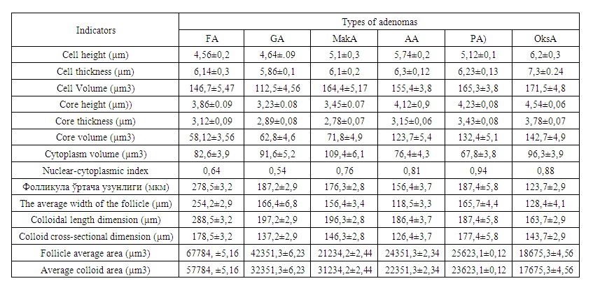 | Table 2. Morphometric indicators of tissue structures of thyroid adenomas |
Since the main tissue structures in thyroid adenomas are follicles, when their sizes were examined by morphometric methods, it was found that even in normal forms of adenoma, the length of the follicles is 278.5±3.2, the width is 254.2±2.9 μm, and the dimensions of the colloid are: length - 196.3±2.8, width - 146.3±2.8 μm, while in atypical and oncocytic forms of adenoma, these dimensions decrease, that is, follicle length - 163.7±2.9, width - 143, It was observed that the long diameter of the colloidal liquid was 7±2.9 μm, 128.4±4.1, and the narrow diameter was 123.7±2.9 μm.
4. Conclusions
1. It was found that the size of the epithelium and nuclei of thyroid gland adenomas is relatively smaller in normal forms, and larger in atypical and oncocytic forms, and accordingly, the cell size has also increased accordingly.2. Since follicles are the main tissue structures in thyroid adenomas, their size was observed to be twice as small as compared to the width in normal forms of adenoma, and in atypical and oncocytic forms.
References
| [1] | Agarwal S, Bychkov A, Jung CK. Emerging Biomarkers in Thyroid Practice and Research. // Cancers (Basel). 2021 Dec 31; 14(1): 204. |
| [2] | Dell'Aquila M, Granitto A, Martini M, Capodimonti S, Cocomazzi A, Musarra T, Fiorentino V, Pontecorvi A, Lombardi CP, Fadda G, Pantanowitz L, Larocca LM, Rossi ED. PD-L1 and thyroid cytology: A possible diagnostic and prognostic marker. // Cancer Cytopathol. 2020 Mar; 128(3): 177-189. |
| [3] | Caulley L, Eskander A, Yang W, Auh E, Zafereo M, Stack BC Jr, Randolph G, Davies L. Trends in Diagnosis of Noninvasive Follicular Thyroid Neoplasm With Papillarylike Nuclear Features and Total Thyroidectomies for Patients With Papillary Thyroid Neoplasms. // JAMA Otolaryngol Head Neck Surg. 2022 Feb 1; 148(2): 99-106. |
| [4] | French B, Hattier G, Mardekian SK. Utility of Tumor Capsule Thickness as a Predictor of Invasion in Encapsulated Follicular Variant of Papillary Thyroid Carcinoma and a Diagnostic Tool for Noninvasive Follicular Thyroid Neoplasm With Papillary-Like Nuclear Features. // Int J Surg Pathol. 2020 Feb; 28(1): 13-19. |
| [5] | Yang H, Chen L, Cheng Z, Yang M, Wang J, Lin C, Wang Y, Huang L, Chen Y, Peng S, Ke Z, Li W. Deep learning-based six-type classifier for lung cancer and mimics from histopathological whole slide images: a retrospective study. // BMC Med. 2021 Mar 29; 19(1): 80. |
| [6] | Zhu X, Chen C, Guo Q, Ma J, Sun F, Lu H. Deep Learning-Based Recognition of Different Thyroid Cancer Categories Using Whole Frozen-Slide Images. // Front Bioeng Biotechnol. 2022 Jul 6; 10: 857377. |
| [7] | Boursier L, Clerc Urmes I, Garon J, Klein M, Demarquet L. Ultrasound and cytological characteristics of non-invasive follicular thyroid neoplasm with papillary-like nuclear features compared to papillary carcinomas. // Ann Endocrinol (Paris). 2020 Feb; 81(1): 28-33. |
| [8] | Rosario PW, Mourão GF. Noninvasive follicular thyroid neoplasm with papillary-like nuclear features (NIFTP): a review for clinicians. // Endocr Relat Cancer. 2019 May 1; 26(5): R259-R266. |
| [9] | Kholová I, Haaga E, Ludvik J, Kalfert D, Ludvikova M. Noninvasive Follicular Thyroid Neoplasm with Papillary-like Nuclear Features (NIFTP): Tumour Entity with a Short History. A Review on Challenges in Our Microscopes, Molecular and Ultrasonographic Profile. // Diagnostics (Basel). 2022 Jan 20; 12(2): 250. |

 Abstract
 Reference
 Full-Text PDF
 Full-text HTML