Mushtariy M. Nuryogdieva1, Sayyora M. Akhmedova2, Umida K. Akhmadalieva3, Khabibulla Kh. Pulatov4
1Doctoral Student, Andijan State Medical Institute, Uzbekistan
2Doctor of Medical Sciences, Associate Professor, Tashkent Medical Academy, Uzbekistan
3Candidate of Medical Sciences, Associate Professor, Andijan State Medical Institute, Uzbekistan
4Candidate of Medical Sciences, Associate Professor, Tashkent Medical Academy, Uzbekistan
Copyright © 2022 The Author(s). Published by Scientific & Academic Publishing.
This work is licensed under the Creative Commons Attribution International License (CC BY).
http://creativecommons.org/licenses/by/4.0/

Abstract
In 160 fetuses the width of the transparent septum, lateral ventricles and IV ventricle prenatally during ultrasound examination were studied. Pregnant women were divided into two groups, the main group consisted of pregnant women in a state of hypothyroidism, the control group included women with uncomplicated pregnancy. The analysis of our data showed that in hypothyroid pregnant women there is an expansion of the cavity of the transparent septum, lateral ventricles and the anteroposterior size of the IV ventricle compared with the control group.
Keywords:
Fetometry, Hypothyroidism, Fetal screening, Brain indicators, Lateral ventricles, Pregnant women
Cite this paper: Mushtariy M. Nuryogdieva, Sayyora M. Akhmedova, Umida K. Akhmadalieva, Khabibulla Kh. Pulatov, Prenatal Change of Certain Brain Indicators with Hypothyroidism, American Journal of Medicine and Medical Sciences, Vol. 12 No. 12, 2022, pp. 1319-1322. doi: 10.5923/j.ajmms.20221212.28.
1. Introduction
In solving the problem of reducing maternal and perinatal morbidity and mortality, early diagnosis of complications of the gestational process, forecasting and choosing rational tactics for managing pregnancy and childbirth in high–risk women are of particular importance. Currently, up to 5% of newborns are born with various congenital and hereditary diseases. About 30% of the bed capacity of children’s hospitals of various profiles are occupied by patients with hereditary pathology, 40% of infant mortality in some other degree is due to hereditary and congenital defects, among which congenital cerebral defects occupy the leading place. Malformations of the nervous system occupies the third place in the structure of developmental anomalies after congenital pathology of the cardiovascular and urinary systems, and about 80% of these diseases are represented by hydrocephalus of various origins. Perinatal lesions of the nervous system in children remain one of the leading problems in medicine [2,5,9]. Perinatal brain lesions are a common cause of morbidity and mortality in newborns [1,2,7,10]. Perinatal lesions vary in etiology and pathogenesis of the brain of the fetus and newborn, arising in an unfavorable course of pregnancy, childbirth and the early neonatal period [3,4,11,12]. In this regard, their prenatal diagnosis is of particular importance, one of the main directions of which is the development of new most informative criteria that could be used for screening ultrasound examination at different stages of pregnancy. During pregnancy, the organs and systems of the fetus grow and develop, including the structures of the brain. The ultrasound picture of the brain in the prenatal period changes. As a result, the criteria for assessing the structures of the brain are changing. Knowing the patterns and characteristics of development allows you to conduct an ultrasound effectively and diagnose various developmental anomalies in time. The possibility of combining congenital malformations of the brain with other anomalies increases the relevance of their timely diagnosis. The presence and observance of the standards for ultrasound examination will allow to optimize the diagnosis of anomalies in the development of the structures of the fetal brain. In modern conditions, there are opportunities for an earlier diagnosis of defects that were previously diagnosed only in late pregnancy or after the birth of a child. Such pathologies, in particular, include anomalies in the development of the cerebral cortex. Thyroid hormones are essential during pregnancy for fetal development, in particular for fetal neurogenesis. Hypothyroidism in the mother, especially in the first trimester, has been associated with delayed neural development in the child.
2. Materials and Methods
To achieve this goal, 160 pregnant women were examined in terms of 16 to 28 weeks. The control group consisted of the fetuses of 65 women with uncomplicated pregnancy, the main group consisted of the fetuses of 95 hypothyroid women. All pregnant women underwent a comprehensive examination, including a thorough collection of somatic and obstetric–gynecological anamnesis, general clinical examination and ultrasound examination. In all fetuses, the width of the transparent septum, lateral ventricles and IV ventricle were studied prenatally during ultrasound examination. Measurements of the main structures of the brain were carried out retrospectively after taking the volumes of the fetal brain image on an ultrasound scanner using LOGIQ C5 ultrasound devices carried out on an ultrasound machine.To achieve the goal of the study, we used the offspring of rats born from 23 control and experimental white laboratory rats – mothers under conditions of hypothyroidism. Group 1 constituted the control group of healthy rats. Group 2 was 1–experimental group, in this group, 20 female white laboratory rats were given Mercazolil at a dose of 0.5 mg per 100 g of body weight for 14 days to induce experimental hypothyroidism. Then the rats were given Mercazolil at a dose of 0.25 mg per 100 g of body weight for 1 month. Female rats continued to be given Mercazolil at a dose of 0.25 mg per 100 g of body weight during lactation, both after pregnancy and after delivery.Female rats in the control group were given 1.0 ml of distilled water and 1.0 ml of 1% starch suspension every morning to reduce the harmful effects of oral gavage on rat stomachs. A subcutaneous catheter was used as a probe. Rat pups were bled under ether anesthesia on days 3, 7, 14, 21, and 30 after birth.At a thickness of 8–10 μm, paraffin histological sections prepared on a rotary microtome were stained with hematoxylin–eosin in the standard way.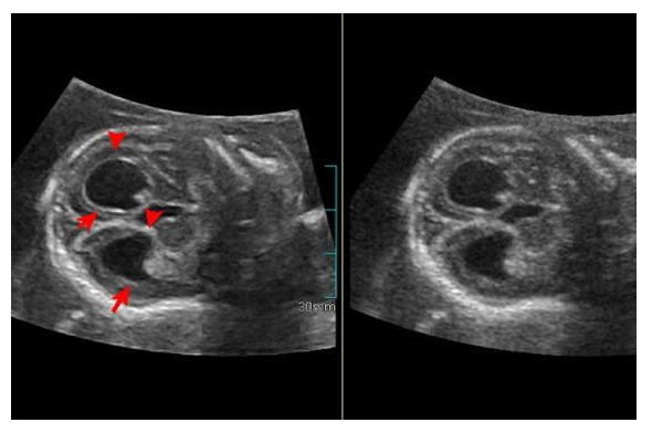 | Figure 1. Axial plane of scanning, measurement of the width of the cavity of the transparent septum. 19–20 weeks of gestation (12.5 mm) |
The results of the study showed that the value of the width of the cavity of the transparent septum of the fetus in healthy pregnant women at the stage of the first screening was not determined in 60% of cases, only 40% of cases averaged 5.7 ± 1.3 mm. In fetuses of pregnant women in a state of hypothyroidism at the stages of the first screening study, this indicator did not significantly differ from fetuses of healthy pregnant women and averaged 6.0 ± 1.4 mm. At the same time, a pattern was established that often in the period from 11 to 13 weeks of pregnancy, an axial section passing slightly below the cavity of the transparent septum creates a visual illusion of its image, although in fact this image corresponds to the legs of the brain. At the same time, a symmetrical image of both hemispheres of the fetal brain at the level of the visual hillocks was strictly observed; the M–echo was clearly visualized throughout and was interrupted only in the projection of the cavity of the transparent septum.At the stage of the second screening study, the width of the transparent septum of the fetuses in pregnant women in a state of hypothyroidism was significantly greater than in fetuses of healthy pregnant women: group and 6,8 ± 1,4 mm in fetuses of pregnant control groups. It should be noted that with an increase in the gestational age, a gradual increase in individual fluctuations in standard values was found. To assess the width of the cavity of the transparent septum, the mode of multiplanar reconstruction of the fetal brain was used to obtain a standard axial section using volumetric echography.The width of the transparent septum of fetuses in healthy pregnant women at the stage of the third screening study averaged 9.4 ± 1.5 mm; in fetuses of mothers in a state of hypothyroidism, this indicator was 1.6 times greater than in the control group and amounted to 15.2 ± 1.4 mm. An analysis of the study of the width of the transparent septum will make it possible to detect the expansion of the cavity of the transparent septum in 12 (80%) fetuses of pregnant women in a state of hypothyroidism, the absence of a transparent septum was diagnosed in 2 fetuses.Thus, in the course of the analysis, it was found that in fetuses of pregnant women in a state of hypothyroidism, an expansion of the cavity of the transparent septum is observed.The analysis of the conducted studies showed that the assessment of the width of the lateral ventricles of the brain was possible in all fetuses of the main group. Analysis of the results obtained showed the presence of a directly proportional relationship between the width of the lateral ventricles of the brain and the gestational age: with an increase in the gestational age, the indicators of the width of the lateral ventricles increase. The indicators of the width of the lateral ventricles of the fetus at the first screening examination in fetuses of healthy pregnant women averaged 4.9 ± 0.9 mm, with an increase in the gestational age, a gradual increase in the studied parameters is observed. It should be noted that at the time of the second screening, the width of the lateral ventricles in fetuses of healthy pregnant women was greater by 1.9 mm; at the stage of the third screening, this indicator increased by 1.6 mm and averaged 8.4 ± 1.2 mm. In 46% of pregnant fetuses in a state of hypothyroidism at the 1st screening period, lateral ventricular enlargement was revealed and this indicator averaged 6.2 ± 0.5 mm, in 54% of fetuses the width of the lateral ventricles did not differ from the control group. At the stage of the 2nd screening in fetuses of pregnant mothers in a state of hypothyroidism, expansion of the lateral ventricles by 1.9 times was noted and averaged 12.9 ± 0.7 mm. In fetuses of pregnant women in a state of hypothyroidism at 30–34 weeks, an expansion of the lateral ventricles by 2.1 mm compared with the control group was revealed and was equal to an average of 17.6 ± 1.6 mm.In the course of our studies, it was found that the IV ventricle of the fetal brain is a fairly easily identifiable structure when using the standard average sagittal scanning plane during screening ultrasound examination at 11–14 weeks of gestation. At the stage of the first screening during transabdominal scanning, the IV ventricle is visualized in 95% of fetuses. In 5% of pregnant women with transabdominal scanning, the IV ventricle of the fetus is not visualized, therefore, these women additionally underwent transvaginal echography during the observed periods of pregnancy.At the same time, at the stages of the first screening examination, the anteroposterior size of the IV ventricle in fetuses of healthy pregnant women was 1.62 ± 0.9 mm; in fetuses of pregnant women in a state of hypothyroidism at the stages of the first screening examination, an expansion of the anteroposterior dimension of the IV ventricle was noted and averaged 2.9 ± 1.4 mm. The average values of this indicator in fetuses of healthy pregnant women at 18-20 weeks were 3.2 ± 2.6 mm, and in fetuses of pregnant women in a state of hypothyroidism, a significant increase in the width of the IV ventricle was found and equal to 5.6 ± 1.4 mm. During the third screening examination, the average values of the anteroposterior size of the IV ventricle in fetuses of healthy pregnant women were 4.5 ± 2.6 mm, while in fetuses of the main group they were 2.2 mm larger and averaged 5.8 ± 2.6 mm.It was found that the number of nerve cells in the control group is 210 per 1 mm2, and the area of nerve cells is 185. In the case of hypothyroidism, a reduction of nerve cells and nuclei per 1 mm2 was observed in the brain. And in the case of hypothyroidism, there was no reliable change in the number of nerve cells in 7–day–old rat pups compared to the control group; it was found that in 14–day–old rats, it was 7% lower than in the control group, and in 21–day–old rats it was 20%, and at the 30th day of the experiment, it was 35% lower than in the control group, having the highest indicator. Morphometric studies showed that the area of nerve cells was also reduced compared to the control group.When studying the morphological structure of histological preparations, signs such as fullness of blood vessels in the brain, dimness, dimness in small blood vessels, and diapedesis hemorrhage appeared. no significant changes were observed on the 7th day of the experiment. On the 14th day of the experiment, initial signs of swelling appeared. The swelling appeared around the capillary and nerve cells. Fibrillation of collagen fibers was observed in the wall of blood vessels (Fig. 2). Dystrophic changes and desquamation were found in endotheliocytes. The nuclei of edotheliocytes are swollen. By the 21st day of the experiment, swelling around the capillaries increased and spread to all parts of the brain. Small spaces filled with swelling fluid appeared (Fig. 3).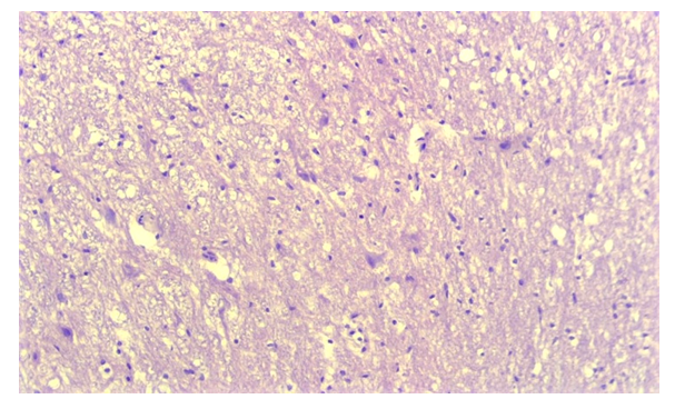 | Figure 2. Day 14 of the experiment. Vessels of the microcirculatory bed are in a state of uneven blood supply: some of them show stasis, while in other vessels the lumen is empty and dormant. Staining: hematoxylin–eosin. X: approx. 10, rev. 20 |
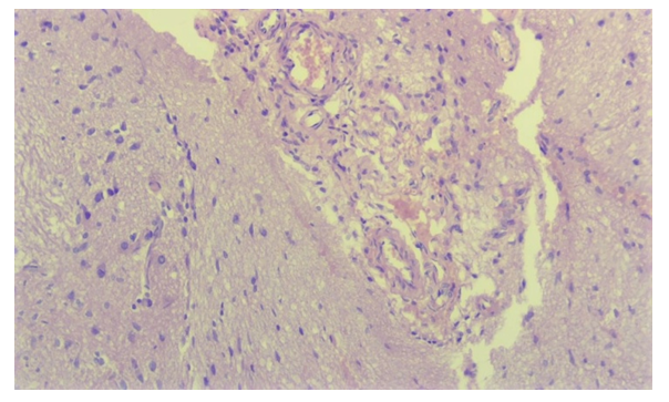 | Figure 3. 21 days of the experiment. Around the inflammatory focus, the brain tissue is loosened, depleted in cells and vacuolated due to edema of the brain substance. Staining: hematoxylin–eosin. X: approx. 10, rev. 20 |
The above–mentioned signs were more common in the white matter of the brain, and less often in the gray matter. By the 30th day, it was found that the swelling increased, and medium–caliber blood vessels increased around it. Structural changes in neurosecretory cells began to be observed in the areas where the tumor accumulated. Around the blood vessels there was a swelling of collagen fibers. During this period of the experiment, it was found that destructive and dystrophic changes in nerve cells increased. A decrease in the size of the neurons was observed. The neurocytes were not swollen, their deformation was detected. Vacuoles appear in the cytoplasm of astrocytes.
3. Conclusions
Thus, the use of a routine fetal screening study during pregnancy to assess the fetal brain allows an extended examination, which is necessary if an abnormal brain structure is suspected. Analysis of our data shows that in pregnant women in a state of hypothyroidism, an expansion of the cavity of the transparent septum is observed in 80%. In 90% of fetuses, the expansion of the lateral ventricles exceeded the standard values by 10 mm. This change was observed starting at 12 weeks. The anteroposterior size of the IV ventricle was larger in the fruits of the main group. According to foreign authors, the expansion of the cavity of the transparent septum allows one to suspect the abnormal development of the corpus callosum. When conducting screening ultrasound examination, the assessment of the cavity of the transparent septum, the width of the lateral ventricles of the brain allows early diagnosis of both complete and partial agenesis of the corpus callosum.
References
| [1] | Albu, D.–F.A Dandy –Walker variant prenatally diagnosed using ultrasound on one of the fetuses of a twin pregnancy obtained through in vitro fertilization / D.–F. Albu, C.–C. Albu, S.–D. Albu // International Journal of Medical Research and Review. Vol. 3, No. 3, 2015. – p. 127–131. |
| [2] | Bar – Yosef, O. Neurodevelopmental outcome of isolated ventriculomegaly: a prospective cohort study / O. Bar – Yosef, E. Barzilay, S. Dorembus [et al.] // Prenat. Diagn. Vol. 37. 2017. – p. 764–768. |
| [3] | Blinov A.Yu. Fundamentals of ultrasonic fetometry / A.Yu. Blinov, M.V. Medvedev. M.: Real Time, 2012. – p. 136. |
| [4] | Carseldine W. Antenatal diagnosis of a fetal dural venous thrombosis / W. Carseldine, F. Park, C. Abel [et al.] // Ultrasound Obstet. Gynecol. Vol. 42, No. 1, 2013. – p. 151. |
| [5] | Chiu T.H. A retrospective study on the course and outcome of fetal ventriculomegaly / T.H. Chiu, G. Haliza, Y.H. Lin [et al.] // Taiwanese Journal of Obstetrics & Gynecology. Vol. 53, 2014. – p. 170–177. |
| [6] | Esetov M.A. Echography of the central nervous system of the fetus. Intermediate sail cavity: own observation and literature review / M.A. Esetov, G.M. Bekeladze, E.M. Huseynova // Prenat. Diagn. Vol. 14, No. 1. 2015. – p. 46–51. |
| [7] | Malanina, E.N. Prenatal diagnosis of a rare syndrome with a wide range of brain malformations: case description, differential diagnosis, literature review / E.N. Malanin, D.R. Kasymova, R.R. Azizova [and others] // Prenat. Diagn. Vol. 10, No. 2. 2011. – p. 138–145. |
| [8] | Medvedev, M.V. Prenatal echography. Differential diagnosis and prognosis / M.V. Medvedev. 3rd ed. M.: Real Time, 2012. – p. 464. |
| [9] | Mirsharopov U.M., Usmonov R.J., Teshaev O.R. Mirzamuhamedov O.Kh. Akhmedova S.M. Morphological change of myocardium in hypothyroidism // Central Asia Journal of Medicine, 2020. – p. 71–83. |
| [10] | Mirzamukhamedov O.Kh., Mirsharopov U.M., Sodikova Z.Sh., Akhmedova S.M., Khatamov A.I., Mirzabekova O.A. Especially the development of myocarditis in hypothyroidism in postnatal ontogenesis // Indian Journal of Forensic Medicine & Toxicology. Vol. 14, № 4, 2020. – p. 7737–7745. |
| [11] | Trofimova T.N. Beam research of the fetal and newborn brain / T.N. Trofimova, A.S. Job, D.V. Voronin [and others] // Ed. Trofimova T.N. SPb.: Baltic Medical Educational Center, 2011. – p. 196. |
| [12] | Voevodin S.M. Differential diagnosis of diseases and malformations of the central nervous system and face in the fetus: dis. ... doct. medical sciences / Voevodin Sergey Mikhailovich. M.: 2012. – p. 312. |

 Abstract
 Reference
 Full-Text PDF
 Full-text HTML