American Journal of Medicine and Medical Sciences

p-ISSN: 2165-901X    e-ISSN: 2165-9036

2022;  12(11): 1156-1161

doi:10.5923/j.ajmms.20221211.13

Received: Nov. 2, 2022; Accepted: Nov. 26, 2022; Published: Nov. 29, 2022


A New Composite Plate for Hemostasis in Parenchymal Organ Surgery

Abdullajanov Bahrom Rustamjanovich, Babadjanov Azam Khasanovich, Khayaliev Rustem Yakubovich, Saliev Gayratbek Zakirovich

Republican Specialized Scientific and Practical Medical Center of Surgery Named after Academician V. Vakhidov, Andijan State Medical Institute, Uzbekistan

Copyright © 2022 The Author(s). Published by Scientific & Academic Publishing.

This work is licensed under the Creative Commons Attribution International License (CC BY).
http://creativecommons.org/licenses/by/4.0/

Abstract

Introduction. During trauma of parenchymal organs and closure of wound defects using special coatings, surgeons are faced with the problem of their functional versatility. A variety of collagen sponges and bands used in clinical practice have only a hemostatic effect. The aim of the study was experimental substantiation of the efficiency of a new polymer band implant for stopping parenchymal bleeding in surgery. Material and methods. The authors have demonstrated the effect of the new hemostatic implant, which was confirmed by laboratory, experimental studies, and morphological evaluation of tissues at the point of contact with the composite plate. Results. The results of in vitro studies have shown that the composite hemostatic band shortens the blood clotting time, which indicates an increase in the total coagulation activity of whole blood. This will allow to use it as a local hemostatic agent. Conclusion. The developed plate has a very high adhesive ability to a wet surface, the degree of which can ensure the stopping of not only capillary, but also mixed bleeding from parenchymal organs.

Keywords: Liver, Bleeding, Hemostasis, Plate, Experiment

Cite this paper: Abdullajanov Bahrom Rustamjanovich, Babadjanov Azam Khasanovich, Khayaliev Rustem Yakubovich, Saliev Gayratbek Zakirovich, A New Composite Plate for Hemostasis in Parenchymal Organ Surgery, American Journal of Medicine and Medical Sciences, Vol. 12 No. 11, 2022, pp. 1156-1161. doi: 10.5923/j.ajmms.20221211.13.

1. Introduction

As a result of abdominal injuries or extended surgical interventions, extensive wound defects are often formed, the restoration of which with the use of local tissues is not possible. It leads to the development of a number of complications such as: bleeding, lymphorrhea, prolonged healing, infection, formation of adhesions, etc. Injuries lead to 10% of all deaths worldwide, and blood loss is the most common preventable death cause after injury. Bleeding and hemorrhagic shock are the cause of 30-40% of traumatic deaths [1-5].
The closure of wound defects using special coatings faces the problem of their functional universality. A variety of collagen sponges and bands used in clinical practice have only a hemostatic effect; biological adhesives are designed to connect tissues; anti-adhesive gels are used only to prevent the adhesive process [6-11].
Effective and rapid hemostasis is critically important in surgical operations and emergency injuries. Hemostatic materials currently available include collagen (Col), gelatin (GE), alginate (AG), chitosan (CS), oxidized cellulose, cyanoacrylic acid-based tissue glue and porous zeolite. All of them have an effective hemostasis function, but they also have disadvantages [12-18].
The efficiency of hemostatic agents based on natural polymers should be based on the fact that they still have a lower potential effect than inorganic hemostatic materials. Advanced products for effective treatment of medical and surgical wounds include hemostatic agents, fibrin and other sealants, high-strength medical adhesives and products preventing postoperative adhesions [19-24].
There is a need to develop unique hemostatic agents that can deliver a hemostatic agent in a few seconds and stop bleeding within two minutes at the site of injury, without any intervention in the form of manual pressure on the wound area or any special preparation for their use. There is also a need to develop more hemostatic agents that can remain functional for a longer time without the need for replacement. Based on this, additional research is needed to analyze performance close to "real world" situations, which often requires unique and personalized injuries and clinical conditions.
The aim of the study was experimental substantiation of the efficiency of a new domestic polymer band implant for stopping parenchymal bleeding in surgery.

2. Material and Methods

We have developed a version of the hemostatic band, which has a complex composition and is a composite material. The main composition of the composite material is cellulose derivatives:
• A layer that provides high adhesion to the wet surface of biological tissues and is a band of Na-CMC (85%) + Ca2+ ions at a concentration of 15% with a thickness of 50 microns;
• The second layer is an enhanced hemostatic layer composed of Na-CMC (63%) + oxidized viscose (12%) + Ca2+ ions (25%). There are fibers of oxidized viscose in the second layerwith a thread thickness of up to 25 microns and a distance between the fibers of 500-1000 microns;
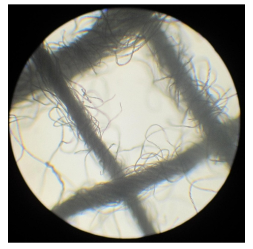
• The outer layer is a hydrophobic band up to 10 microns thick from vegetable oil (cottonseed oil) (Fig. 1).
Figure 1. Hemostatic band. The structure of the band of the mesh component from the OC. х40. SM
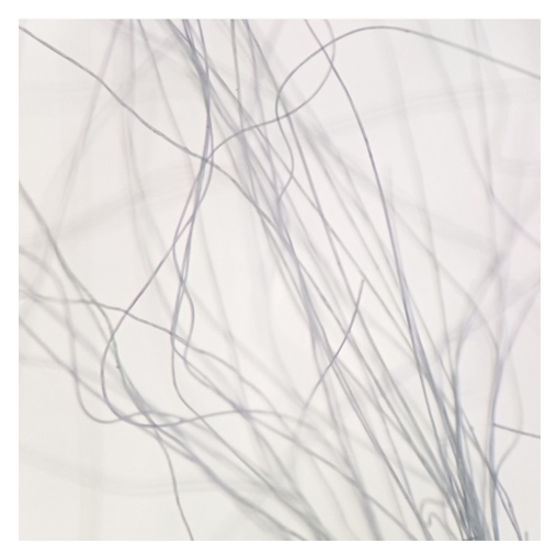
Viscose fiber is extracted by processing natural plant cellulose and is considered a modification of cellulose fiber called hydrate cellulose (Fig. 2). In our study, the native state of both fibers was evaluated using a stereomicroscope (MSP-2 LOMO).
Figure 2. Viscose fiber. х40. SM
At the same time, viscose, when visually examined, is free of defects, smooth, transparent, the thickness of the fibers is the same. Cotton cellulose fiber consists of many small fibers and is not smooth. The thickness is not the same along the length of the thread and varies unevenly.
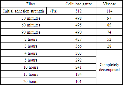
When studying the effect of oxidants (oxidizer - 12% sodium hypochlorite solution) on the breaking strength of the fibers that make up the composite coating, it was found that the breakage of the viscose fiber increases over time, and the effort spent on breaking decreases compared to cellulose fiber (Tab. 1).
Table 1. Breaking strength of the fibers included in the composite coating
     
Both fibers were changed in different ways under the influence of oxidants. Cotton cellulose showed high resistance, the degree of fiber breakage decreased only by 1.5 times in 4 hours. Viscose fiber lost strength 3 times in 3 hours, completely disintegrated by 4 hours.
The adhesion of the fibers was also evaluated. As a result of the tests, it was found that the adhesive ability of the viscose composite material is equal to 961 pascal (Pa).
Visual and stereomicroscopic evaluation of the new composite coating shows that the fibers were distributed in the center of the composite band and form a single unit with other components of the band. When examined under a stereomicroscope, the gauze fibers completely retained their mesh structure. The band thickness was 2 mm.
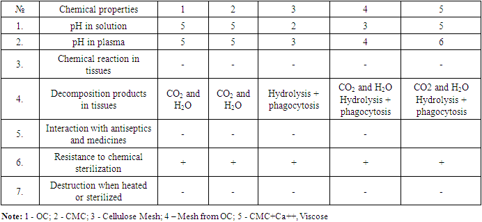
The physico-chemical properties of the composite hemostatic band in comparison with the control samples are shown in Table 2.
Table 2. The physico-chemical properties of the composite hemostatic band
     

3. Results

Adhesion of composite material. Taking into account the properties of the composite band, we have developed a device for studying the adhesion to various materials and fabrics.
The results obtained were calculated using the following formula: adhesive strength (F): F= mg. Here m - weight, g - acceleration of gravity 9.8 m²/c. Then the stickiness was calculated: F/S. Ra (Pascal) was chosen as the unit of measurement. The test substance, cut into 1 cm * 1 cm on the surface of the device, was evenly mixed with 100 μl of water. Lifting threads were attached to the attached cover glass with a small groove. We waited one minute, then attached it to the clamp, conducted an experiment to change the weight and studied it by adding water to the wetted part of the equipment. In this case, the density of water was assumed to be 1 g/cm3.
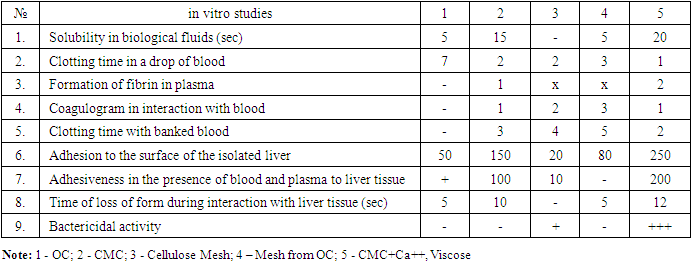
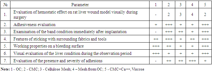
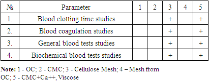
Table 3-7 shows the results on the chemical properties of the implant, In vitro, In vivo – morphological, In vivo – biochemical and toxicological studies.
Table 3. Chemical properties of composite hemostatic band
     
Table 4. Results of in vitro studies
     
Table 5. Results of In vivo studies - morphological
     
Table 6. Results of In vivo studies - biochemical
     
Table 7. Results of toxicological studies
     

4. Discussion

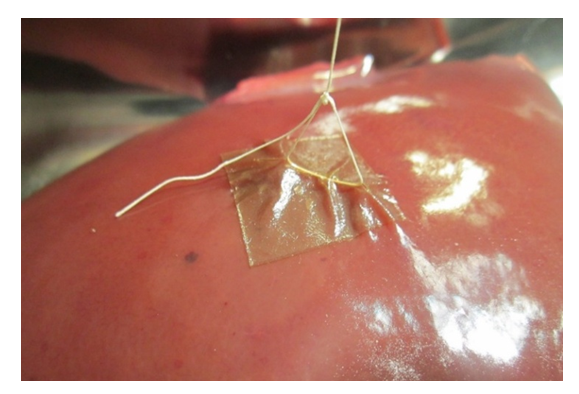
Eх vivo studies. Isolated sheep liver with a shelf life of no more than 1 hour in saline solution was used to evaluate adhesion to biological tissues. 1 cm2 of a hemostatic band was attached to the liver surface and the adhesion strength was measured using the technique described above (Fig. 3).
Figure 3. The beginning of the band shrinkage with increasing load
As a result of the tests, it was found that the adhesiveness of the prepared composite material using oxidized viscose was equal to 961 Ra (Pascal), which was a sufficient indicator for covering the wound surface, tight fixation and non-separation from it. In general, in a comparative aspect, among the various variants of cellulose-based biomaterials, it was found that the inclusion of oxidized viscose in the composition improved the physico-chemical properties of the composite hemostatic band, including the best indicators of tensile strength, adhesion to the surface of the isolated liver, as well as in the presence of blood and plasma, hygroscopicity and bactericidal activity.
A composite hemostatic band reinforced with viscose fibers was used as a material for experimental studies. The control was a plate reinforced with cotton cellulose fibers.
The study of blood clotting time according to the Lee-White method. A composite hemostatic band in the amount of 1 cm2 was added to a test tube with 1 ml of venous blood. A test tube with blood was placed on a water bath at 37°C, and a stopwatch was detected at the same time. 2 minutes after placing the blood, and then every 30 seconds, the tube was gently tilted at an angle of 60-45°. Blood flowed freely along the tube wall at the beginning of the study. The determination was carried out until a dense clot was formed. The time of blood clotting was considered to be the time from the moment of immersion of the hemostatic band into the test tube to the appearance of a clot, which made up 5.27 ± 0.50 seconds. Donation whole blood served as the study control. According to this study, the time of blood clotting in the control group was 11.2±0.54 sec. Normally, according to this method of investigation, the blood clotting time ranges from 4 min 55sec to 11 min 44sec.
The results of in vitro studies have shown that the composite hemostatic band shortens the blood clotting time, which indicates an increase in the total coagulation activity of whole blood.

5. Conclusions

In a comparative aspect, among the various variants of cellulose-based biomaterials, it was found that the inclusion of oxidized viscose in the composition provided an improvement in the physico-chemical properties of the composite hemostatic band, including the best indicators for tensile strength, adhesion to the surface of the isolated liver, as well as in the presence of blood and plasma, hygroscopicity and bactericidal activity.
Thus, the developed plate has a very high adhesive ability to a wet surface, the degree of which can ensure the stopping of not only capillary, but also mixed bleeding from parenchymal organs.
The possibility of having a hemostatic property was justified by the results of in vitro studies, which showed that the composite hemostatic band shortens the clotting time of blood, which, therefore, indicates an increase in the total coagulation activity of whole blood. This will allow it to be used as a local hemostatic agent.
The authors declare no conflict of interest.
This study does not include the involvement of any budgetary, grant or other funds.
The article is published for the first time and is part of a scientific work.

References

[1]  Sadykov R.A., Ismailov B.A., Kim O.V. A new band coating from cellulose derivatives for local hemostasis. In Russian // News of surgery.2019. №3. URL: http://985.so/b0v0e.
[2]  Holcomb J.B., Moore E.E., Sperry J.L., Jansen J.O., Schreiber M.A., et al. Evidence-Based and Clinically Relevant Outcomes for Hemorrhage Control Trauma Trials. Ann. Surg. 2021; 273: 395. doi:10.1097/SLA.0000000000004563.
[3]  Kalkwarf K.J., Drake S.A., Yang Y. Bleeding to Death in a Big City: An Analysis of All Trauma Deaths from Hemorrhage in a Metropolitan Area during 1 Year. J. Trauma Acute Care Surg. 2020; 89: 716-722. doi:10.1097/TA.0000000000002833.
[4]  Pourshahrestani, S., Kadri N. A., Zeimaran, E., Towler, M. R. Well-ordered mesoporous silica and bioactive glasses: promise for improved hemostasis. Biomaterials Sci, 7(1): 31-50 (2018). doi: 10.1039/c8bm 01041b.
[5]  Zhang S, Li J, Chen S, Zhang X, Ma J, He J. Oxidized cellulose-based hemostatic materials. Carbohydr Polym. 2020 Feb 15; 230: 115585. doi: 10.1016/j.carbpol.2019.115585.
[6]  Lipatov V.A., Kudryavtseva T.N., Severinov D.A. Application of carboxymethylcellulose in experimental surgery of parenchymal organs/ In Russian // Nauka molodykh (Eruditio Juvenium). -2020. -Vol. 8, №2.- P. 269-283.
[7]  Boerman MA, Roozen E, Sánchez-Fernández MJ, Keereweer AR, Félix Lanao RP, Bender JCME, Van Hest JCM. Next Generation Hemostatic Materials Based on NHS-Ester Functionalized Poly(2-oxazoline)s. Biomacromolecules. 2017 Aug 14; 18(8): 2529-2538. doi: 10.1021/acs.biomac.7b00683.
[8]  Khan MA, Mujahid M. A review on recent advances in chitosan based composite for hemostatic dressings. Int J Biol Macromol 2019; 124: 138-47. 10.1016/j.ijbiomac.2018.11.045.
[9]  Li D, Chen J, Wang X, Zhang M, Li C, Zhou J. Recent Advances on Synthetic and Polysaccharide Adhesives for Biological Hemostatic Applications. Front Bioeng Biotechnol. 2020 Aug 14; 8: 926. doi: 10.3389/fbioe.2020.00926.
[10]  Mndlovu H, du Toit LC, Kumar P, Choonara YE, Marimuthu T, Kondiah PPD, Pillay V. Bioplatform Fabrication Approaches Affecting Chitosan-Based Interpolymer Complex Properties and Performance as Wound Dressings. Molecules. 2020 Jan 6; 25(1): 222. doi: 10.3390/molecules25010222.
[11]  Yin M.L., Wang Y.F., Zhang Y., Ren X.H., Qiu Y.Y., Huang T.S. (2020). Novel quaternarized N-halamine chitosan and polyvinyl alcohol nanofibrous membranes as hemostatic materials with excellent antibacterial properties. Carbohyd. Polym. 232: 115823. 10.1016/j.carbpol.2019.115823.
[12]  Belozerskaya G.G., Bychichko D.Yu., Kabak V.A. et al. Creation of new hemostatic coatings of local action based on sodium alginate. In Russian // Clinical physiology of blood circulation. - 2018. - Vol. 15. - № 3. - P. 222-229.
[13]  Kholturaev B.Zh., Atakhanov A.A., Sarymsakov A. A. Hemostatic films based on carboxymethylcellulose. In Russian // Universum: chemistry and biology: electron. scientific. Journal /. 2021. 9(87).
[14]  Gu BK, Park SJ, Kim MS, et al. Fabrication of sonicated chitosan nanofiber mat with enlarged porosity for use as hemostatic materials. Carbohydr Polym 2013; 97: 65-73. 10.1016/j.carbpol.2013.04.060.
[15]  Jiang X, Wang Y, Fan D, Zhu C, Liu L, Duan Z. A novel human-like collagen hemostatic sponge with uniform morphology, good biodegradability and biocompatibility. // J Biomater Appl. 2017 Mar; 31(8): 1099-1107.
[16]  Lan G., Lu B., Wang T., Wang L., Chen J., Yu K., et al. (2015). Chitosan/gelatin composite sponge is an absorbable surgical hemostatic agent. Colloids Surf. B 136 1026-1034. 10.1016/j.colsurfb.2015.10.039.
[17]  Nakielski P., Pierini F. Blood interactions with nano- and microfibers: Recent advances, challenges and applications in nano- and microfibrous hemostatic agents. Acta Biomaterialia, 84, 63–76 (2019). doi: 10.1016/j.actbio.2018.11.029.
[18]  Sung YK, Lee DR, Chung DJ. Advances in the development of hemostatic biomaterials for medical application. Biomater Res. 2021; 25(1): 37. Published 2021 Nov 12. doi:10.1186/s40824-021-00239-1.
[19]  Budko E.V., Chernikova D.A., Yampolsky L.M., Yatsuk V.Ya. Local hemostatic means and ways of their improvement/ In Russian // Russian med.-biol. vestn. named after. akad. I.P. Pavlov. 2019. №2. URL: https://cyberleninka.ru/article/n/mestnye-gemostaticheskie-sredstva-i-puti-ih-sovershenstvovaniya.
[20]  Lempert A.R., Belozerskaya G.G., Makarov V.A. et al. Hemostatic activity of a new compound based on fibrin monomer during intravenous administration in the experiment // Experimental and clinical pharmacology. - 2018. - Vol. 81. - № 11. - P. 14-17.
[21]  Chen Y, Qing J, Chang Q, Zhao Y, Jing Y, Ding J, Guo H. Preparation and evaluation of porous starch/chitosan composite crosslinking hemostatic. European Polym. J. 2019; 118: 17-26. doi:10.1016/j.eurpolymj.2019.05.039.
[22]  Hazra C., Kundu D., Chatterjee A. Stimuli-responsive nanocomposites for drug delivery. In: Inamuddin A.A.M., Mohammad A., editors. Applications of Nanocomposite Materials in Drug Delivery. 1st ed. Elsevier Inc; Amsterdam, The Netherlands: 2018. pp. 823-841.
[23]  Jiang X, Wang Y, Fan D, Zhu C, Liu L, Duan Z. A novel human-like collagen hemostatic sponge with uniform morphology, good biodegradability and biocompatibility. // J Biomater Appl. 2017 Mar; 31(8): 1099-1107.
[24]  Xie Y, Gao P, He F, Zhang C. Application of Alginate-Based Hydrogels in Hemostasis. Gels. 2022 Feb 10; 8(2): 109. doi: 10.3390/gels8020109.