American Journal of Medicine and Medical Sciences

p-ISSN: 2165-901X    e-ISSN: 2165-9036

2022;  12(10): 1068-1073

doi:10.5923/j.ajmms.20221210.13

Received: Sep. 25, 2022; Accepted: Oct. 8, 2022; Published: Oct. 21, 2022


The Analysis of Surgical Treatment Results of Patients with Intracerebral Hemorrhages

L. B. Maksudova1, B. G. Gafurov2, M. K. Makhkamov1

1Republican Research Center of Emergency Medicine, Tashkent, Uzbekistan

2Center for the Development of Professional Qualifications of Medical Employees, Tashkent, Uzbekistan

Copyright © 2022 The Author(s). Published by Scientific & Academic Publishing.

This work is licensed under the Creative Commons Attribution International License (CC BY).
http://creativecommons.org/licenses/by/4.0/

Abstract

The authors carried out a comparative analysis of surgical treatment results of 80 patients with hypertensive intracerebral hemorrhage (ICH). There were 22 females and 58 males. The mean age was 58.8±14.0 years. The results of the study showed that a comprehensive analysis of MSCT morphometric parameters were necessary in order to achieve high rates of 30- and 90-day survival in patients with intracerebral hemorrhage. This, in turn, allows the surgeon to choose the optimal and adequate method of surgical intervention in each specific case.

Keywords: Intracerebral hemorrhage, Intraventricular hemorrhage, Surgical treatment

Cite this paper: L. B. Maksudova, B. G. Gafurov, M. K. Makhkamov, The Analysis of Surgical Treatment Results of Patients with Intracerebral Hemorrhages, American Journal of Medicine and Medical Sciences, Vol. 12 No. 10, 2022, pp. 1068-1073. doi: 10.5923/j.ajmms.20221210.13.

1. Introduction

There has been an increase in the prevalence of intracerebral hemorrhages (ICH) in recent years which rank second according to the World Health Organization. The frequency of ICH is on average 20 cases per 100,000 population, the fatal outcome exceeds 40% [27]. There are discussions about the surgical treatment of ICH to this day. Most specialists in this field offer a lot of different types of techniques of surgical treatment. Despite the existing various methods of surgical interventions on improving the treatment results and survival of patients with ICH, the mortality and disability rates with this formidable pathology still remain high [1-6,28]. There are still no clear standards or algorithms for surgical interventions in relation to hemorrhagic stroke. Postoperative mortality in the world's leading ICH surgery clinics reaches 48 – 50% [2,11,15,26]. The high mortality rate is justified by the fact that hemorrhagic stroke is a consequence not only of an isolated cerebral lesion, but also a complication of medically uncontrolled hypertension [16,21,32]. High mortality and disability of patients determine the relevance and detailed study of this issue in the field of ICH surgery.
The aim of this research is to study the 30- and 90-day survival in patients with ICH after surgery depending on MSCT morphometric parameters.

2. Material and Methods

We conducted a retrospective analysis of 80 patients with brain ICH. There were 22 females and 58 (72,5%) males aged 15 to 88 years The mean age was 58.8±14.0 years. All patients were performed neurological examination and assessment of the condition severity according to the National Institutes of Health Stroke Scale (NIHSS) and the modified Rankin scale (mRs). Evaluation of the consciousness level violation was carried out using the Glasgow Coma Scale (GCS).
After studying the anamnesis and physical examination, all patients were performed a multi-slice computed tomography (MSCT). MSCT-morphometry included the assessment of the following indicators: localization and volume of the hematoma, the presence of a breakthrough of blood into the ventricular system, the condition of the bypass cistern, deformation or expansion of the ventricular system and its sizes, displacement of the median structures of the brain, perifocal edema around the hematoma at its presence and secondary brain changes.
We used a modified Graeb scale (mGS) when assessing the spread of intraventricular hemorrhage. The 30- and 90-day survival of patients using the Kaplan-Mayer method was estimated based on the analysis of MSCT-morphometric parameters.

3. Results

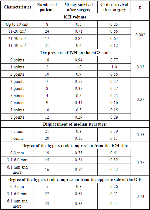
An analysis of the results of 80 patients showed the following: in 22.5% (n=18) cases, lobar localization of ICH took place, in 13.8% (n=11) cases - medial localization, 15% (n=12) cases - lateral localization, in 10% (n=8) cases - localization in the thalamic area, in 27.5% (n=22) cases - mixed localization and in 11.2% (n=9) cases hemorrhage was diagnosed in the posterior cranial fossa. The analysis of 30- and 90-day survival of patients with ICH showed the following results: in 10% (n=8) cases the ICH volume was up to 10 cm3 (breakthrough into the ventricular system with a high gradation on the mGS scale), in 30% (n=24) cases, the volume of ICH ranged from 11 to 20 cm3: in 9 of them hemorrhage was localized in the posterior cranial fossa, and in the remaining 11 cases there was a medial localization with a breakthrough of blood into the ventricular system with occlusion of the liquor pathways. Endoscopic removal of the ICH was performed in 4 cases with lateral hemorrhage with an ICH volume of 20 cm3. In 21.2% (n=17) cases the volume of ICH ranged from 21 to 30 cm3 and in 51.7% (n=31) cases it varied from 31 to 40 cm3 (Table 1).
Table 1. Characteristics of 30- and 90-day survival of patients with ICH, depending on clinical, CT-morphometric and ultrasound parameters after surgery
     
The results of the large-scale study "STICH II" showed strong evidence in support of the hypothesis of improved outcomes after early surgical intervention, the advantage of which was justified by the fact that after the removal of ICH, the perfusion of the medulla around the bed was allegedly improved due to the elimination of mass effect and local vascular compression [12,14,17,18,22,23,29]. But other numerous randomized studies contradict the above mentioned theory, as the surgical intervention itself is an additional factor of aggression, causes an aggravation of vascular spasm, an increase in edema around the hematoma bed, which in turn leads to a persistent unsatisfactory functional outcome [8,10,30]. However, there was a statistically significant positive result towards early surgery in compare with conservative therapy in patients with a level of consciousness according to GCS >13 points without signs of IVH and lobar localization [19].
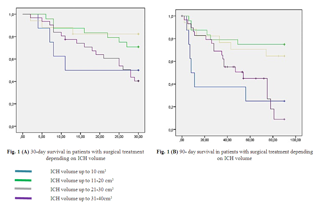
We conducted a comparative analysis of the 30- and 90-day survival rates of patients with ICH, depending on the volume of hemorrhage. The analysis showed a tendency towards an increase of these indicators as the volume of ICH increased, which specified the efficiency of surgical intervention. Thus, the 30-day survival rate with an ICH volume up to 10 cm3 was 0.5, with a volume from 11 to 20 cm3 – 0.71 and with a volume from 21 to 30 cm3, this indicator was 0.81, respectively. There was also a dynamic increase of 90-day survival rates by rising of ICH volume – 0.15, 0.66 and 0.63, respectively. Particular attention was drawn to the category of patients with an ICH volume from 31 to 40 cm3: there was a contradictory picture of survival rates in this group of patients, i.e. a decrease in 30-day survival rates to 0.4 and 90-day to 0.12, respectively (Figure 1 A and B).
Figure 1
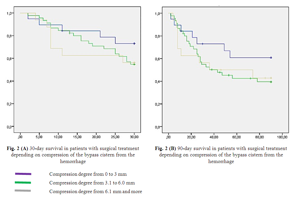
Consequently, there is a question and contradictory data in favor of surgical intervention. Possible reasons for this phenomenon may be other MSCT morphometric parameters, such as the presence of a pronounced compression of the bypass cistern, a pronounced displacement of the median structures, and a high gradation of intraventricular hemorrhage (IVH) according to the mGS scale. Thus, the analysis of the bypass cistern compression degree showed that at the most pronounced compression of the bypass cistern, this indicator ranged from 0 to 3 mm (the most pronounced cerebral edema), which was observed in 23.7% (n=19) cases, in 56.3% (n=45) cases – from 3.1 to 6.0 mm and in 20% (n=16) cases – 6.1 mm or more. The 30- and 90-day survival rates were highest in patients with severe cerebral edema – 0.73 and 0.61, respectively. This was the result of adequate and optimal surgical tactics aimed not only to the evacuation of the ICH, but also decompressive trepanation for the elimination of the bypass cistern compression from the hemorrhage, since the evacuation of the hematoma itself did not eliminate the compression factor of the brain bypass cistern of the due to the formation of cerebral edema around the ICH because of hemorrhage and surgical manipulation. Thus, it can be summarized that hematoma evacuation itself does not eliminate the problem of cerebral compression and thus requires an individual approach in each specific case. With sizes from 3.1 to 6.0 mm and from 6.1 mm or more, the 30-day survival rate was 0.54 and 0.56, respectively. The 90-day survival rate was 0.39 and 0.42, respectively, which also testifies in favor of the results we obtained (Figure 2 A and B).
Figure 2

4. Discussion

When analyzing the world literature on the basis of multiple large-scale studies conducted by leading experts in this field, it was noted that one of the main predictors of an unfavorable outcome was IVH as a result of the breakthrough of ICH into the ventricular system of the brain [7,13,20]. It was proved in the STICH study, that intraventricular hemorrhage due to an ICH breakthrough reduced the frequency of favorable outcomes from 31% to 15% [9]. Trifan G et al. in his researches proved that the indicators of the mGS scale were the most important predictors of the treatment outcome in patients with ICH complicated by a breakthrough of blood into the ventricular system. Thus, it was noted in this study that in patients with mGS >5 points, the probability of an unfavorable outcome was high. In patients with mGS <5 points, the probability of a favorable outcome was high [24]. Tuhrim et al. showed in his studies that IVH volume was associated with lower 30-day survival rates [25]. Also Young et al. determined in their studies that an IVH volume >20 ml was a poor prognostic factor [31]. Analysis of the results of 30- and 90-day survival of patients with surgical treatment, depending on the degree of grade according to the mGS scale, showed the following: with zero grade by the mGS scale, 12.8% (n=18) cases were observed (the 30-day survival rate was 0.94); with the first grade by the mGS scale, there was 1.4% (n=2) - the 30-day survival rate was 1.0; with the second grade - (7.1% (n=10)) – 0.9; at the third grade - (5% (n=7)) – 0.57; with the fourth one - (5.7% (n=8)) – 0.37; with the fifth grade - (2.8% (n=4)) – 0.5; with the sixth one - (6.4% (n=9)) – 0.44; with the seventh grade - (7.1% (n=10)) – 0.3; 8.6% (n=12) cases were observed with the eighth grade (30-day survival was 0.29) (Figure 3 A and B).
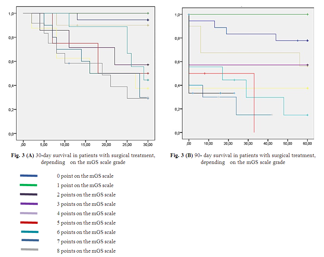
Figure 3

5. Conclusions

Thus, we can conclude based on our study that 30- and 90-day survival rates in patients with ICH who were performed surgery depend on MSCT morphometric parameters: localization of ICH, volume of hemorrhage, degree of intraventricular hemorrhage grade on the mGS scale and the degree of compression of the bypass cistern of the brain.
A comprehensive analysis of MSCT morphometric parameters in patients with ICH allows the surgeon to choose the optimal and adequate method of surgical intervention in each case, which in turn increases the survival rates of ICH patients and thereby reduces the degree of mortality.
The authors declare no conflict of interest.
This study does not include the involvement of any budgetary, grant or other funds.
The article is published for the first time and is part of a scientific work.

References

[1]  van Asch CJJ, Luitse MJA, Rinkel GJE, van der Tweel I, Algra A, Klijn CJM. Incidence, case fatality, and functional outcome of intracerebral haemorrhage over time, according to age, sex, and ethnic origin: a systematic review and meta-analysis. // Lancet Neurol 2010; 9: 167–76.
[2]  Zorin N.A., Grigoruk S.P., Sirko A.G., Garus D.V., Plushev E.I. The use of frameless CT navigation and local fibrinolysis in surgery for non-traumatic intracerebral hemorrhages of deep localization. In Russian // Ukrainian neurosurgical journal.2010 – No 2.P. 20-26.
[3]  Krylov V.V., Dashyan V.G., Godkov I.M. Endoscopic surgery for hemorrhagic stroke. In Russian - M.: Binom, 2014. – P.96.
[4]  Likholetova N.V., Kovalev V.V., Gorbachev V.I., Petrova I.L. Predictors of the outcome of hemorrhagic stroke in intensive care practice. In Russian // Siberian medical journal. Irkutsk. 2011 – No 6; P. 105-108.
[5]  Piradov M.A. Hemorrhagic stroke: new approaches to diagnosis and treatment. In Russian // Nervous diseases. 2005 – No 1; P. 17-19.
[6]  Yarikov A.V., Balyabin A.V. Analysis of the causes of mortality and prognostically unfavorable factors of surgical treatment of hemorrhagic stroke. In Russian // Medial. 2015 – No3; P. 148-151.
[7]  Al Shardan, M.M. Factors that predict hydrocephalus following intraventricular hemorrhage // Brit j neurosurg. - 2015. -Vol. 29, № 2. - P. 225-228.
[8]  Xi G, Keep RF, Hoff JT. Mechanisms of brain injury after intracerebral haemorrhage. // Lancet Neurol 2006; 5: 53–63.
[9]  Feigin VL, Lawes CMM, Bennett DA, Anderson CS. Stroke epidemiology: a review of population-based studies of incidence, prevalence, and case-fatality in the late 20th century. // Lancet Neurol 2003; 2: 43–53.
[10]  Lee H.S., Lee K.S., Bae H.G. et al. Clinical course of spontaneous gangliotalamic hemorrhage in the acute period - who requires surgical removal? // J. Korean Med. Sci. - 1991. - Vol. 6 (2). - P. 103-111.
[11]  van Asch CJJ, Luitse MJA, Rinkel GJE, van der Tweel I, Algra A, Klijn CJM. Incidence, case fatality, and functional outcome of intracerebral haemorrhage over time, according to age, sex, and ethnic origin: a systematic review and meta-analysis. // Lancet Neurol 2010; 9: 167–76.
[12]  Mendelow AD. Mechanisms of ischemic brain damage with intracerebral hemorrhage. // Stroke 1993; 24 (suppl I): I115–I7.
[13]  Nehls DG, Mendelow DA, Graham DI, Teasdale GM. Experimental intracerebral hemorrhage: early removal of a spontaneous mass lesion improves late outcome. // Neurosurgery 1990; 27: 674–82.
[14]  Zuccarello M., Brott T., Derex L. et al. // Early Surgical Treatment for Supratentorial Intracerebral Hemorrhage // Stroke. - 1999. - Vol. 30. – P. 1833-1839.
[15]  Herrick, D.B. Determinants of external ventricular drain placement and associated outcomes in patients with spontaneous intraventricular hemorrhage / D.B. Herrick, N. Ullman, S. Nekoovaght-Tak et al. // Neurocrit care. - 2014. - Vol. 21, № 3. - P. 426-434.
[16]  Kuo Lu-Ting, Chen Chien-Min, Li Chien-Hsun et al. Early endoscope- assisted hematoma evacuation in patients with supratentorial intracerebral hemorrhage: case selection, surgical technique, and longterm results // Focus Neurosurg. - 2011. - Vol. 30 (4). - P. 1-8.
[17]  Mendelow AD, Bullock R, Teasdale GM, Graham DI, McCulloch J. Intracranial haemorrhage induced at arterial pressure in the rat: part 2. Short term changes in local cerebral blood flow measured by autoradiography. // Neurol Res 1984; 6: 189–93.
[18]  Mendelow DA, Gregson BA, Rowan EN, Murray GD, Gholkar A, Mitchell PM // Early surgery versus initial conservative treatment in patients with spontaneous supratentorial lobar intracerebral haematomas (STICH II): a randomised trial // Lancet 2013; 382: 397–408.
[19]  Siddique MS, Fernandes HM, Arene NU, Wooldridge TD, Fenwick JD, Mendelow AD. Changes in cerebral blood fl ow as measured by HMPAO SPECT in patients following spontaneous intracerebral haemorrhage. // Acta Neurochir Suppl 2000; 76: 517–20.
[20]  Trifan G., Arshi B., Testai F.D. Intraventricular Hemorrhage Severity as a Predictor of Outcome in Intracerebral Hemorrhage // Frontiers in Neurology 2019 10: 217. doi:10.3389/fneur.2019.00217.
[21]  Xi G, Wagner KR, Keep RF, et al. Role of blood clot formation on early edema development after experimental intracerebral hemorrhage. // Stroke 1998; 29: 2580–86.
[22]  Bhattathiri, P.S. Intraventricular hemorrhage and hydrocephalus after spontaneous intracerebral hemorrhage: results from the STICH trial // Acta neurochirurg. - 2006. - Vol. 96, suppl. - P. 65-68.
[23]  Cho D.Y., Chen C.C., Chang C.S. et al. Endoscopic surgery for spontaneous basal ganglia hemorrhage: comparing endoscopic surgery, stereotactic aspiration, and craniotomy in noncomatose patients //Surg. Neurol. - 2006. - Vol.65. - P. 547-555.
[24]  Young W.B., Lee K.P., Pessin M.S., Kwan E.S., Rand W.M., Caplan L.R. Prognostic significance of ventricular blood in supratentorial hemorrhage: a volumetric study. // Neurology. 1990 40:616–9. doi: 10.1212/WNL.40. 4.616.
[25]  Moradiya, Y. Intraventricular thrombolysis in intracerebral hemorrhage requiring ventriculostomy: a decade-long real-world experience // Stroke. - 2014. - Vol. 45, № 9. - P. 2629-2635.
[26]  Auer LM, Deinsberger W, Niederkorn K, et al. Endoscopic surgery versus medical treatment for spontaneous intracerebral hematoma: a randomized study. // J Neurosurg 1989; 70: 530–35.
[27]  Keep RF, Xi G, Hua Y, Hoff JT. The deleterious or beneficial effects of different agents in intracerebral hemorrhage: think big, think small, or is hematoma size important? // Stroke 2005; 36: 1594–96.
[28]  Morgenstern L.B., Hemphill III J.C., Anderson C. et al. Guidelines for the management of spontaneous intracerebral hemorrhage: a guideline for healthcare professionals from the american heart association/American stroke association // Stroke. - 2010. - Vol. 41. - P. 2108-2129.
[29]  Bhattathiri PS, Gregson B, Prasad KS, Mendelow AD, STICH Investigators. Intraventricular hemorrhage and hydrocephalus after spontaneous intracerebral hemorrhage: results from the STICH trial. // Acta Neurochir Suppl 2006; 96: 65–68.
[30]  Tuhrim S., Horowitz D.R., Sacher M., Godbold J.H. Volume of ventricular blood is an important determinant of outcome in supratentorial intracerebral hemorrhage. // Crit Care Med. 1999 27: 617–21. doi: 10.1097/00003246-199903000-00045.
[31]  Smith EE, Shobha N, Dai D, Olson DM, Reeves MJ, Saver JL, Hernandez AF, Peterson ED, Fonarow GC, Schwamm LH. A risk score for in-hospital death in patients admitted with ischemic or hemorrhagic stroke. J Am Heart Assoc. 2013; 2: e005207.