American Journal of Medicine and Medical Sciences

p-ISSN: 2165-901X    e-ISSN: 2165-9036

2022;  12(10): 1034-1038

doi:10.5923/j.ajmms.20221210.05

Received: Sep. 8, 2022; Accepted: Oct. 2, 2022; Published: Oct. 14, 2022


Clinical-Immunological Methods of Clinical-Immunological Research of Atopic Dermatitis in Children Permanently Residing in the Area of Oil Refining Enterprises

Achilova Donokhon Nutfullayevna , Khushvaktova Madina Farkhodovna

Bukhara State Medical Institute Named after Abu Ali Ibn Sina, Uzbekistan

Copyright © 2022 The Author(s). Published by Scientific & Academic Publishing.

This work is licensed under the Creative Commons Attribution International License (CC BY).
http://creativecommons.org/licenses/by/4.0/

Abstract

To date, a number of factors have been identified that aggravate the course of atopic dermatitis - food sensitivity, contact with pets, psychological discomfort and emotional stress, a history of intrauterine infections, skin colonization with Staphylococcus aureus. But the information received is not always reliable from the point of view of evidence-based medicine. However, an integrated one has not been developed; systems for predicting the nature of the disease in children. Accession of a secondary skin infection adversely affects the severity of the disease and the quality of life of patients; the frequency of complex forms of atopic dermatitis in children is on average 25-34%.

Keywords: Atopic dermatitis, Сhildren's age, Young child, Inflammation, Bifalak-Zinkum+S+D3, Prevention, Treatment

Cite this paper: Achilova Donokhon Nutfullayevna , Khushvaktova Madina Farkhodovna , Clinical-Immunological Methods of Clinical-Immunological Research of Atopic Dermatitis in Children Permanently Residing in the Area of Oil Refining Enterprises, American Journal of Medicine and Medical Sciences, Vol. 12 No. 10, 2022, pp. 1034-1038. doi: 10.5923/j.ajmms.20221210.05.

1. Introduction

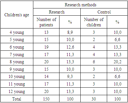
Children from 4 to 12 years of age living in the territory of the Bukhara Oil Refinery, Karavulbazar District, Bukhara Region, were examined. During the research, according to the tasks, 180 children were under our control, 150 of them were children with atopic dermatitis and 30 were healthy children (Table 1).
Table 1. Examined age levels of children in the study
     
To select a group of sick children, we used the following criteria for inclusion and exclusion of sick children during the study period.
Research criteria:
1. Male and female patients with atopic dermatitis aged 4 to 12 years.
2. Mental health.
3. Written consent of parents to participate in the study.
Exclusion criteria:
1. In our opinion, the presence of any medical condition, which, in our opinion, could create an unreasonable risk of harm to the child's health during his participation in the study, as well as seriously affect the reliability of the data, was obtained during the study.
2. The primary episode of the disease.
3. Hypersensitivity to any components of the studied drugs.
4. Presence of common secondary skin infection.
5. Reception of immunosuppressants, corticosteroids, treatment with various drugs in the last 4 weeks, as well as the planned course of therapy indicated above during the study period.
6. Therapy with topical glucocorticoids one week before the study.
7. Presence of serious somatic pathology.
8. Participation in any other clinical treatment within the next 3 months.
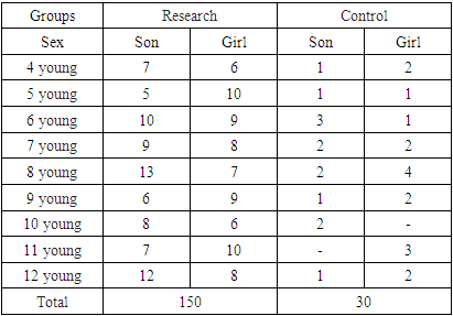
During the study, we selected children whose age ranged from 4 to 12 years. When analyzing the gender of children, it was determined what the ratio of boys and girls was (Table 2).
Table 2. Gender ratio of children in the study
     
The selection of patients in the age group was influenced by the fact that they are the most vulnerable group to the chronic effects of toxic substances, as well as their permanent residence in this area. Patients were examined and treated at the Department of Skin and Genitourinary Diseases and Allergology of the Bukhara State Medical Institute. A group of healthy children was selected during a clinical examination at the polyclinic of the Karavulbazar district of the Bukhara region from 2020 to 2021.
Children in the control group did not suffer from atopic dermatitis. They had no genetic predisposition to bronchial asthma, allergic rhinitis and other allergic diseases, no other chronic somatic diseases, no abnormalities in clinical and biochemical blood tests, no infectious diseases at least one month before the study. The study contingent was selected to have a certain predominance of girls from 59% to 41%. Table 2 shows the distribution of children by gender and age.
The study was conducted based on the clinical features of the diagnosis of atopic dermatitis. To solve the specified tasks, we used a set of special research methods (Table 3).
Table 3. Verification methods used during the research
     

2. Methods of General Examination of Children with Atopic Dermatitis

Dermatologic Examination (SCORAD)
The severity of atopic dermatitis was evaluated using the SCORAD scale (scoring of atopic dermatitis) during the study. When evaluating the SCORAD index, the objective characteristics of rashes, as well as the severity of itching and sleep disturbances are taken into account. Objective signs (erythema, edema / papules, crusting, excoriations / lichenification, pruritus, dry skin) were scored on a 4-point scale.
The maximum spread of rashes is 100 points with common skin areas. Each subjective symptom was evaluated on a scale from 0 to 10 points. The maximum score for subjective marks is 20.
The formula for calculating the SCORAD index:
SCORAD=A/5+7B/2+C
where A is the area of skin damage,
V is the sum of strength values of clinical signs of atopic dermatitis,
C is the sum of the scores of subjective complaints on the visual analogue scale. The maximum value of the SCORAD index - 103 - is considered a severe form of atopic dermatitis.

3. Clinical Blood Test

During the study, blood tests were performed at the same time under the same conditions, before meals. Blood for research was taken in a certain order: EChT, determination of hemoglobin, then - counting of leukocytes and erythrocytes.

4. Biochemical Examination of Blood

Biochemical examination of blood during the study includes the study of the following indicators: total protein, albumin, total bilirubin, transaminases (AlAt and ACT), alkaline phosphatase, glucose, antistreptolysin-O, potassium magnesium, sodium, calcium. It was carried out on the "Konelab 30i" autoanalyzer (firmy Thermoelectron, Switzerland).

5. Clinical and Pharmacological Recommendations in Children with Atopic Dermatitis

During the study, we divided the patient children into 3 parallel groups for all research groups. This was the main purpose of this study. Comparison of the effectiveness of basic therapy and therapy in combination with drugs that help to remove xenobiotics from the body of sick children.
The therapy carried out in groups of sick children is the traditional therapy for the acute period of atopic dermatitis in groups 1 and 3: hypoallergenic diet, antihistamines, sedatives (pustirnik, valerian), local treatment (fucorcin solution, topical glucocorticosteroids). The duration of treatment in the acute period is on average 5-7 days. For three weeks, during this period, with the elimination of acute inflammatory events on the skin, the following were used: elimination measures to eliminate the causative allergens; antihistamines; external therapy (topical glucocorticosteroids of weak activity, the first 3-5 days, calcineurin inhibitors, then switching to active ointments or pastes). The duration of treatment during the period of agitation was from 10 to 20 days. In addition to the above therapy, all patient children in group 2 (n=64) are started. During the acute period, sorbents and antidotes are given.
Bifalak-Zinkum+S+D3.,0.5g preparation was used as a sorbent.
Bifalak-Zinkum+S+D3., consists of 0.5g of polyphane and lactulose. Polyphane is a complex natural organic compound, a product processed by hydrolysis, an enterosorbent. Due to its large surface area and developed system, it has a high resorption capacity and is able to remove toxins and pathogenic microflora from the body. Lactulose is a carbohydrate consisting of a galactose residue and a fructose residue. Lactulose is an ideal environment for the development of bifidobacteria and lactobacteria in the large intestine, it helps to normalize the metabolism of proteins, fats and carbohydrates, promotes the proper absorption of vitamins, macro- and microelements, and also stimulates non-specific immunity. Lactulose is not absorbed by the human body. Bifalak-Zinkum+S+D3.,0.5g has a dual effect: sorbent and probiotic. The choice of this drug is influenced by the fact that sorbents have a rough surface and, when ingested, often irritate the mucous membrane of the gastrointestinal tract and cause irritation and vomiting in children with pharyngeal reflexes. The form of Bifalak-Zinkum+S+D3.,0.5g provides the most convenient way of taking this sorbent.
Bifalak-Zinkum+S+D3.,0.5g is taken 1-2 tablets 3 times a day for 2 weeks. The daily dose depends on the age and weight of patients:
Children from 4 to 6 years old - 1 tablet, 3 times a day,
Children from 7 to 12 years old, 3 tablet 3 times a day.
Unitiol was selected as an antidote to remove heavy metal ions. The drug binds to thiol poisons and forms non-toxic compounds that can be excreted from the body through the kidneys. Blocked by heavy metals, sulfur-containing groups in enzymes and other biologically active proteins and cofactors are released, and this is the enzyme system that leads to the restoration of their activity. Unitiol is known to have activity against arsenic, mercury, chromium, bismuth, cadmium and other metal ions known as thiol poisons. Unitiol is also used in the treatment of diseases caused by an overdose of cardiac glycosides, and in the complex treatment of chronic alcoholism. Unitiol is administered orally to children with atopic dermatitis at the rate of 1 ml of a 5% solution per 10 kg of body weight per day for 10 days. The use of the drug is divided into three doses - taken 30 minutes before meals, with a small amount of water.
The statistical analysis of the research results was carried out after selecting the central characteristics of the studied quantitative data and extracting their distribution form. We calculated the mean, standard deviation, and standard error. The normality criterion was tested using the one-sample Kolmogorov-Smirnov method. Student's t-test was used for parametric tests. As non-parametric methods, we chose Fisher's exact test. Using Pearson's correlation coefficient, the strongest relationship between the severity of microlosis and the SCORAD index was found in the group of children living near, Karavulbazar district, Bukhara region (correlation coefficient r=0.8 at the significance level of r<0.05).
Information about sick children was entered into a special coder, which included clinical, laboratory data, dynamics of the SCORAD index, and indicators of micronutrient status. All calculations were made using the STAYISTICA mathematical package.

References

[1]  Мачарадзе Д.Ш. 2005, 2007, Плаксина И.А., 2007; Горланов И.А., 2012).
[2]  Аллергология и клиническая иммунология. Клинические рекомендации / Под ред. Р.М. Хаитова, Н.И. Ильиной. – М.: ГЭОТАР-Медиа, 2019. – 336 с.
[3]  Prescott S.L. Early-life environmental determinants of allergic diseases and the wider pandemic of inflammatory noncommunicable diseases // Journal of Allergy & Clinical Immunology. – 2013. – Vol. 131. – P. 23 - 30.
[4]  Platts-Mills T. The allergy epidemics: 1870-2010 // Journal of Allergy & Clinical Immunology. - 2015. – Vol. 136. – P. 3 - 13.
[5]  DaVeiga S.P. Epidemiology of atopic dermatitis: a review // Allergy & Asthma Proceedings. – 2012. – Vol. 33, N. 3. – P. 227 - 234.
[6]  Альбанова В.И., Пампура А.Н. Атопический дерматит. – М.: ГЭОТАРМедиа, 2016. - 128 с.
[7]  Wollenberg A., Barbarot S. et al. Consensus-based European guidelines for treatment of atopic eczema (atopic dermatitis) in adults and children: part I.J Eur Acad Dermatol Venereol. 2018; 32 (5): 657–682.
[8]  Кубанов А.А., Богданова Е.В. Организация и результаты оказания медицинской помощи по профилю дерматовенерология в Российской Федерации. Итоги 2018 года. Вестник дерматологии и венерологии. 2019; 95 (4): 8–23.
[9]  Вишнева Елена Александровна – доктор медицинских наук, член Российской ассоциации аллергологов и клинических иммунологов, член Союза педиатров России. Конфликт интересов отсутствует.
[10]  Данилычева Инна Владимировна – кандидат медицинских наук, член Российской ассоциации аллергологов и клинических иммунологов. Конфликт интересов отсутствует.
[11]  Kim BJ, Hong SJ. Ambient air pollution and allergic diseases in children. Korean J Pedatr, 2012, 55: 185-92.
[12]  Takizawa H Impact of air pollution on allergic diseases. Korean J Intern Med, 2011, 26: 262-73.
[13]  S. S. Arifov teri tanosil kasalliklari darslik. 2010. 560-86.
[14]  Белан Э.Б., Гавриков А.С., Касьянова А.А, Панина А.А., Гутов М.В., Садчикова Т.Л. Перинатальные факторы риска атопического дерматита у детей в зависимости от наличия аллергических заболеваний у матери // Аллергология и иммунология. – 2014. - №15 (1). – С. 41 – 42.
[15]  DaVeiga S.P. Epidemiology of atopic dermatitis: a review // Allergy & Asthma Proceedings. – 2012. – Vol. 33, N. 3. – P. 227 - 234.
[16]  Flohr C., Mann J. New insights into the epidemiology of childhood atopic dermatitis // Allergy. – 2014. – Vol. 69. – P. 3 - 16.
[17]  Nomura T., Kabashima K. Advances in atopic dermatitis in 2015 // Journal of Allergy & Clinical Immunology. – 2016. – Vol. 138, N. 6. – P. 1548 - 1555.
[18]  Скрипкина П.А., Матушевская Е.А, Григорьев В.С., Свирщевская Е.В. Иммуномодулирующая и противовирусная терапия атопического дерматита.// Росс. журнал кожных и венерических болезней. 2008. № 2. С. 30–37.
[19]  Салахутдинова Е.А., Монахов К.Н., Петрова Г.Н. Факторы, влияющие на течение атопического дерматита. // Журнал дерматовенерологии и косметологии. 2002. № 2. С. 22–25.
[20]  Атопический дерматит. Руководство для врачей. // Под ред. Ю.А. Сергеева: Медицина для всех. — Москва. 2002. С. 15–36.
[21]  Экология и здоровье детей; под ред. М.Я. Студеникина, А.А. Ефимовой. — М.: Медицина, 1998. — 384 с.
[22]  Зайцева С.В. Атопический дерматит у детей // Лечащий врач. — 2003. — № 2. — С. 40–44.
[23]  Hamid Q., Boguniewicz M., Leung D.Y. Differential in situ cytokine gene expression in acute versus chronic atopic dermatitis. J Clin Invest 1994 Aug; 94 (2): 870—6.
[24]  Homey B., Steinhoff M., Ruzicka T., Leung D.Y. Cytokines and chemokines orchestrate atopic skin inflammation. J Allergy Clin Immunol 2006 Jul; 118 (1): 178—89.
[25]  Kunz B., Oranje A.P., Labre`ze L., Stalder J.F., Ring J., Taieb A. Clinical validation and guidelines for the SCORAD index: consensus report of the European Task Force on Atopic Dermatitis. Dermatology 1997; 195: 10—19.
[26]  Schram M.E., Spuls P.I., Leeflang M.M., Lindeboom R., Bos J.D., Schmitt J. EASI, (objective) SCORAD and POEM for atopic eczema: responsiveness and minimal clinically important difference. Allergy 2012; 67: 99—106.
[27]  Samsonov V.A. Neyrodermatit va bakterial allergiya. d.m.s. M. 1984 yil.
[28]  Mazitov L.P. Bolalarda allergik dermatozlarning patogenezi va davolashning zamonaviy jihatlari. Rossiya tibbiyot jurnali 2001 yil. 9-jild. № 11 p. 457-459.
[29]  Fedenko E.S. - Atopik dermatit: terapiyaga bosqichma-bosqich yondashuv uchun asos. Consilium Medicim 2001 yil v.3 No 4, 176-183-betlar.
[30]  Мокроносова М.А., Максимова А.Е., Батуро А.П., Федоров С.М., Селисский Г.Д., Левин М.М. Значение колонизации кожи Staphylococcus aureus для дифференциалной диагностики атопического дерматита // Вестник дерматологии и венерологии. 1997. № 5. с. 37–39.
[31]  Leyden J.J., Marples R.R., Kligman A.M. Staphylococcus aureus in lesions of atopic dermatitis. Br J Dermatol 1974. 90: 525–527.
[32]  Neuber K., Konig W., Ring J. Staphylococcus aureus und atopisches Ekzem. Hautarzt 1993. 44: 135–142.
[33]  Willims R.E.A., Heathy carriage of S.aureus, its prevalence and importance. Bac Rev 1963. 27:1 — 2: 56–71.
[34]  Lio P, Lee M., LeBovidge J., Timmons K., Schneider L. Clinical Management of Atopic Dermatitis: Practical Highlights and Updates from the Atopic Dermatitis Practice Parameter 2012. J. Allergy Clin. Immunol. Pract. 2014; 2 (4): 361-369.
[35]  Williams H., Flohr C. How epidemiology has challenged 3 prevailing concepts about atopic dermatitis. J Allergy Clin Immunol 2006; 118: 1: 209–213.
[36]  Novak N., Leung D.Y. Advances in atopic dermatitis. Curr Opin Immunol 2011; 23: 6: 778–783.
[37]  Тренева М.С., Пампура А.Н. Новорожденные Москвы: востребованность первичной диетопрофилатики аллергии. Рос аллергол журн 2013; 3: 25–29. (Treneva M.S., Pampura A.N. Newborns Moscow: relevance of primary prevention of allergy. Ros allergol zhurn 2013; 3: 25–29.)
[38]  Gdalevich M., Mimouni D., David M. Breast-feeding and the onset of atopic dermatitis in childhood: a systematic review and meta-analysis of prospective studies. J Am Acad Dermatol 2001; 45: 520–527.
[39]  Clyne P.S., Kulczycki A.Jr. Human breast milk contains bovine IgG. Relationship to infant colic? Pediatrics 1991; 87: 439–444.
[40]  Vadas P., Wai Y., Burks W., Perelman B. Detection of peanut allergens in breast milk of lactating women. JAMA 2001; 285: 1746–1748.
[41]  Arroyave W.D., Rabito F.A., Carlson J.C. et al. Impermeable dust mite covers in the primary and tertiary prevention of allergic disease: a meta-analysis. Arch Dermatol 2007; 143: 12: 1570–1577.
[42]  Langan S.M., Flohr C., Williams H.C. The role of furry pets in eczema: a systematic review. Ann Allergy Asthma Immunol 2014; 112: 3: 237–248.
[43]  Flohr C., Mann J. New insights into the epidemiology of childhood atopic dermatitis. Allergy 2014; 69: 3–16.
[44]  Harris J.M., Williams H.C., White C. et al. Early allergen exposure and atopic eczema. Br J Dermatol 2007; 156: 698–704.
[45]  Макаренко Л.А. Неинвазивная диагностика в дерматологии. Рос журн кожн и венерич бол 2013; 2: 40–45. [Makarenko L.A. Non-invasive diagnostics in dermatology. Ros zhurn kozhn venerich bol 2013; 2: 40–45 (in Russ)].
[46]  Дмитерко С.В. Перспективи застосування дерматоскопії при іхтіозі. Вісник Вінницького національного медичного університету 2013; 1: 81–86. [Dmyterko S.V. Prospects of dermatoscopy for ichthyosis. Vistnyk Vinnytskoho natsionalnoho medychnoho universytetu 2013; 1: 81–86 (in Ukr)].
[47]  Зубейко К.А., Курлович М.В., Смысленова М.В., Привалова Е.Г. Ультразвуковое исследование кожи. Радиология – практика 2014; 6(48): [Zubeiko K.A., Kurlovych M.V., Smyslenova M.V. Privalova E.G. Ultrasound skin research. Radiolohiya – Praktika 2014; 6(48): 40–49 (in Russ)].
[48]  Кубанов А.А., Богданова Е.В. Организация и результаты оказания медицинской помощи по профилю дерматовенерология в Российской Федерации. Итоги 2018 года. Вестник дерматологии и венерологии. 2019; 95 (4): 8–23.
[49]  Авдеенко Н. В. Экологические факторы и аллергия у детей: Автореф. дис... канд. мед.наук. М. 1989, с. 21.
[50]  Хаитов Р. М., Лусс Л. В., АриноваТ. У., Лысикова И. В., Ильина Н. И. Распространенность бронхиальной астмы, аллергического ринита и аллергодерматозов у детей по критериям ISAAC // Аллергия, астма и клиническая иммунология. 1998. № 9. С. 58-69.
[51]  Dotterund L. K., Kvammen B., Boile R., Falk E. S. A survey of atopic diseases among school chidren in Sos-Varangar community: possible effects of subartic climate and industrial polution from Russia // Actadermatol. Venerol. Scand. 1994/ N74. P. 124-128.
[52]  Cantani A.PediatricAllergy. AsthmaandImmunology / A. Cantani. - Berlin: Springer, 2008. - 1619 p.
[53]  Балаболкин И. И. Современная концепция патогенеза и терапии атопического дерматита у детей. В cб.: Современные проблемы аллергологии, клинической иммунологии и иммунофармакологии. М., 1998., с. 113-119.
[54]  Ревякина В. А. Атопический дерматит у детей: Автореф. дис... докт. мед.наук. М., 1993.
[55]  Е Олисова О. Ю. Значение иммунного статуса в диагностике атопического дерматита: (Тез.докл. VII Рос.съезда дерматол. и венерол. Казань. 1996. Ч. 1. С. 57.
[56]  Сергеев Ю. В. Иммунные механизмы патогенеза и обоснование дифференцированных подходов к иммунокорригирующему лечению и профилактике атопического дерматита: Автореф. дис... докт. мед.наук. М., 1990.
[57]  Ситкевич А. Е., Казеко А. Г. Профилактика и лечение аллергических заболеваний кожи у детей: Cправ. пособие. Минск, 1997.
[58]  Потемкина А. М. Диагностика и лечение аллергических заболеваний у детей. Казань. 1990. 320 с.
[59]  Короткий Н.Г. Современная наружная терапия дерматозов (с элементами физиотерапии) / Н.Г. Короткий. - Тверь: «Губернская медицина», 2001. - 528с.
[60]  Пампура А.Н. Атопический дерматит у детей / А.Н. Пампура, М.О. Смирнова // Медицинский вестник. - 2009. - № 20 (489). - С. 9-10. 70.