-
Paper Information
- Next Paper
- Previous Paper
- Paper Submission
-
Journal Information
- About This Journal
- Editorial Board
- Current Issue
- Archive
- Author Guidelines
- Contact Us
American Journal of Medicine and Medical Sciences
p-ISSN: 2165-901X e-ISSN: 2165-9036
2022; 12(10): 1026-1029
doi:10.5923/j.ajmms.20221210.03
Received: Sep. 22, 2022; Accepted: Sep. 30, 2022; Published: Oct. 12, 2022

Dopplerometric Changes in Uterine Vessels and Appendages in Women Who have Undergone Ectopic Pregnancy: Before and after Surgical and Conservative Treatment
Akhmedova B. T.
Bukhara City Hospital, Republic of Uzbekistan
Correspondence to: Akhmedova B. T. , Bukhara City Hospital, Republic of Uzbekistan.
Copyright © 2022 The Author(s). Published by Scientific & Academic Publishing.
This work is licensed under the Creative Commons Attribution International License (CC BY).
http://creativecommons.org/licenses/by/4.0/

The main group consisted of 69 patients who underwent ectopic pregnancy. The control group included 30 healthy women of reproductive age, comparable in age, with an unchanged menstrual cycle, having spontaneous ovulation, who did not use hormonal contraceptive drugs for 6 months before the examination. All studies were performed during the second phase of the menstrual cycle.
Keywords: Ectopic pregnancy, Color and pulse-wave dopplerography, Arterial perfusion index, Maximum blood flow rate, Resistance index, Pulsation index, Volumetric blood flow
Cite this paper: Akhmedova B. T. , Dopplerometric Changes in Uterine Vessels and Appendages in Women Who have Undergone Ectopic Pregnancy: Before and after Surgical and Conservative Treatment, American Journal of Medicine and Medical Sciences, Vol. 12 No. 10, 2022, pp. 1026-1029. doi: 10.5923/j.ajmms.20221210.03.
Article Outline
1. Introduction
- Ectopic pregnancy (EP) occurs in 1-6% of gynecological patients admitted to the hospital [1,2]. In recent years, the number of patients with EP has been increasing, due to a large number of abortions, inflammatory diseases of the female genital organs, neuroendocrine disorders, and the use of assisted reproductive technologies [3,4,7]. Despite the introduction of high technologies in the diagnosis of EP, the problem of its detection at early stages remains to date not completely solved. Therefore, the search for new methods of timely diagnosis of EP remains of current interest. Ultrasound examination (US) with color Doppler mapping of blood flow (DMBF) is considered promising in this direction, which allows visualizing active vascularization in the ectopic trophoblast zone [1,2,5]. Currently, there are not enough published works in both foreign and domestic literature demonstrating a variety of blood flow options in various vessels in the early stages of EP.
2. The Purpose of the Study
- Dopplerometric assessment of changes in hemodynamics, vascular pattern in the uterus and appendages in those who have undergone ectopic pregnancy.
3. Material and Methods of Research
- In order to assess the hemodynamics of the uterus and its appendages before and after an ectopic pregnancy, 69 women of reproductive age who were admitted to the department of emergency gynecology of the multidisciplinary hospital of the Tashkent Medical Academy and the department of emergency gynecology of the Bukhara branch of the republican scientific and practical center of emergency medical care (RSPCEMC) for the period 2018 – 2020 were examined. The age of the patients ranged from 18 to 40 years.Depending on the frequency of ectopic pregnancy (EP), the age of patients is conditionally divided into 2 groups: group 1 patients aged 18-30 years and group 2 31-40 years, the average age of which was 29 ± 0.5 years. In the first group, EP was 21 (30.4%) cases, in the second group this figure was 48 (69.6%). The duration of the disease with EP ranged from 3 weeks to 8 weeks, the duration with the manifestation of the clinical picture revealed from 2 hours to 8 weeks, the average duration of the disease was 3 ± 0.5 weeks.The study examined the effectiveness of the applied methods of laboratory, clinical, instrumental studies: complex ultrasound examination included abdominal, transvaginal examination, Dopplerography with color Doppler mapping of the vessels of the pelvic organs; hormonal, (testing for qualitative and quantitative beta HCG) and surgical interventions in emergency medical care. (There were 60 (86.9%) patients under dynamic supervision, who underwent an average of 2-3 studies).The comparison group consisted of 30 healthy women of the same age who were examined in the second phase of the menstrual cycle.Ultrasound examination with DMBF was performed in all using a series of longitudinal and cross sections on the Resona7 Mindrey apparatus (China), equipped with a Doppler pulsing wave unit and DMBF function with multi-frequency transabdominal (3.5 Mhz) and transvaginal sensors (7.0 Mhz), allowing to obtain a three-dimensional image. A comprehensive ultrasound examination was performed, which included an assessment of the volume of the uterus, the thickness of the endometrium, the parameters of pulse-wave dopplerography of the uterine arteries (maximum blood flow rate, pulsation index and resistance index) before and after surgical and conservative treatment.The listed parameters were compared with the data of healthy women of the same age. The results were also compared among operated women (66) and women undergoing conservative therapy. Diagnosis of ectopic pregnancy was also carried out by clinical and laboratory methods. In addition, 66 (95.6%) women were operated on, in these cases, a histological examination was the verification of the diagnosis.
4. Research Results and Discussion
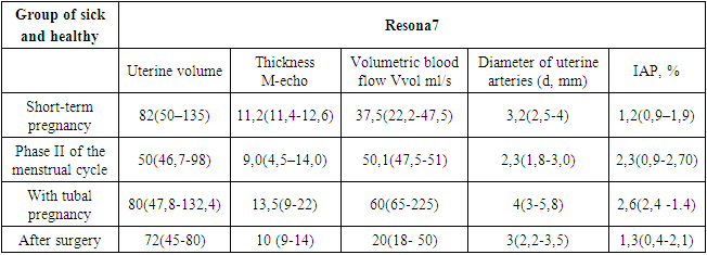
- The analysis of the reproductive function with EP revealed the presence of childbirth in 23 (46.0%), of which: 14 patients had one birth, 4 (6.6%) of 14 patients had previously undergone artificial abortions, 9 (15%) had two or more births.Tubal pregnancy in the anamnesis was noted in 3 (4.4%) patients. 2 (3.0%) suffered from secondary infertility. Attention is drawn to the high frequency of inflammatory diseases of the uterus and its appendages - 36 (52.2%), complicated childbirth, cesarean section 11 (16.0%), IUD (regardless of the duration of use 9 (13.0%), abortions 4 (5.8%), operations on the fallopian tubes, including previous surgical treatment tubal pregnancy 3 (4.4%), ovulation induction, IVF 1 (1.4%), endocrine disorders (obesity 1-2 art. 14 (20%), endometriosis 1 (1.4%), infantilism, congenital anomalies of the uterus 1 (1.4%).The revealed maximum increase in M-echo in short-term uterine pregnancy (14.8 ± 0.7 mm) did not significantly exceed the average values of this parameter in patients in the second phase of the menstrual cycle of the control group and tubal pregnancy, the average size of M-echo was 14.4 ± 0.6 mm, p < 0.05. In 3 (10.0%) women with a short-term uterine pregnancy, 4 (8.0%) with EP had a thickened endometrium with smooth contours identified on echograms, the values of the anteroposterior size of the median structure of the uterus did not exceed 18-20 mm.In 28 (56.6%) patients with tubal pregnancy, the structure of the endometrium was homogeneous, in 13 (26.0%) there was a "spongy" structure of the M-echo with hypoechoic inclusions up to 0.5 mm, in 9 (18%) ultrasound symptoms of a "false" fetal egg were detected. Dopplerometric studies indicated a significant decrease in the values of the arterial perfusion index in the 2nd phase of the menstrual cycle (P < 0.05 for all complications). Arterial perfusion index values in the subgroup of operated women – 0.004 (0.002–0.005) median (5th–95th percentiles), in the subgroup of women on conservative therapy – 0.011 (0.006–0.018) (P < 0.05).In operated patients, compared with those on conservative treatment, there was a significant decrease in the values of the maximum blood flow velocity of 26.8 (16.4–36.8) and 36.0 (19.9–52.5) cm/s) and a significant increase in the pulsation index of 3.07 (1.84–3.98) and 2.23 (1.33–3.36) and the resistance index (0.91 (0.85–1.00) and 0.85 (0.71–1.00)) in the uterine arteries (P < 0.05 for all comparisons).A comparative analysis of echograms and DMBF indicators was carried out in all observed patients. The comparison parameters were the data of the volume of the uterus, the thickness of the M-echo.Color and pulse-wave Doppler echography make it possible to trace the main branches of the uterine vascular network, from the main uterine arteries to the arcuate, radial and, ultimately, to the spiral arteries of the endometrium. Each of the vessels is recognized by a characteristic localization area within the uterine body and a specific shape of the profile of the blood flow velocity curve (BFVC). As we approach the endometrium, IR decreases by about 0.1 in each of the links of the vascular tree without a significant difference from the phase of the menstrual cycle, which corresponds to the published data of domestic researchers. Spiral arteries undergo structural changes in the early stages of pregnancy, which leads to the appearance of a special shaped blood flow profile, which in this regard is called peritrophoblastic blood flow. Such blood flow is detected only during uterine pregnancy (normal or undeveloped) and is localized near the fetal egg (if it is determined), as well as in the thickness or along the outer contour of the endometrium. Compared with blood flow in radial and spiral arteries outside pregnancy, peritrophoblastic blood flow is characterized by higher maximum systolic and diastolic velocities, which reflects the presence of lower vascular resistance. The maximum systolic rate increases with an increase in the size of the fetal egg, reaching the range of speeds observed during early pregnancy.One of the most significant echographic signs that make it possible to suspect a tubal pregnancy in the absence of a fetal egg in the uterine cavity is a change in the endometrium characteristic of its decidual transformation. For the discussed nosological forms, the size of the uterus is decisive in assessing changes in the myometrium and/or endometrium. With a delay in menstruation from 5 to 10 days, the size of the uterus did not significantly differ. The volume of the uterus ranged from 80 to 132.4 cm3, which is comparable to the control group (50-98) cm3. Also, the total vascularization of the uterus during the physiological course of early gestation of pregnancy practically does not increase and is very similar to that for a non-pregnant uterus. With a normal menstrual cycle in the second phase, the uterine arteries have a larger diameter (see Table No. 1). Calculations show that from the early proliferative phase to ovulation, the volumetric blood flow in the aggregate along the two arteries increases by more than 2 times, on average from 22.2 to 47.5 ml/sec. For the entire period of functioning of the corpus luteum, this indicator remains at about 50 ml / c and sharply decreases by menstruation. Between the ages of 18 and 30, the maximum blood filling occurs in all phases of the cycle and, accordingly, the maximum indicators of IAP are. On average, the IAP of the luteal phase of the cycle was 2.3 s-1 (p<0.05). In our observations, in the age group from 31 to 40 years, those who underwent EP had a volume blood flow and IAP significantly less than 18-30-year-olds on 3-4 e. In this group, there were mainly obese women and women with a history of inflammatory and infectious diseases, adhesive processes after surgical interventions, etc. The increase in blood flow in the endometrium strongly depends on the blood flow in the uterine artery, arched arteries and radial arteries. and have spiral arteries detect lower blood flow rates (p 0.05) and lower vascular resistance (p 0.05). The parameters of blood flow in healthy patients in different phases of the menstrual cycle are directly related not only to the production of central and peripheral sex hormones in a certain ratio, but also to the entire neuroendocrine-biochemical complex of the female body. As we approach the endometrium, IR decreases by about 0.1 in each of the links of the vascular tree without a significant difference from the phase of the menstrual cycle, which corresponds to the published data of domestic researchers. It should be noted that when working on expert-class devices, intraendometrial vessels are absent in the early proliferative phase, but after the 8th-10th day of the cycle, basal arteries are registered in 65.7%, spiral arteries - in 29.4% of cases. In phase II of the cycle, the frequency of detection of small uterine vessels increases: basal - 84.0%, spiral - 46.7%. The frequency of vascular detection is also affected by the mapping method. When using the energy doppler option, the number of vessels is determined more than with standard color mapping.
5. Conclusions
- 1. In ectopic pregnancy, the degree of total uterine vascularization varies from mild to moderate, peritrophoblastic blood flow is not detected, venous blood flow around the endometrium is minimal, and luteal arterial blood flow is determined in one or both ovaries.2. Even if there is a picture of a "false" fetal egg in the uterine cavity, peritrophoblastic blood flow is not recorded.3. Low-resistance changes in arterial blood flow in both ovaries in almost 25% of cases during early pregnancy, one of the ovaries usually has higher blood flow rates. This ovary will determine the probable localization side of the ectopic fetal egg.4. On the localization side of the ectopic fetal egg, in 95% of cases, the yellow body is determined, so it can serve as a guide when searching for a pathological formation in the tube. The size of the corpus luteum less than 2.0 cm in diameter was noted in 6 out of 30 patients of the control group who were in the second phase of the menstrual cycle, which was regarded as the presence of a corpus luteum in the regression stage. In 18 out of 30 subjects, the diameter of the corpus luteum varied from 20 to 30 mm, of which 9 had corpus luteum persistence, 8 had a short-term uterine pregnancy and 1 with EP.
References
| [1] | Volkova A.E. Ultrasound diagnostics in obstetrics and gynecology. Practical guide. Rostov-on-Don: Phoenix, 2006. 480 p. |
| [2] | Ozerskaya I.A. Echography in gynecology. 2nd edition, revised and expanded. LLC Publishing house Vidar – M., 2013. 551 p. |
| [3] | Florensova E.V., Apartsin M.S., Chertovskikh M.N. The role of echography in the diagnosis of ectopic pregnancy at the hospital stage. Echography 2001; 3 (4); 344 – 348. |
| [4] | Baltarowich O.H., Scoutt L.M. Ectopic Pregnancy. In: Norton M.E., Scoutt M.L., Feldstein V.A. Callen's Ultrasonography in Obstetrics and Gynecology. 6th ed. California: Elsevier Health Sciences, 2016: 967–998. |
| [5] | Tulandi T. (ed.). Ectopic Pregnancy. A Clinical Casebook. Springer International Publishing Switzerland, 2015. 162 p. |
| [6] | Karimov A.H., M.A. Talipova, L.F. Ruzieva. Modern etiological factors, methods of diagnosis and treatment of women with menstrual disorders. Journal No.1-2. 2021. "News of dermatovenerology and reproductive health". |
| [7] | Rakhimov A.Ya., Qurbonov O.M., Sagdullayeva G.U. Transcutaneous oximetry as the choice of the research for determination of level of amputation of the crus at critical ischemia of the lower extremities at patients with the diabetes mellitus. Asian Journal of Multidimensional Research. AJMR, Vol 8, Issue 12, December 2019, p. 120-125. Impact Factor: SJIF 2018 = 6.053. |
| [8] | Khamdamova M. T., Barotova М.М. Сlinical aspects of the use of laser photodynamic therapy in cervical pathology. American Journal of Medicine and Medical Sciences 2021, 11(4): 353-355. DOI: 10.5923/j.ajmms.20211104.19 Р.353-355. |
| [9] | Khamdamova M. T., Barotova М. Laser photodynamic therapy in the treatment of cervical pathology. Academicia: An International Multidisciplinary Research Journal https://saarj.com. ISSN: 2249-7137. Vol. 11, Issue 3, March 2021. Р.2499-2504. |
| [10] | Khamdamova M. T., Akhmedov F.Kh. A study of ultrasound examination in the prevention of complications of operations on the biliary tract. Asian Journal of Multidimensional Research (AJMR) https://www.tarj.in. ISSN: 2278-4853 Vol 10, Issue 9, September, 2021 Impact Factor: SJIF 2021 = 7.699. Р.212-214. |
 Abstract
 Reference
 Full-Text PDF
 Full-text HTML