Bobur Sobir Ugli Turonov1, Malika Alisherovna Iskandarova2, Alisher Iskandarovich Iskandarov3
1Doctoral Student, Republican Scientific and Practical Center for Forensic Medical Expertise
2Doctor of Medical Science, Associate Professor, Tashkent Pediatric Medical Institute
3Professor, Director of the Republican Scientific and Practical Center for Forensic Medical Expertise
Copyright © 2022 The Author(s). Published by Scientific & Academic Publishing.
This work is licensed under the Creative Commons Attribution International License (CC BY).
http://creativecommons.org/licenses/by/4.0/

Abstract
Iridological diagnosis has very ancient roots. Even more than five to seven thousand years ago in India, China, Asia Minor, this diagnostic method was used. The experience of ancient doctors in “eye diagnostics” was not completely lost and was partially used in Eastern and European medicine for centuries. In our time, doctors of many specialties use various eye symptoms in the diagnosis of a number of diseases.In recent years, the morphological (forensic) direction in iridological diagnosis has been developing. Hundreds of known pathological changes in the iris – signs have a specific interpretation that allows you to determine the nature and severity of pathological changes in the body. Signs of a general nature provide information about changes at the level of the whole organism, local iridosigns – about the pathology of specific organs. In addition, the unique capabilities and speed of assessing predisposition to hereditary diseases and human heredity by iridological diagnosis can be used to determine disputed paternity (or motherhood) in forensic practice, as well as to assess suicidal tendencies and drug addiction of the subject under study. In this work, we have made an attempt using the method of iridological diagnosis to explain and give an expert assessment of morphological changes in the internal organs during sudden death and some types of violent death, as well as in determining disputed paternity.
Keywords:
Iridological diagnosis, Forensic histological examination, Forensic–medical examination, Medical forensic examination, Forensic chemical examination, Histological analysis
Cite this paper: Bobur Sobir Ugli Turonov, Malika Alisherovna Iskandarova, Alisher Iskandarovich Iskandarov, Forensic Medical Significance of Systemic Iridological Diagnosis, American Journal of Medicine and Medical Sciences, Vol. 12 No. 9, 2022, pp. 983-986. doi: 10.5923/j.ajmms.20221209.27.
1. The Purpose
The purpose of this study was to explore the possibilities of iridological diagnosis for forensic medicine.
2. Materials and Methods
The material for the study was 203 corpses of people who died suddenly from various chronic diseases and 230 cases of violent death from traumatic brain injury as a result of a traffic accident (RTA), 43 cases of death from mechanical asphyxia as a result of hanging (suicide) and 16 forensic medical examinations regarding the determination of controversial paternity. All cases were verified by the material of the investigation, forensic medical research, including forensic histological, toxicological and medico–criminalistic.In the work were used forensic–chemical, histological, forensic and statistical methods of research.
3. Results and Discussion
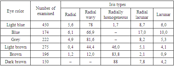
The results of the research showed that out of an infinite number of structural combinations of the iris, reflecting the constitutional features of a person, in this work we used several of the simplest types. In total, five types are distinguished: radial, radial–wavy, radial–homogeneous, radial–lacunary and lacunar.According to our data, the radial type of the iris is more common in the corpses of people who died suddenly as a result of cardiovascular diseases: coronary heart disease (36.8%), myocardial infarction (23.7%), hypertensive heart disease and atherosclerosis (43.3%). This type of iris occurs 10 times more often in people with light eyes than in dark–eyed people (Table No. 1).Table 1. The frequency of occurrence of iris types in people with different eye colors (%) (according to E.S. Velkhover)
 |
| |
|
In the radially wavy type of iris, the appearance of radially extending and somewhat flattened trabeculae creates some undulation in the trabecular fibers. This is the so–called neurogenic type of constitution, which is characterized by estenographic manifestations and a tendency to spasms.In our observations, neurological diseases and lung diseases (chronic pneumonia, pneumosclerosis, sclerosis of cerebral vessels) were found in the corpses of persons who died suddenly. The third type of iris is radially homogeneous, characterized by a combination of a radial pattern in the pupillary girdle with a dense, homogeneously colored celiac circle. This type of iris is observed almost exclusively in dark-eyed people. Just like the radial type of the iris, it is a sign of a good constitution and is observed in practically healthy people.According to our observations, we distinguished this type of iris in the corpses of persons who died in road accidents (89.9%), mainly at a young age.The fourth type of iris – radial–lacunar – is presented in the form of a thinned stroma with scattered leaf–shaped depressions – lacunae, occupying up to 30% of the surface of the iris. This type of iris is typical for people with a weakened constitution and a tendency to dysfunction of internal organs and chronic diseases.In our observations, this type of iris occurs in the corpses of people who died suddenly from chronic heart pathologies against the background of concomitant diseases, such as diabetes mellitus (24.6%), chronic coronary heart disease (18.4%), hypertension (16.3% ) and cerebral atherosclerosis (52.6%), hepatic (12.6%) and renal failure (8.7%).The fifth type of iris is lacunar, characterized by a thin, sometimes broken stroma with a chaotic pattern of trabeculae and a large number of lacunae. This is the weakest type of the human constitution, indicating the pronounced inferiority of many organs and systems. It is more common in people with light eyes than in people with brown eyes.According to our observations, this type of iris was found in the corpses of persons who died both suddenly (62.4%) and during mechanical asphyxia by hanging (suicide) at a relatively young age (16.4%).Along with architectonics or the type of iris, iridology attaches great importance to the density of iridescent structures (Fig. No. 1).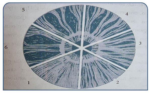 | Figure 1. Various gradations of iris density (according to V. Lensen, 1964) |
It is generally accepted that the cleaner and denser the iris of the eye, the healthier and stronger the body.V. Lensen (1964) distinguishes between several degrees of density of the iris. He compares it to the density of hard, medium and soft wood.Density 1 is the ideal iris type with dense stroma and clear coloration. Its surface is smooth, homogeneous, the trabeculae are very tightly adjacent to each other.Such an iris occurs in people with very good heredity and excellent health. In our observations, there was not a single case with such an iris.Density 2 – the color of the iris can be different. The stroma is quite dense, but not so homogeneous. You can easily see the radial threads in it. The iris looks like a light transparent veil is thrown over its entire surface. It occurs in healthy people with good heredity. In our observations, there was a single case of a traumatic brain injury as a result of a car accident.Density 3 – the color of the iris is different, its stroma is very dense. Trabeculae are stretched, weakened and tortuous. It can immediately be assumed that the organs have lost their tone. The owners of such a density of irises have increased fatigue, low resistance, and a tendency to many diseases of a functional nature.In our observations, we met with such an iris in the corpses of people who died suddenly (6.7%), mainly from respiratory diseases (pneumonia) in children at an early age.Density 4 – the color of the eyes of different density is satisfactory, consists of separate long thinned trabeculae, between which gaps are visible. These cracks are numerous, most often oval. Carriers of such an iris are people with poor health, suffering from neurological diseases.In our observations, we mainly recorded this type of iris density in the corpses of people who died from acute cardiovascular diseases, most often from myocardial infarction (42.8%), as well as in people who committed suicide (7.4%).A density of 5.6 is a very weak iris. The stroma of the iris is dotted with many depressions and pits that change their color and shape.Pronounced voids deform the small circle of the iris and do not allow localizing the site of the lesion. Such irises indicate severe hereditary and acquired diseases, poor constitution, and a decrease in the body’s defenses.In our studies, the corpses of persons who committed suicide by hanging (43), as well as 12 living persons with incomplete suicide, had irises with weak and very weak density.Studies of the density of the iris are directly related to both the prognosis in severe diseases and the identification of predisposition to any genetic characteristics of the individual. The assessment of these features is important not only in forensic and clinical practice, but also in the work of various kinds of medical expert commissions.Interesting information in terms of morphogenesis and pathogenesis of various diseases is provided by differences in the reliefs of the iris. The study of the iris by relief characteristics gives us the opportunity to correctly interpret the data on the protective and reserve capabilities of the human body.The surface of the iris does not look smooth or flat, but is a conglomeration of bulges and depressions. From the center of the pupil (iris), the surface of the iris rises to the elevation of an autonomous ring, the shape of which is very variable. There are several types of relief (Fig. No. 2).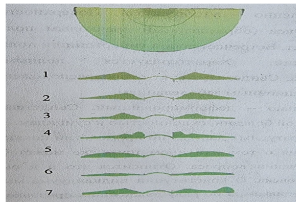 | Figure 2. Relief views of the iris in the interpretation of G. Jansas (1974): 1–normal, 2–bowl–shaped, 3–flattened–lateral, 4–crater–shaped, 5–rounded thickened, 6–flat, 7–locally deformed |
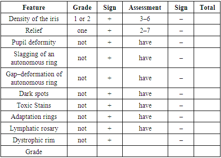
Normal – characterized by the average size of the top of the autonomous ring and uniform internal and external slopes. Indicates good heredity.Bowl–shaped – characterized by compression of the pupillary belt and the middle part. It occurs in people with a tendency to hypertension, type II diabetes and cardiovascular disorders.Flattened–lateral is characterized by the compression of the slope of the ciliary belt. Persons with this type of relief of the iris are more prone to hypofunction of the sympathetic nervous system.Crater–shaped – characterized by a steep slope of the pupillary belt protruding forward. It occurs in people prone to endocrine and humoral disorders.The rounded–thickened relief is characterized by a swollen surface, the Fuchs angle (formed by the pupillary belt and autonomous ring) is absent. It is more common in people prone to hypertension.Flat–characterized by the complete disappearance of the autonomous ring. This type of relief of the iris indicates a predisposition to various chronic infections.Locally deformed – indicates severe chronic diseases, as well as a tendency to suicide.It should be noted that the most complete information about a particular feature can be obtained through a comprehensive study of the issue. This also applies to the assessment of human genetic characteristics.We propose to judge the constitution of an individual not by one or two signs, but by a number of the most important signs proposed by E.S. Velkhover (1992).As follows from Table No. 2, good morphogenetic traits are evaluated with a sign (+), bad (–). When deriving the final grade, which can range from 0 to 10 points, only positive signs are taken into account. Ideally, if there are 10 positive signs, the constitution is estimated at 10 points. However, such individuals are extremely rare. Exceptionally rare are also observed persons with a constitution of 0–1 points.Table 2. A ten-point system for assessing the constitutional features of a person (according to E.S. Velkhover, 1992)
 |
| |
|
Such an accelerated scoring system can be used in the study of genetic predisposition to various suicidal acts, chronic diseases, or to a tendency to inappropriate actions during forensic medical commission examinations.B. Jensen (1982) on the basis of many years of research believes that the iris is the only structure that displays congenital defects that are inherited up to the fourth generation inclusive.Based on the needs of a forensic medical examination, the above iridoscopic hereditary patterns can be used in the examination of disputed paternity or motherhood (when replacing children in maternity hospitals) as additional objective evidence criteria (Fig. 3).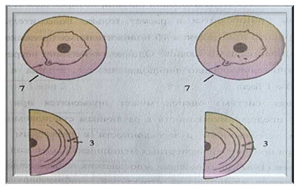 | Figure 3. Examination of iridoscopic signs: A–swelling of the autonomous ring of the right iris at “7” o’clock, due to a large lacuna. 1–A.Yu.N. –36 years old (father), 2–A.N.Yu. –9 years old (daughter), B–pigment spot in the left iris at “2.55” 1–R.I.M. –34 years old (father), 2–R.M.V. –18 years old (son) |
Thus, from the forensic and forensic positions, the above studies could provide invaluable assistance to experts in determining the individual characteristics of the iris in identifying a person, establishing a predisposition to various diseases, to suicide, drug addiction, and also in determining disputed paternity or motherhood as one of objective signs of evidence, which will undoubtedly increase the objectivity and evidentiary value of forensic medical conclusions.
References
| [1] | Abramov M.S., Abramov G.S., Kulikova S.V., Petrova R.P. Algorithms for the differential diagnosis of iris biomicroscopy. Collection of materials of the All–Union Association of Iridologists. M., 1990, No. 2, – p. 14–15. |
| [2] | Amosov N.M. Medical information system. – Kyiv. Science thought. – p. 1971–178. |
| [3] | Amosova E.A. Iridology of hypertension. Materials of the I All–Union Conference of Iridologists. M. 1990, – p. 50–53. |
| [4] | Baevsky R.M. Forecasting states on the verge of norm and pathology. – M., Medicine, 1979. – p. 295. |
| [5] | Velkhover E.S., Shulkina N.B., Alieva Z.A. Fundamentals of iridology. – Baku: Azerneshr., 1982. – p. 188. |
| [6] | Vorobyov E.I., Kitov A.I. Introduction to medical cybernetics. – M:. Medicine, 1977. – p. 277. |
| [7] | Gubler E.V. Computational methods for the analysis and recognition of pathological processes. – The medicine. 1978. – p. 300. |
| [8] | Drozdetsky S.I., Miroshnichenko N.B., Rumyantsev T.I. Possibilities of iridology in the recognition of diseases of internal organs. Materials of the I All–Union Conference of Iridologists. – M:. 1990, – p. 15–17. |
| [9] | Zelensky V.A. Diagnosis of diseases of the stomach with the help of computers. – Abstract. diss. M. 1969. – p. 16. |
| [10] | Ershov A.P. Programming is the second literacy. – Novosibirsk: 1981. |
| [11] | Romashov F.N., Velkhover E.S. Possibilities and errors of iridology. – Experiment. Surgery and anesthesiology, 1973, No. 2, – p. 49–56. |
| [12] | Shulgina R.B., Vints A.A. On the possibility of using iridology in clinical practice. – Herald oftalmol. No. 3, 1986. |
| [13] | Deck I. Grundlangen der iridiagnostik. Ettlingen, 1955. – p. 68. |
| [14] | Jensen B. The Sciense and practice of iridological diagnosis. Escondido 1964, 1970. – p. 178. |
| [15] | Kriege T. Grundbegriffe der iris–diagnostic. Osnabruk, 1981. – p. 1029. |
| [16] | Schumanu E. Auglu diagnose. Berlin. 1961. – p. 829. |


 Abstract
 Reference
 Full-Text PDF
 Full-text HTML

