Akhmedov F. K., Negmatullaeva M. N.
Bukhara State Medical Institute, Uzbekistan
Copyright © 2022 The Author(s). Published by Scientific & Academic Publishing.
This work is licensed under the Creative Commons Attribution International License (CC BY).
http://creativecommons.org/licenses/by/4.0/

Abstract
To assess the significance of hemodynamic parameters in the prediction and early diagnosis of preeclampsia. The main patterns of the central hemodynamics of the mother, utero-placental-fetal blood flow in the I and III trimesters of pregnancy have been established. The characteristic features of hemodynamics in these terms of gestation make it possible to predict the development of PE and its progress. Only careful control over the indicators of central, regional hemodynamics in the form of continuous monitoring allowed us to timely decide on the tactics of managing patients.
Keywords:
Preeclampsia, Dopplerometry, L-argenin, Hemodynamic parameters, Uterine artery
Cite this paper: Akhmedov F. K., Negmatullaeva M. N., Hemodynamic Markers for Predicting Preeclampsia, American Journal of Medicine and Medical Sciences, Vol. 12 No. 9, 2022, pp. 934-937. doi: 10.5923/j.ajmms.20221209.18.
1. Introduction
Preeclampsia (PE) is a serious medical and social problem worldwide, as it remains one of the main causes of perinatal and maternal mortality. Arterial hypertension (AH) is by far the most serious pathology among cardiovascular diseases and occupies a significant place, humanity suffers from this pathology from 15 to 25%. The female population has gender-specific risk factors for the development of this pathology, respectively, hypertensive disorders during pregnancy are one of the leading problems in the field of maternal and child health [1,3].The main objectives of the demographic policy of the Republic of Uzbekistan to continue depopulation, reduce the birth rate and mortality, including maternal and infant mortality, are aimed at improving the health of the entire population, including women of reproductive age [6,8].But it should be noted that its diagnosis causes certain difficulties due to the polymorphism of the clinical picture and the discrepancy between the severity of clinical symptoms and the severity of organ-systemic disorders, in this regard, accurate diagnosis, assessment of the severity and prediction of preeclampsia is of great importance for practical healthcare.In order to save the life of the mother, the issue of termination of pregnancy is often resolved, and therefore, the proportion of premature births in patients with hypertensive disorders significantly exceeds the corresponding figures for a physiologically proceeding pregnancy, which undoubtedly affects the health of newborns [9].To date, according to the world literature and the results of scientific research, a different concept of the nature and essence of preeclampsia is presented not only as a hypertensive state during gestation in combination with structural and functional disorders of the placenta, but as a development of a general pathological, specific generalized endothelial dysfunction of the vascular system. This idea of preeclampsia allows us to think about a new etiopathogenetic approach to the diagnosis, management and treatment of patients with this severe pathology of pregnancy. In recent years, ideas about the physiology and pathology of pregnancy as a process occurring against the background of different types of systemic hemodynamics in the female body have significantly expanded [2,4].At the same time, the criteria for the significance of certain parameters of blood circulation for the gestational process have not yet been sufficiently outlined. The relationship between the features of central hemodynamics and the dominant type of neurovegetative regulation in the body of pregnant women has been little studied. These issues are important for managing a woman at the outpatient stage, since the organization of adequate preventive and therapeutic measures and, ultimately, the outcome of pregnancy depends on their solution [7].Purpose of the study: To assess the significance of hemodynamic parameters in the prediction and early diagnosis of preeclampsia.
2. Materials of the Study
In-depth studies were carried out starting from the I-II trimester of pregnancy on a contingent of 100 women with anamnestic data on the risk of developing PE 1A-group, 1B-group, 50 patients with the physiological course of pregnancy.The study of the parameters of central and regional hemodynamics, starting from the first trimester of gestation, is of paramount importance in predicting and early diagnosis of PE, assessing its severity, choosing rational obstetric tactics, antihypertensive therapy, and reducing perinatal morbidity and mortality. To assess the functional state of the mother-placenta-fetus system, an ultrasound Doppler study of blood flow in the uterine arteries was performed. In our observations, dopplerometry was carried out using the Mindray DC-3 ultrasonic diagnostic device.The studies were carried out in terms of 10-14 weeks of pregnancy in the Bukhara Regional Perinatal Center and in the Department of Pathology of Pregnant Women of the Maternity Complex of the Zhondor District Medical Association. To assess the uteroplacental, fetal-placental blood flow, the curves of blood flow velocities (BFRs) in the uterine arteries were recorded.Analysis of the profile of the blood flow spectrum in the uterine vessels was carried out on the basis of studying its shape, determining the maximum systolic and end-diastolic velocity, as well as the average velocity during one cardiac cycle. With the calculation of peripheral resistance indices, systole-diastolic ratio (SDR), resistance index (IR).
3. Research Results
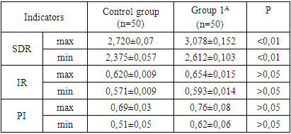
The Doppler ultrasound method gives us information about changes in the uterus-placenta and placenta-fetus system, with the help of which complications can be predicted. It is known that for dynamic observation in order to prevent complications, Doppler monitoring should be carried out. Dopplerometry performed at 10-11 weeks indicates the processes of implantation and the development of the chorion, at 20-24 weeks of pregnancy it shows the state of blood flow in the uterus-placenta and fetus-placenta system, in addition, it can be used to predict possible complications from the mother and fetus, which manifests itself in the form of structural and functional changes in the placenta. The primary study was conducted at 10-14 weeks of gestation, because it is in this week of gestation that some complications are possible.Table 1. Comparative dopplerometric data of the uterine artery in the first trimester of pregnancy
 |
| |
|
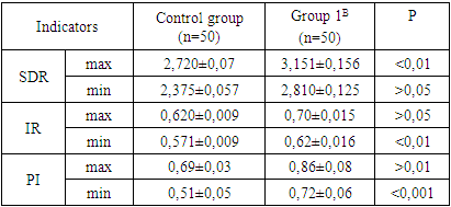
Conducted Doppler assessment of blood circulation in the uterine arteries at gestational age 10-14 weeks of pregnancy found that the indicators in the group of patients with the risk of developing preeclampsia in group 1-A, the indicators of SDR were significantly higher than in the control group. As a result of our research, it was found that women at risk of developing PE had their own peculiarities of blood circulation in the uterine arteries, which manifested themselves with a tendency to increase IR by 6.2% more than in the control group. Such changes in IR may be due to impaired remodulation of the spiral arteries of the uterus and invasion of the trophoblast at the early stages of the formation of the placental complex. Thus, an increase in uterine blood flow (SDR and IR) in the first trimester and early gestation can be used as markers indicating the formation of blood flow disorders in the mother-placenta-fetus system and predictive factors for the development of PE.Violations of trophoblast invasion into the spiral arteries of the myometrium, due to which the uterine arteries will not be able to provide adequate blood flow, which contributes to the development of ischemia in the placenta, which can progress with an increase in the gestation period and is the starting point for the formation of endothelial dysfunction, and subsequently the development of organ-systemic disorders.Based on the above, we can conclude that Doppler assessment of the blood flow of the uterine arteries from early gestation is a key factor in the development of preventive measures necessary to prevent the development of pregnancy pathology, in particular PE, and improve its outcome.Based on the data of Doppler studies, revealed violations of blood flow in the uterine artery, the examined contingent of women belonging to the I-A group was started pathogenetic treatment aimed at improving blood flow with dipyridamole 75 mg / day. in three doses, since acetylsalicylic acid is contraindicated in the early stages of gestation. The effect of therapy was assessed after 10-15 days from its initiation by changes in Doppler parameters. During the control of this method of research in all examined patients of group I-A, the indicators of SDR and IR tended to improve and no significant differences from the indicators of the control group of patients were found. In this connection, the drug of choice as an antiplatelet action can be considered chimes at a dosage of 75 mg / day. with violations of utero-placental perfusion.At the next stage, a comparative assessment of the indicators of uteroplacental blood flow was carried out in pregnant women registered at a later gestational age - in the second trimester, who also have a risk of developing PE.Table 2. Comparative dopplerometric data of the uterine artery in the second trimester of pregnancy
 |
| |
|
As can be seen from the above table, it was taken for three pregnant women and was observed in a later pregnancy, with significant marked blood flow and uterine artery disorders, only showing SDR max. In this group, patients with higher SDR min than 15.5%, correspondingly IR max than 11.5%, IR min than 17.8%, compared to the control group, showed no blood flow disorder and uterine-placental system. Our data confirm the primacy of involvement in the pathogenesis of the development of morphological and functional disorders of the placenta, which dictates a high risk of developing endothelial dysfunction, layering of severe obstetric pathology - PE and dictates the implementation of pathogenetic preventive therapy.Recently, the number of publications on the use in obstetric practice of various treatment regimens and correction of uteroplacental blood flow disorders, which is carried out in several directions, however, pharmacological "aggression" can lead to an increase in the frequency of side effects on the part of the mother and fetus. From this point of view, the need for the use of endogenous metabolites is generated. In modern obstetrics, it has become possible to supplement the tactics of treating placental dysfunction with NO donors (L-argenin), which fully meets these requirements - gravid protector - Tivortin. This drug has a positive effect on the function of the vascular endothelium, improves endothelial dependent vasodilation.In order to improve uteroplacental blood flow in the form of an antioxidant, the drug tivortin-asparganate 5 ml 3 times a day orally, antiplatelet agent aspirin 75 mg per day, trace elements magnesium B6 - forte 1 table 3 times / day, calcium D3 - nycomed according to 1 tablet 2 times a day and dydrogesterone 10 mg 2 times a day - for 14 days. The effectiveness of therapy was monitored under the monitoring of Doppler parameters. Of the 50 women in the study group, 40 received a positive effect of therapy, manifested by an improvement in blood flow in the uterine artery corresponding to the normal values of a given gestation period, and in 10 patients, no visible improvements in the parameters we studied were found, and against the background of an increase in IR, clinical symptoms of PE began to appear, manifested by worsening indicators of systemic and central hemodynamics of the mother (increase in SBP, diastolic arterial blood pressure, decrease in BCC, impact volume, total peripheral vascular resistance and ejection fraction). These 10 pregnant women were transferred to group 2 with clinical manifestations of PE; in total, this group consisted of 60 pregnant women with an established diagnosis of PE.The study of the parameters of CH and regional blood flow is a valuable method for assessing the nature of the course of pregnancy. However, the issue of its diagnostic effectiveness is far from being finally resolved. Recent studies have convincingly shown that adaptive hemodynamic processes in a single functional system mother - placenta - fetus are designed to ensure the physiological course of pregnancy, growth and development of the fetus.Blood circulation in the mother-placenta-fetus system, which is formed with the development of the gestational process, is one of the main factors determining the normal development of the fetus [5]. The fetoplacental complex suffers in any form of arterial hypertension.Hemodynamic disturbances in the mother-placenta-fetus system are the leading pathogenetic mechanism for the violation of the state and development of the fetus in PE [6].Thus, the main regularities of the central hemodynamics of the mother, utero-placental-fetal blood flow in the I and II trimesters of pregnancy have been established. The characteristic features of hemodynamics in these terms of gestation make it possible to predict the development of PE and its progress. Only careful control over the indicators of central, regional hemodynamics in the form of continuous monitoring allowed us to timely decide on the tactics of managing patients.
References
| [1] | Akhmedov F.K. Peculiarities of cardiac hemodynamic in pregnant women with mild preeclampsia // Europen Science Review. - 2015. - №4-5. - С. 56 -58. |
| [2] | Akhmedov F.K. Features of renal function and some indicators of homeostasis in women with mild preeclampsia// Europen Science Review. - 2015. - №4-5. - С. 58 - 60. |
| [3] | Akhmedov F.K., Negmatullaeva M.N., Kurbanova Z.Sh. Modern views on the problem of preeclampsia // A new day in medicine. - 2018. - № 1 (21). - S. 180-185. |
| [4] | Akhmedov F.K., Negmatullaeva M.N., Features of the state of central hemodynamics and hemostasis in pregnant women with preeclampsia of varying degrees and severity // New Day of Medicine. - 2020. - No. 1 (29) - S. 147-150. |
| [5] | Negmatullaeva M.N., Akhmedov F.Q., Tuksanova D.I. Modern diagnostics of markers of preeclampsia // Vestnik Tashkentskoy meditsinskoy akademii. - 2020. -№2 (94). - S. 145 - 147. |
| [6] | Negmatulleva M.N., Tuksanova D.I., Nosirova M.Sh., Akhmedov F.K. Features of the state of the circulatory system mother and fetus in the second trimester ofpregnancy in women with mitral stenosis ofrheumatic etiology // European Journal of Biomedical and Pharmaceutical sciences. - 2020. - №7(6). - P. 100 - 103. |
| [7] | Tuksanov D.I. Features of changes in indicators of systemic and organ blood flow in women with severe preeclampsia // Tibbiyotda yangi kun. – Tashkent, 2018. 1(21). - S. 134-137. |
| [8] | Tuksanov D.I. Features of the state of parameters of homeostasis and cardiohemodynamics in women with a physiological course of pregnancy // New Day of Medicine. - 2019. - No. 1 (25). - S. 159-163. |
| [9] | Tuksanov D.I. Features of the state of systemic and organ blood flow in women with physiological pregnancy // News of dermatology and reproductive health. - Tashkent, 2017. - No. 3-4 (I). - S. 135-136. |

 Abstract
 Reference
 Full-Text PDF
 Full-text HTML