American Journal of Medicine and Medical Sciences

p-ISSN: 2165-901X    e-ISSN: 2165-9036

2022;  12(9): 878-885

doi:10.5923/j.ajmms.20221209.05

Received: Aug. 4, 2022; Accepted: Aug. 28, 2022; Published: Sep. 15, 2022


The Relationship of Chronic Kidney Disease with the Condition of the Oral Cavity

Rizaev J. A., Husanbaeva F. A.

Samarkand State Medical University, Samarkand, Uzbekistan

Correspondence to: Rizaev J. A., Samarkand State Medical University, Samarkand, Uzbekistan.

Email:

Copyright © 2022 The Author(s). Published by Scientific & Academic Publishing.

This work is licensed under the Creative Commons Attribution International License (CC BY).
http://creativecommons.org/licenses/by/4.0/

Abstract

The article describes the prevalence and intensity of dental diseases in patients with CKD in Uzbekistan. The study involved 100 people, including 68 patients with CKD, of which 15 patients received hemodialysis. 32 practically healthy people made up the control group. The results obtained after the application of standard dental treatment are presented: data on the intensity and prevalence of caries, the dynamics of the hygienic condition, periodontal status and the state of the oral mucosa, assessment of the condition of fillings in patients with CKD.

Keywords: Caries, Prevalence, Intensity, Non-carious lesions, Periodontal disease, COPD, Dentistry, Chronic kidney disease, CKD

Cite this paper: Rizaev J. A., Husanbaeva F. A., The Relationship of Chronic Kidney Disease with the Condition of the Oral Cavity, American Journal of Medicine and Medical Sciences, Vol. 12 No. 9, 2022, pp. 878-885. doi: 10.5923/j.ajmms.20221209.05.

1. Introduction

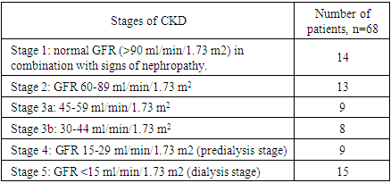
Almost all chronic diseases of the body are associated with poor oral health, which leads to the need for better dental care. This is especially evident in patients with chronic kidney diseases, where oral diseases are a potential cause of deterioration of their already fragile health [18,19].
Existing data on the prevalence and severity of oral diseases in patients with chronic kidney disease are limited to small samples. Studies show various cases of oral diseases in such patients. However, based on these scant data, it is estimated that almost 90% of patients with chronic kidney diseases show some symptoms of oral diseases, especially gum hyperplasia, xerostomia and changes in salivation and saliva composition [4].
Patients with chronic kidney diseases require special attention from the dentist due to multiple manifestations in the oral cavity, side effects and treatment features of the underlying disease [4].
The treatment of CKD is the treatment of a polymorbid patient, simultaneously aimed at slowing down the progression of kidney dysfunction (renoprotection) and preventing the development and progression of cardiovascular pathology (cardioprotection) in order to improve the prognosis. The commonality of the causes and mechanisms of damage to the kidneys and the cardiovascular system (hyperactivation of the renin-angiotensin system, expression of inflammatory mediators and fibrogenesis factors) gives grounds for recommending drugs with reno- and cardioprotective effects - blockers of the renin-angiotensin-aldosterone system: angiotensin-converting inhibitors enzyme or angiotensin II receptor blockers. In addition, these drugs have antioxidant and anti-inflammatory activity.
The need to study the manifestations of CKD in the oral cavity in patients in Uzbekistan served as the purpose of this study.
Materials and methods of research. The study involved 100 people, including 68 patients with CKD, of which 15 patients received hemodialysis. 32 practically healthy people made up the control group. The age of the patients was 45-56 years. Men - 58, women - 42 people. The study took place in 2020-2022 on the basis of Samarkand State Medical University, Tashkent State Dental Institute.
Criteria for inclusion in the study: therapeutic patients with inflammatory diseases of the endo- and periodontium (acute and chronic pulpitis, chronic generalized catarrhal gingivitis and chronic generalized periodontitis) with concomitant chronic kidney pathology. Exclusion criteria: patients with a decrease in glomerular filtration rate <15 ml/min/1.73 m2.
The patients were divided into the following groups:
1. A group of people who do not have pathology from the urinary system - 32 people (group A);
2. Patients with chronic kidney disease who are not being treated on hemodialysis - 53 people (group B);
3. Patients with chronic kidney disease undergoing hemodialysis treatment - 15 people (group B).
Table 1. Examined patients with CKD, depending on the stage of the disease [21]
     
The dental status was assessed using a clinical examination of the oral cavity: patients' complaints were collected, the condition of the hard tissues of the teeth (the presence of plaque, carious spots and cavities, non-carious lesions), periodontal disease (tooth mobility, bleeding, the presence of pathological pockets), the condition of the SOPR were revealed. A set of dental instruments was used for the examination: a mirror, tweezers, a graduated probe.
To assess the prevalence and intensity of periodontal diseases, the need for treatment of periodontal diseases, the CPITN index was used.
Dental treatment was carried out in all groups according to the recommended protocols. Modern composite materials were used in the treatment of caries and non-carious lesions of the hard tissues of the teeth, modern fillings and medications in the treatment of complications of caries and periodontal diseases and COPD.
In the treatment of periodontal diseases, taking into account their course and severity, as well as to prevent their recurrence, professional hygiene was carried out every six months. Therapeutic and prophylactic manipulations were carried out for the treatment and prevention of diseases of the SOPR.
After 6 and 12 months, the safety of fillings, the effectiveness of endodontic treatment, and the hygienic status of patients were checked.
Patients with CKD are prone to developing lesions of the oral cavity due to a pathological process or therapy, or both. Lesions of the oral cavity and caries are non-traditional risk factors for the progression of CKD.
Table 2. The volume of therapeutic and preventive dental care for the examined persons
     

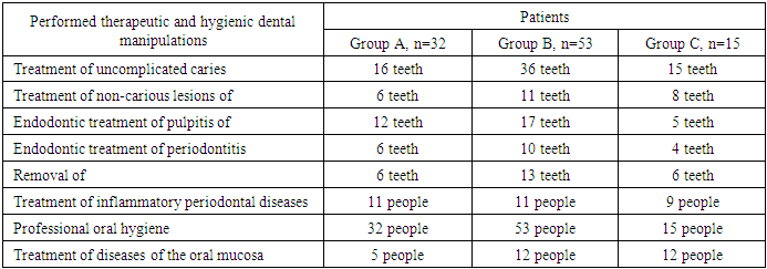
2. Results of the Dental Status Study

Table 3. Analysis of complaints and objective indicators of examination of patients with CKD
     
Table 4. Change in the prevalence (%) and intensity of caries in group A
     
Group A patients were treated for carious teeth, non-carious lesions, endodontic treatment and removal of untreated teeth. In total, 22 teeth were cured for carious and non–carious lesions in this group, 18 teeth were treated endodontically for pulpitis and periodontitis, and 6 teeth were removed (the main cause was complications of pulpitis and periodontitis – 33.33%). At the initial examination, the number of filled teeth was 3.21 ± 0.24, at the examination after 6 months, the number of filled teeth increased to 3.64 ± 0.33, after 12 months - 3.68± 0.43. The number of teeth affected by the carious process decreased from 1.59± 0.13 at the first examination to 1.28± 0.13 at the examination a year later (reduction by 24%). The increase in the prevalence of caries in 1 year is 2.8%.
In this group, the component "P" is predominant: 3.21±0.24- 3.68±0.43, against "K" - 1.19±0.14-1.59±0.13 and "Y" - 1,16±0,11-1,24±0,18. We associate this with the medical and hygienic measures carried out.
An assessment of the condition of the seals after six months revealed their safety in 98.1% of cases, a year later – in 95.7%.
The X-ray method revealed the absence of periapical lesions in endodontically treated teeth in 97.3% after 6 months and 95.2% after 12 months.
Table 5. The reason for tooth extraction in group A
     
Table 6. Changes in the prevalence (%) and intensity of dental caries in group B patients
     
Group B patients were treated for carious teeth, endodontic treatment and removal of untreated teeth. In total, 47 teeth were cured for carious and non–carious lesions in this group, 27 teeth were endodontically treated for pulpitis and periodontitis, and 13 teeth were removed (the main cause is periodontal disease – 46.15%). At the initial examination, the number of filled teeth was 3.43 ± 0.33, at the examination after 6 months, the number of filled teeth increased to 3.89± 0.35, after 12 months - 3.94± 0.53. The number of teeth affected by the carious process increased from 1.27 ± 0.17 at the first examination to 1.7± 0.15 at the examination a year later (an increase of 33.8%). The increase in the prevalence of caries in 1 year is 5.2%.
In this group, also, the component "P" is predominant: 3.43±0.33- 3.89±0.35 (with a reduction in a year by 13.4%), against "K" - 1,27±0,17-1,7±0,15 ( growth in a year by 33.8%) and "Y" - 1.24± 0.15-1.32±0.15. An assessment of the condition of the seals after six months revealed their safety in 87.2% of cases, a year later - in 81.0%.
The X-ray method revealed the absence of periapical lesions in endodontically treated teeth in 89.1% after 6 months and 77.1% after 12 months.
Table 7. Reasons for tooth extraction in group B
     
Table 8. Changes in the prevalence (%) and intensity of dental caries in group B patients
     
Group B patients were treated for carious teeth, endodontic treatment and removal of untreated teeth. In total, 23 teeth were cured for carious and non–carious lesions in this group, 9 teeth were treated endodontically for pulpitis and periodontitis, and 6 teeth were removed (the main cause is periodontal disease – 50.0%). At the initial examination, the number of filled teeth was 4.28 ± 0.35, at the examination after 6 months, the number of filled teeth increased to 4.33 ± 0.53, after 12 months - 43.77 ± 0.33. The number of teeth affected by the carious process increased from 1.4 ± 0.17 at the first examination to 1.87± 0.15 at the examination a year later (an increase of 33.57%). The increase in the prevalence of caries in 1 year is 4.6%.
And in this group, the component "P" prevails: 3.77±0.33- 4.28±0.35 (with a reduction of 13.5% in a year), against "K" - 1,4±0,17-1,87±0,15 ( an increase of 33.6% in a year) and "Y" - 1,24±0,15-1,62±0,15 (an increase of 30.6%).
An assessment of the condition of the seals after six months revealed their safety in 78.6% of cases, a year later – in 72.3%.
The X-ray method revealed the absence of periapical lesions in endodontically treated teeth in 78.2% after 6 months and 74.4% after 12 months.
Table 9. Reasons for tooth extraction in group B
     

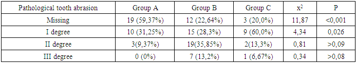
3. Prevalence of Non-Carious Dental Lesions

Table 10. Prevalence of pathological erasability by degrees
     
Non-carious enamel lesions (wedge-shaped defects and erosion) in group A occur in 32.1% of cases, in patients of group B - in 21.3% of cases, in group B - in 47.4%, (p=0.049).
Erosion of hard tissues of teeth in patients with CKD occurs in 25.4% of cases, and among healthy individuals - in 9.5% of cases (p=0.078). In patients of group B, erosion of hard tissues of teeth is observed in 35.4% of cases (p= 0.067), and in group B - in 19.3% of cases. In patients with CKD, non-carious lesions are located on the labial/ buccal and lingual sides, the depth almost reaches the pulp chamber (its translucency is observed).
Wedge-shaped defects are found in 28.2% of cases in patients with CKD and in 25.3% of group A (p>0.1). In group B, wedge-shaped defects occur in 17.2% of cases, which is significantly less than among healthy individuals (p=0.023). In the group of patients receiving hemodialysis, this indicator is equal to 37.2% of cases (p>0.1). A combination of wedge-shaped defects and erosions is very often observed in patients with CKD (86.3% of cases).
Dynamics of the hygienic state of the oral cavity of patients with CKD.
During the initial examination, an unsatisfactory (high OHI-S index) hygienic condition was revealed in all groups: in healthy individuals, this indicator was 1.81± 0.12, in patients of group B - 2.3± 0.18, in group B - 2.52± 0.23 (p<0.001).
When studying a week after hygienic measures (cleaning of soft and hard dental deposits, AirFlow, polishing teeth, training in dental cleaning techniques) in healthy individuals, the index value significantly (p<0.001) decreased (to 0.7±0.14) and corresponded to a satisfactory level of hygiene. In group B, the index was 1.2±0.23, (p<0.001), and in group B – 1.35±0.31, (p<0.001), which also corresponded to the average level of hygiene.
An examination six months after hygiene measures revealed the presence of plaque on all teeth. The OHI-S indices in healthy individuals were 1.13±0.29, in group B – 1.3±0.3, in group B – 1.5±0.19 (p<0.001).
A year later, in the group of people without CKD, the index value was equal to 1.15± 0.12, in group B – 1.8± 0.16, in group B - 2.1± 0.17 (p<0.001).
Table 11. Changes in the hygienic index OHI-S in the examined patients
     

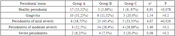
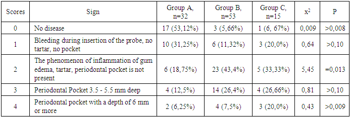
4. Study of the Periodontal Condition

In patients with CKD, inflammatory processes of periodontal tissues are common. Their occurrence is shown in Table 12.
Table 12. Prevalence of inflammatory periodontal diseases
     
According to the data obtained, periodontal diseases are more common among patients with CKD than in group A individuals (p<0.007). The prevalence of periodontal diseases in patients with CKD is 94.34% in group B and 93.33% in group B, and among healthy individuals - 46.88%. At the same time, in the group of patients receiving hemodialysis, inflammatory processes in the periodontium occur to a slightly milder degree than in those who do not receive it, however, severe periodontitis is observed among them 2.67 times more often (p=0.014). Among group B individuals, gingivitis occurs in 11.3% of cases, periodontitis of mild severity occurs in 43.4%, moderate severity occurs in 26.4%, and severe severity occurs in 7.5%. In group B: gingivitis - 13.33% of cases, mild periodontitis - 33.33%, moderate periodontitis - 26.66%, severe periodontitis — 20.0%.
Objectively, periodontal diseases in the presence of CKD are inactive – there is no exudation, the gums are dense, pale, which is due to a decrease in the activity of immunity in patients. Often, the radiological and clinical pictures do not coincide.
Gum recession in the examined individuals; it occurred quite often: in 63.0% of cases in group B, in 100.0% in group B (p=0.02). Gum recession is the result of dystrophic processes in the periodontium.
When assessing the periodontal CPITN index, the prevalence and intensity of individual signs of periodontal pathology (bleeding, tartar, periodontal pocket) were established and, depending on this, the need for periodontal care of the examined patients was determined. The results of this study are presented in table 13;
Table 13. Results of the study of the periodontal condition (CPITN)
     
Patients with CKD to a greater extent (94.34% in group B and 93.33% in group C) need to improve individual and professional oral hygiene (p= 0.034), to eliminate factors contributing to plaque retention, and up to 33.33% of cases in curettage.
Differences in the prevalence and intensity of individual signs of periodontal pathology (bleeding, tartar, periodontal pocket) were revealed (p=0.076). Periodontal pockets with a depth of 3.5 mm or deeper were more often observed in group B.
The degree of tooth mobility in patients with CKD does not differ (p>0.1). An increase in the degree of tooth mobility was found among patients with a total protein content in the blood below 60 g/l (p=0.045).
Inflammatory periodontal lesions in patients with CKD occur against the background of dystrophic phenomena, which are significantly more common in patients receiving hemodialysis (p=0.027). Signs of periodontal disease are found in 37% of cases in patients with CKD and in 56% of cases in patients of group B. Deterioration of the hygienic condition of the oral cavity aggravates the degree of inflammation (p= 0.004). Elevated blood creatinine leads to the development of osteodystrophic phenomena and dystrophy in periodontal tissues (p= 0.03).

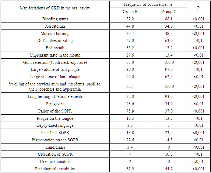
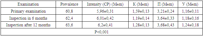
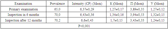
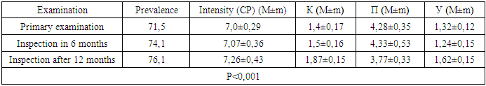
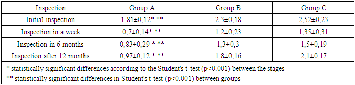
5. Discussion of the Results of the Study of Dental Status

We found that in all groups of patients with CKD, the prevalence and intensity of caries were higher than in those without urological diseases. The data obtained by us indicate that all patients with CKD have an unsatisfactory hygienic level of the oral cavity, which coincides with the research data of many authors. As a result, after the preventive measures carried out a year later, the prevalence of caries in group A was 63.6% (an increase of 2.8% compared to the initial examination), the intensity of caries changed this time period from 5.96 ± 0.31 to 6.2±0.41 (p<0.01), the increase in caries was 4.02%;. Examination after 12 months revealed the number of carious teeth equal to an average of 1.28 ± 0.13.
When examining patients in group B a year later, the increase in the prevalence of caries was 5.2%, with an intensity of 6.6 ± 0.43, the increase in the intensity of caries was 3.6%.
In group B, the increase in the prevalence of caries after 12 months was 5.4%. The intensity was 7.26 ± 0.43, the increase in the intensity of caries was equal to 3.71%.
Minor changes in the prevalence and intensity of dental caries clearly demonstrate to us that in patients with CKD, these indicators are greatly affected by an increased level of urea in saliva, even despite a decrease in the mineralizing properties of oral fluid and a deterioration in the hygienic condition of the oral cavity.
So, if during the examination a week after cleaning, the hygiene index in group B was detected at the level of 1.2 ± 0.23 (during the initial examination, before cleaning - 2.3 ± 0.18), then after 12 months it was 1.8± 0.16 (an increase of 50%).
We can observe similar results in group B. At the initial examination, the hygiene index was 2.52 ± 0.23, a week after cleaning - 1.35± 0.35, and a year later it increased to 2.1± 0.17 (an increase of 55.55%).
While in group A, an increase in the hygiene index occurred by 38.5% - from 0.7± 0.14 a week after cleaning to 0.97±0.12 a year later.
The deterioration of the hygienic condition in groups with CKD after a year is the result of deterioration of the processes of natural self-purification, a decrease in the activity of local immunity. In this regard, we believe that doctors should motivate patients to perform individual hygiene procedures.
Pathological erasability of hard dental tissues (about 80% of patients with CKD) may be associated with uremia, which usually occurs in patients with CKD. It has been suggested that hypocalcemia, secondary to chronic kidney disease, which contributes to renal osteodystrophy, is one of the causes of non-carious lesions of the hard tissues of the teeth [25].
Hyperpigmentation (27.6% and 14.3% in groups B and C) may probably be associated with insufficiency of beta-melanocytostimulating hormone secreted by the kidneys. As a result, excess melanin is deposited in the basal layer of the epithelium of the oral cavity [23].
Swelling and pasty gums (62.1 and 100% in groups B and C, respectively) can be caused by drugs taken by patients, which can be divided into three main groups: anticonvulsants, immunosuppressants and calcium channel blockers. However, the exact cause of drug-induced gum hyperplasia is unknown; however, it is believed that this condition is associated with some risk factors that contribute to gum inflammation, such as poor oral hygiene, the presence of plaque, the dose and duration of the drug used [24].
High levels of urea (16.34±0.88 and 27.24±0.83 mmol/L in groups B and C), dimethyl- and trimethylamines, and low levels of zinc may be associated with reduced taste perception in patients with uremia [25]. The increased concentration of urea, which is split by salivary urease into ammonia and carbon dioxide, gives a metallic, unpleasant taste [25]. The mechanisms underlying the changes in taste perception in patients with uremia are unknown, but they are probably related to the effect of uremic toxins on the central nervous system and peripheral nervous system (taste receptors) [14]. Gum bleeding was observed in 87.9 and 88.1% of patients with CKD and was associated with poor oral hygiene, periodontal inflammation [15].
The burning sensation in the mouth, which was significantly higher in patients with CKD (30.4% in group B and 48.3% in group C), was associated with dry mouth, damage to peripheral nerves by urinary toxins and the effect of drugs [3,16].
Complaints of xerostomia in patients with CKD (44.8 and 34.0% in groups B and C, respectively) are associated with fluid restriction, electrolyte imbalance, the use of certain medications such as furosemide and hydrochlorothiazide (antihypertensive agents), oral respiration, gland alteration (atrophy of the parenchyma of the small salivary glands), leading to a decrease in secretion saliva [20].
35.2% and 17.2% of the participants in this study complained of halitosis (groups B and C, respectively). The uremic fetid odor or bad breath reported by patients with CKD is an ammonia odor that is caused by a high concentration of urea in saliva and is broken down to ammonia [22]. This is due to a reduced kidney function for the excretion of urea from the body, therefore, the concentration of urea in the blood (uremia) increases, as well as in saliva. In addition, patients with CKD often neglected oral hygiene [20].
Pallor of the SOPR (75.9 and 37.0% in groups B and C) petechiae (13.8 and 23.0%) may be associated with anemia, be the result of anticoagulant therapy and/or platelet dysfunction [26].
The prevalence of periodontitis (31.2 and 47.0% in groups B and C) in this patient population increases, most likely due to immunosuppression in uremia. This suppresses the inflammatory reaction of the gums when plaque accumulates [1]. Periodontitis is associated with increased values of other components of the acute phase of inflammation, including lower concentrations of high-density lipoproteins [12,13], increased amounts of low-density lipoproteins [14-16] and neutrophils [17]. Inflammatory periodontal lesions in patients with CKD occur against the background of dystrophic phenomena, which are significantly more common in patients receiving hemodialysis (p=0.027). Signs of periodontal disease are found in 37% of cases in patients with CKD and in 56% of cases in group B patients.
Deterioration of the hygienic status of the oral cavity affects the aggravation of the degree of inflammation (p= 0.004). Elevated blood creatinine leads to the development of osteodystrophic phenomena and dystrophy in periodontal tissues. (p=0.03).
Chronic kidney disease and paradontopathy are also associated with common risk factors such as diabetes mellitus [2], age and tobacco smoking [6]. It is believed that periodontal diseases are an unconventional risk factor for chronic kidney disease due to systemic changes caused by periodontal inflammation. Due to the periodontal presence of bacteria, inflammatory mediators such as interleukin-1, interleukin-6, prostaglandin 2 and tumor necrosis factor-alpha are locally produced, and their antigens can enter the bloodstream [9]. Studies have shown that compared with healthy people, patients with periodontitis may have an increased level of C-reactive protein and, as a consequence, a weakly expressed systemic acute-phase inflammatory reaction. Apparently, there is a mutual influence between periodontal diseases and chronic kidney diseases. Since the affected periodontal tissue is susceptible to chronic inflammation, it is likely that oral bacteria can affect the course of chronic kidney diseases. In patients with chronic kidney diseases, a greater number of periodontal red complex bacteria (P. gingivalis, T. forsythia, T. denticola) and C. albicans were detected, as well as significant destruction of periodontal tissue [18], as well as a greater number of periodontal bacteria (P. gingivalis, T. forsythia, P. intermedia and P. nigrescens, A. actonomycemconcomitans) in periodontal pockets in these patients [19]. Therefore, regular assessment and prevention of periodontitis are of particular importance for patients with chronic kidney diseases.
The lesions detected in uremic stomatitis (2% in group B) were very painful and were most often localized on the lower surface of the tongue (81%). It manifested itself in the form of a gray pseudomembrane covering painful spots of erythema or red ulcers with a "purulent" cover.
Studies on the state of periodontal disease in patients with CKD indicate poor oral hygiene (in patients with CKD of both groups, a large amount of soft and hard plaque was found) and gingivitis [4,6-10]. This is probably a consequence of a pronounced uremic syndrome associated with impaired immune function, as well as altered activity of lymphocytes and monocytes [11].
The results obtained give us reason to assert that there is a need to develop a scheme for more effective treatment of dental diseases in patients with CKD, for which our research will continue.

References

[1]  Lomova A.S., Moroz P.V., Prokhodnaya V.A. Features of antimicrobial immunity of the oral cavity in pregnant women with primary and recurrent caries // Scientific review. Medical sciences. – 2014. – No. 2. – pp. 44-45.
[2]  Malyshev M.E., Lobeyko V.V., Iordanishvili A.K. Immune indicators of saliva in people of different ages living in St. Petersburg and the Leningrad region // Successes of gerontology, 2015. Vol. 28, No. 2. pp. 294-298.
[3]  Abdinian M, Mortazavi M, Jandaghian Z. Comparison of skeletal changes related to patients with chronic kidney disease and healthy individuals in digital panoramic radiography. Indian J Dent Res 2019; 30: 358-62.
[4]  Anuradha B.R., Katta S., Kode V.S., Praveena C., Sathe N., Sandeep N., et al: Oral and salivary changes in patients with chronic kidney disease: A clinical and biochemical study: J Indian Soc Periodontal. 2015; 19: 297-3.
[5]  Brotto R.S., Vendramini R.C., Brunetti I.L., Marcantonio R.A., Ramos A.P., Pepato M.T.: Lack of correlation between periodontitis and renal dysfunction in systemically healthy patients. Eur J Dent. 2011; 5: 8-18.
[6]  Chambrone L., Chambrone D., Pustiglioni F.E., Chambrone L.A., Lima L.A.: The influence of tobacco smoking on the outcomes achieved by root-coverage procedures. A systematic review. J Am Dental Assoc. 2009; 140: 294–6.
[7]  Chambrone L, Foz AM, Guglielmetti MR, Pannuti CM, Artese HP, Feres M, Romito GA. Periodontitis and chronic kidney disease: a systematic review of the association of diseases and the effect of periodontal treatment on estimated glomerular filtration rate J Clin Periodontol 2013, 40: 443-456.
[8]  Costantinides F., Castronovo G., Vettori E., Frattini C., Artero M.L., Bevilacqua L., et al: Dental Care for Patients with EndStage Renal Disease and Undergoing Hemodialysis. International Journal of Dentistry Volume. 2018| doi.org/10.1155/2018/9610892.
[9]  Craig RG. Interactions between chronic renal disease and periodontal disease Oral Dis 2008; 14: 1-7.
[10]  Dağ A., Firat E.T., Kadiroğlu A.K. et al. Significance of elevated gingival crevicular fluid tumor necrosis factor-alpha and interleukin-8 levels in chronic hemodialysis patients with periodontal disease. J Periodontal Res 2010; 45:4: 445—450.
[11]  Davidovich E., Schwarz Z., Davidovitch M. et al. Oral findings and periodontal status in children, adolescents and young adults suffering from renal failure. J Clin Periodontol 2005; 32: 10: 1076—1082.
[12]  Dioguardi M., Caloro G.A., Troiano G., Giannatempo G., Laino L., Petruzzi M., et al: Oral manifestations in chronic uremia patients. Ren Fail. 2016; 38(1): 1-6.
[13]  Dirschnabel A.J., Martins Ade S., Dantas S.A. et al. Clinical oral findings in dialysis and kidney-transplant patients. Quintessence Int 2011; 42: 2: 127-133.
[14]  Ellis J.S.: New year challenges: major challenges facing today's leaders of dental education. Eur J Prosthodont Restor Dent. 2004; 12: 143.
[15]  Gaál Kovalčíková A, Pančíková A, Konečná B, Klamárová T, Novák B, Kovaľová E, Podracká Ľ, Celec P, Tóthová Ľ. Urea and creatinine levels in saliva of patients with and without periodontitis. Eur J Oral Sci. 2019 Oct; 127(5): 417-424. doi: 10.1111/eos.12642.
[16]  Garcez J., Limeres Posse J., Carmona I.T. et al. Oral health status of patients with a mild decrease in glomerular filtration rate. Oral Surg Oral Med Oral Pathol Oral Radiol Endod 2009; 107: 2: 224-228.
[17]  Gavalda C, Bagan J, Scully C, Silvester F, Milian M, Jimenez Y. Renal hemodialysis patient; oral, salivary, dental and periodontal findings in 105 adult cases. Oral Dis. 1999; 5: 299–302.
[18]  Granitto M, Fall-Dickson J, Norton C, Sanders C. Review of therapies for the treatment of oral chronic graft-versus-host disease. Clin J Oncol Nurs. 2014; 18: 76–81.
[19]  Grubbs V., Plantiga L.C., Tuot D.S., Powe N.R.: Chronic kidney disease and use of dental services in a United States public healthcare system: a retrospective cohort sudy. BMC Nephrol. 2012; 13: 16.
[20]  Kaswan S, Patil S, Maheshwari S, Wadhawan R. Prevalence of oral lesions in kidney transplant patients: A single center experience. Saudi J Kidney Dis Transpl 2015; 26: 678-683.
[21]  Levey A, Eckkardt K, Tsukamoto Y, Levin A, Coresh J, Rossert J, et al. Definition and classification of chronic kidney disease; a position statement from kidney disease improving global outcome. Kidney Int. 2005; 67: 2089–100.
[22]  Lucas V.S., Roberts G.J. Oro-dental health in children with chronic renal failure and after renal transplantation: a clinical review. Pediatr Nephrol 2005; 20: 10: 1388—1394.
[23]  Nylund KM, Meurman JH, Heikkinen AM, Furuholm JO, Ortiz F, Ruokonen HM. Oral health in patients with renal disease: a longitudinal study from predialysis to kidney transplantation. Clin Oral Invest. 2017; doi 10.1007/s00784-017-2118-y.
[24]  Oyetola EO, Owotade FJ, Agebelusi GA, Fatusi OA, Sanusi AA. Oral findings in chonic kidney disease: implications for management in developing countries. BMC. 2015; 15: 24-31.
[25]  Proctor R, Kumar N, Stein A, Moles D, Porter S. Oral and dental aspect of chronic renal failure. J Dent Res. 2005; 84: 199–208.
[26]  Simpson T.C., Needleman I., Wild S.H., Moles D.R., Mills E.J.: Treatment of periodontal disease for glycaemic control in people with diabetes. Cochrane Database Syst Rev. 2010; 12 (5): CD004714.