American Journal of Medicine and Medical Sciences

p-ISSN: 2165-901X    e-ISSN: 2165-9036

2022;  12(9): 872-877

doi:10.5923/j.ajmms.20221209.04

Received: Aug. 4, 2022; Accepted: Aug. 29, 2022; Published: Sep. 15, 2022


Checking the Effectiveness of the Proposed Treatment Regimen for Dental Diseases in Patients with CKD

Rizaev J. A., Husanbaeva F. A.

Samarkand State Medical University, Samarkand, Uzbekistan

Correspondence to: Rizaev J. A., Samarkand State Medical University, Samarkand, Uzbekistan.

Email:

Copyright © 2022 The Author(s). Published by Scientific & Academic Publishing.

This work is licensed under the Creative Commons Attribution International License (CC BY).
http://creativecommons.org/licenses/by/4.0/

Abstract

The article describes the proposed treatment regimen for dental diseases in patients with chronic kidney disease(CKD), tested in 68 patients in Uzbekistan; its application is justified. The study involved 100 people, including 68 patients with CKD, of which 15 patients received hemodialysis. 32 practically healthy people made up the control group. The results obtained after the application of this scheme are presented: data on the intensity and prevalence of caries, the dynamics of the hygienic state, periodontal status and the state of the oral mucosa, assessment of the condition of fillings in patients with CKD.

Keywords: Caries, Prevalence, Intensity, Non-carious lesions, Periodontal disease, COPD, Dentistry, Chronic kidney disease, CKD

Cite this paper: Rizaev J. A., Husanbaeva F. A., Checking the Effectiveness of the Proposed Treatment Regimen for Dental Diseases in Patients with CKD, American Journal of Medicine and Medical Sciences, Vol. 12 No. 9, 2022, pp. 872-877. doi: 10.5923/j.ajmms.20221209.04.

1. Introduction

CKD is associated with clinical and radiological changes in the mouth. Radiological changes include thinning of the cortical plate, which is manifested during X-ray examination by transparent areas of the upper and lower jaw lesion. Clinical data in patients with CKD are basically the same as for oral cavity lesions in other systemic diseases. Unfortunately, attention to the oral cavity in CKD is not properly paid.
A causal relationship between oral infections and systemic diseases has not yet been established; however, significant improvements in major systemic diseases have been reported after treatment of associated oral lesions. This indicates the importance of oral care in the treatment of systemic diseases. Insufficient attention is paid to the needs of oral care in this group of patients, which further worsened the prognosis of the underlying disease. A contributing factor is the poor awareness of patients and some doctors of other specialties of dental aspects [1].
The state of organs and tissues of the oral cavity is closely interconnected with the general state of the body. Therefore, with various concomitant general somatic pathologies, teeth, periodontium and mucous membranes often suffer. Numerous studies have proven a change in salivation, microbiocenosis, the state of factors of nonspecific and specific protection in the oral cavity, and other indicators in this pathology. However, in the scientific literature there is practically no information about the state of acid-base balance in the oral cavity in general somatic diseases of the body.
Materials and methods of research. The study involved 100 people, including 68 patients with CKD, of which 15 patients received hemodialysis. 32 practically healthy people made up the control group. The age of the patients was 45-56 years. Men - 58, women - 42 people. The study took place in 2020-2022 on the basis of Samarkand State Medical University, Tashkent State Dental Institute.
Criteria for inclusion in the study: therapeutic patients with inflammatory diseases of the endo- and periodontium (acute and chronic pulpitis, chronic generalized catarrhal gingivitis and chronic generalized periodontitis) with concomitant chronic kidney pathology. Exclusion criteria: patients with a decrease in glomerular filtration rate <15 ml/min/1.73 m2.
The patients were divided into the following groups:
1. A group of people who do not have pathology from the urinary system - 32 people (group A);
2. Patients with chronic kidney disease who are not being treated on hemodialysis - 53 people (group B);
3. Patients with chronic kidney disease who are being treated on hemodialysis - 15 people (group B).

2. Research Results

Our study revealed that in people with CKD, diseases of the hard tissues of the teeth, periodontal and chronic obstructive pulmonary disease (COPD) occur more often and more intense than in people without diseases of the urinary system. In patients with CKD, periodontal diseases are significantly more often detected 92.45% in group B and 93.33% in group B, and among healthy individuals - 59.38%. Moreover, if periodontitis occurs in 37.5% of cases in healthy individuals, then in patients with CKD – in 77.3% and 80.0% (groups B and C, respectively), they need 89%-97% to remove deposits of soft plaque and up to 92% of tartar - with unsatisfactory individual oral hygiene oral cavity (OHI-S - 1.8 ±0.16 and 2.1± 0.17 in groups B and C), and COPD diseases in patients with CKD are detected, on average, in 20% of cases.
Table 1. Methods used in the study
     
Before the start of therapeutic and preventive measures, the study of dental health in patients of groups A, B and C showed that the prevalence of caries was 60.8%, 65.0% and 71.5%, respectively, non-carious lesions (wedge-shaped defects and erosion) in group A – in 32.1% of cases, in patients of group B - in 21.3% observations, in group B - 47.4%, endodontic treatment (for pulpitis and periodontitis) was carried out in 18, 27 and 9 teeth, respectively. The hygienic condition of the oral cavity was unsatisfactory: OHI-S - 1.81±0.12, 2.3±0.18 and 2.52±0.23 - in three groups. When assessing the periodontal CPITN index, the prevalence and intensity of individual signs of periodontal pathology (bleeding, tartar, periodontal pocket) were established and, depending on this, the need for periodontal care of the examined patients was determined.
An analysis of the effectiveness of dental treatment and preventive measures carried out six months later revealed their safety in 98.1% of cases, a year later - in 95.7% of patients in group A; in group B, an assessment of the condition of fillings in six months revealed their safety in 87.2% of cases, a year later – in 81.0%; in group B, an assessment of the condition of fillings in six months revealed their safety in 78.6% of cases, a year later – in 72.3%.
The X-ray method revealed the absence of periapical lesions in endodontically treated teeth in 97.3% after 6 months and 95.2% after 12 months in group A; in 89.1% after 6 months and 77.1% after 12 months in group B; and in 78.2% after 6 months and 74.4% after 12 months in group B. In group And six months after the oral sanitation measures, the hygienic condition of the oral cavity improved somewhat - 0.83± 0.29 (p≤0.05), and in groups B and C it was still unsatisfactory (1.3± 0.3 and 1.5± 0.19). After a year, the hygienic condition worsened in all groups - 0.97±0.12, 1.8±0.16 and 2.1±0.17 – but in healthy individuals it remained at a satisfactory level.
Therefore, the unsatisfactory hygienic condition of the oral cavity in persons with CKD, lesions of the hard tissues of the teeth, as well as common inflammatory periodontal diseases, require the development of special recommendations for oral care for such patients.
Also, based on such X–ray data - osteodystrophy was observed in patients with CKD - revealed a significant difference in the mandibular cortical index in patients with CKD and the control group (P = 0.038). Patients with CKD had more defects in the cortical layer of the mandible. C3 was observed in patients of groups B and C (3.9% and 8.6%, respectively), but was not detected in any of the healthy individuals. The number of healthy people with C1 was significantly higher than with C2 and C3. Thus, 83.3% of healthy people in group A belonged to class C1, the STC in patients with CKD was more porous than in the control group (P = 0.001). In group A, there was no pattern of loose calcification and loose calcification in the form of frosted glass, while these two patterns were observed in 8.6% and 17.7% of patients in groups B and C, respectively. A dense pattern was observed in the majority of healthy individuals (87.7%) - they indicate the need to improve dental treatment in patients with CKD, especially those receiving hemodialysis treatment, we proposed a treatment regimen for dental diseases in patients with CKD.
The proposed scheme:
1. Dental examination of patients with CKD and professional cleaning of the oral cavity should be carried out every 4 months.
2. In connection with the processes of demineralization of hard tissues of teeth, it is recommended to use low-abrasive fluoride-containing dental gels. Rinsing with 0.05% sodium fluoride solution was prescribed 2 times a day. Mandatory replacement of toothbrushes every 3 months. The use of floss and tongue scrapers is mandatory.
3. Dental examination necessarily includes X-ray examination (orthopantomography) to analyze the indices of the mandibular cortical index and the structure of the trabecular bone in order to assess the degree of osteodystrophy; to identify hidden lesions of the hard tissues of the teeth and evaluate the effectiveness of endodontic treatment.
4. It is possible to reduce the amount of anesthetic administered, which is associated with osteodystrophy of the jaws.
5. When forming cavities for filling carious and non-carious lesions of hard tissues of teeth, it is necessary to create support points in the preserved walls of the cavity, to form a cavity with a gradual narrowing towards the entrance hole, when localizing the carious cavity according to class II according to Black - to bring the cavity to the chewing surface, for better fixation of the seal, in some cases – to form an additional a cavity with cavities of class III and IV according to Black. Thinned edges of enamel should be excised.
6. In the absence of the effectiveness of endodontic treatment of periodontitis - removal of foci of periapical infection - apicotomy or removal of the causal tooth itself. With deep periodontal pockets - removal of the causal tooth.
7. To maintain periodontal health, Parodium gel was prescribed (applied 3 times a day for 2-3 weeks), Eludril rinse aid (2 teaspoons per ½ cup of water for 1-1.5 minutes, 3-4 times a day. For the treatment of periodontal pockets, "Cygerol" was used.
8. For the treatment of mucosal lesions, Lugol's glycerin solution and rosehip oil were used. At home, an infusion of chamomile flowers was used for rinsing (1 tablespoon of the collection was poured with 150 ml of water, heated in a water bath for 15 minutes, infused, filtered, topped up with boiled water to the original volume. Also, patients rinsed their mouth with propolis tincture – 25 drops per 1 glass of warm water.
9. The appointment and use of drugs – only after consultation with a nephrologist.
For its testing, 15 people were selected in group B, and group B was fully included in the study.
Table 2. Patients selected for testing of the scheme, according to the stages of CKD
     
Results of approbation of the treatment regimen of dental diseases in patients with CKD.
12 months after the application of the new protocol, the following patient complaints were identified:
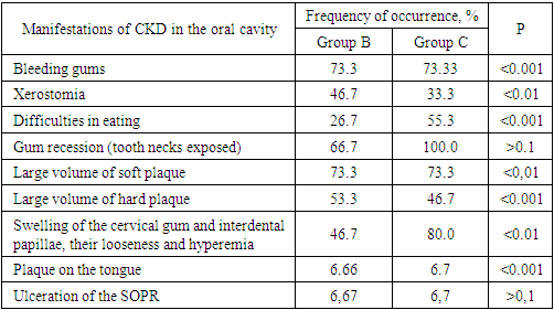
Table 3. Analysis of complaints and objective indicators of examination of patients with CKD
     
Gum bleeding decreased by 10.6-13.2%; difficulty eating decreased in 2.2-7.7% of patients; the amount of soft plaque decreased by 15.58-21.1%; tartar – by 41% and 15.2% in groups B and C, respectively; swelling of the cervical gum and interdental papillae, their looseness and hyperemia decreased by 18.9-21.3%.
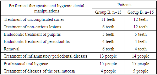
Table 4. The volume of therapeutic and preventive dental care for the examined persons
     
Table 5. Changes in the prevalence (%) and intensity of dental caries in group B patients
     
Table 6. Causes of tooth extraction in group B patients
     
In total, 17 teeth were cured for carious and non-carious lesions in this group, 9 teeth were endodontically treated for pulpitis and periodontitis, and 6 teeth were removed (the reason was ineffective endodontic treatment and periodontal lesions). The number of filled teeth was 3.91±0.65 (an increase of 14.0%). The number of teeth affected by the carious process increased from 1.7 ± 0.15 to 1.81± 0.12 (an increase of 6.47%, earlier in the same period the increase was 33.8%). The increase in the prevalence of caries for 1 year is 0.2%.
The assessment of the condition of the fillings revealed their safety after a year – in 87.9% (earlier for the same period - 81.0%).
The X-ray method revealed the absence of periapical lesions in endodontically treated teeth in 78.3% after 12 months (previously 77.1% for the same period of time).
Table 7. Changes in the prevalence (%) and intensity of dental caries in group B patients
     
In total, 24 teeth were cured for carious and non-carious lesions in this group, 9 teeth were treated endodontically for pulpitis and periodontitis, and 4 teeth were removed (the reason is ineffective endodontic treatment and periodontal lesions). When examined a year later, the number of filled teeth was 4.57 ± 0.26 (an increase of 21.22% compared to the same period). The number of teeth affected by the carious process decreased from 1.87± 0.15 to 1.6± 0.13 (reduction of caries growth by 16.87%). The increase in the prevalence of caries for 1 year is 0.6%.
An assessment of the condition of the seals a year later revealed their safety at 76.7% (an increase of 4.4%).
The X-ray method revealed the absence of periapical lesions in endodontically treated teeth in 77.3% after a year (previously 74.4% for the same period of time).
Table 8. Causes of tooth extraction in group B patients
     
Thus, it can be argued that the effectiveness of endontic treatment in patients with CKD has practically not changed, but more effective treatment of carious and non-carious lesions will, in our opinion, reduce the need for endodontic treatment in this category of patients.
Dynamics of the hygienic state of the oral cavity of patients with CKD.
Table 9. Changes in the hygienic index OHI-S in the examined patients
     
Examination after 12 months revealed that OHI-S in group B patients was 1.34±0.17 (p<0.001) (a decrease of 34.32% compared to the previous examination), in group B - 1.52±0.12 (p<0.001) (a decrease of 38.16% compared to the previous examination).

3. Study of the Periodontal Condition

In patients with CKD, inflammatory processes of periodontal tissues are common. Their occurrence is shown in table 10.
Table 10. Prevalence of inflammatory periodontal diseases in patients with CKD
     
After applying the proposed treatment regimen, the prevalence of periodontal diseases in patients with CKD is 86.67% in group B (a decrease of 7.67%) and 93.33% in group B (unchanged). Among group B individuals, gingivitis occurs in 20.0% (an increase of 8.68%), periodontitis of mild severity occurs in 40.0% (a decrease of 3.4%), moderate severity occurs in 20% (a decrease of 6.4%), severe severity occurs in 6.67% (a decrease of 0.83%). In group B: gingivitis – 20.0% (unchanged) of observations, mild periodontitis - 26.67% (decreased by 6.66%), moderate periodontitis - 33.33% (decreased by 6.67%), severe periodontitis - 13.33% (decreased by 6.67%).
When evaluating the periodontal CPITN index, individual signs of periodontal pathology (bleeding, tartar, periodontal pocket) are still detected.
Table 11. Results of the study of the need for treatment of periodontal diseases
     
Differences in the prevalence and intensity of individual signs of periodontal pathology (bleeding, tartar, periodontal pocket) were revealed (p=0.082). Bleeding of the gums during the introduction of the probe in group B increased by 8.68%, in group B remained unchanged; swelling of the gums and periodontal pockets was no longer detected in 93.3% of patients in group B and 86.67% of patients in group B. Periodontal pockets with a depth of 3.5 mm or deeper were twice as often observed in group B.
Lesions of the
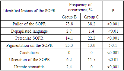
Table 12. Identified lesions of the SOPR
     
The analysis of the lesions of the SOPR in patients with CKD showed that the changes, compared with the data of the previous examination, are very small. The reason for this, in our opinion, is that the lesions of the COPD in CKD are not independent, but are symptoms of a systemic disease.
Based on the above data, it can be concluded that the proposed protocol for the treatment of dental diseases in patients with CKD has shown effectiveness in the treatment of carious and non-carious lesions of the hard tissues of the teeth, in maintaining a satisfactory hygienic condition of the oral cavity. Also, the proposed protocol has shown satisfactory results in maintaining periodontal health, but in our opinion, it is impossible to completely rid a patient with CKD of periodontal diseases by dental measures alone and requires coordinated, comprehensive work of a dentist and a nephrologist.

References

[1]  Lomova A.S., Moroz P.V., Prokhodnaya V.A. Features of antimicrobial immunity of the oral cavity in pregnant women with primary and recurrent caries // Scientific review. Medical sciences. – 2014. – No. 2. – pp. 44-45.
[2]  Malyshev M.E., Lobeyko V.V., Iordanishvili A.K. Immune indicators of saliva in people of different ages living in St. Petersburg and the Leningrad region // Successes of gerontology, 2015. Vol. 28, No. 2. pp. 294-298.
[3]  Abdinian M, Mortazavi M, Jandaghian Z. Comparison of skeletal changes related to patients with chronic kidney disease and healthy individuals in digital panoramic radiography. Indian J Dent Res 2019; 30: 358-62.
[4]  Anuradha B.R., Katta S., Kode V.S., Praveena C., Sathe N., Sandeep N., et al: Oral and salivary changes in patients with chronic kidney disease: A clinical and biochemical study: J Indian Soc Periodontal. 2015; 19: 297-3.
[5]  Brotto R.S., Vendramini R.C., Brunetti I.L., Marcantonio R.A., Ramos A.P., Pepato M.T.: Lack of correlation between periodontitis and renal dysfunction in systemically healthy patients. Eur J Dent. 2011; 5: 8-18.
[6]  Chambrone L., Chambrone D., Pustiglioni F.E., Chambrone L.A., Lima L.A.: The influence of tobacco smoking on the outcomes achieved by root-coverage procedures. A systematic review. J Am Dental Assoc. 2009; 140: 294–6.
[7]  Chambrone L, Foz AM, Guglielmetti MR, Pannuti CM, Artese HP, Feres M, Romito GA. Periodontitis and chronic kidney disease: a systematic review of the association of diseases and the effect of periodontal treatment on estimated glomerular filtration rate J Clin Periodontol 2013, 40: 443-456.
[8]  Costantinides F., Castronovo G., Vettori E., Frattini C., Artero M.L., Bevilacqua L., et al: Dental Care for Patients with EndStage Renal Disease and Undergoing Hemodialysis. International Journal of Dentistry Volume. 2018| doi.org/10.1155/2018/9610892.
[9]  Craig RG. Interactions between chronic renal disease and periodontal disease Oral Dis 2008; 14: 1-7.
[10]  Dağ A., Firat E.T., Kadiroğlu A.K. et al. Significance of elevated gingival crevicular fluid tumor necrosis factor-alpha and interleukin-8 levels in chronic hemodialysis patients with periodontal disease. J Periodontal Res 2010; 45:4: 445—450.
[11]  Davidovich E., Schwarz Z., Davidovitch M. et al. Oral findings and periodontal status in children, adolescents and young adults suffering from renal failure. J Clin Periodontol 2005; 32: 10: 1076—1082.
[12]  Dioguardi M., Caloro G.A., Troiano G., Giannatempo G., Laino L., Petruzzi M., et al: Oral manifestations in chronic uremia patients. Ren Fail. 2016; 38(1): 1-6.
[13]  Dirschnabel A.J., Martins Ade S., Dantas S.A. et al. Clinical oral findings in dialysis and kidney-transplant patients. Quintessence Int 2011; 42: 2: 127-133.
[14]  Ellis J.S.: New year challenges: major challenges facing today's leaders of dental education. Eur J Prosthodont Restor Dent. 2004; 12: 143.
[15]  Gaál Kovalčíková A, Pančíková A, Konečná B, Klamárová T, Novák B, Kovaľová E, Podracká Ľ, Celec P, Tóthová Ľ. Urea and creatinine levels in saliva of patients with and without periodontitis. Eur J Oral Sci. 2019 Oct; 127(5): 417-424. doi: 10.1111/eos.12642.
[16]  Garcez J., Limeres Posse J., Carmona I.T. et al. Oral health status of patients with a mild decrease in glomerular filtration rate. Oral Surg Oral Med Oral Pathol Oral Radiol Endod 2009; 107: 2: 224—228.
[17]  Gavalda C, Bagan J, Scully C, Silvester F, Milian M, Jimenez Y. Renal hemodialysis patient; oral, salivary, dental and periodontal findings in 105 adult cases. Oral Dis. 1999; 5: 299–302.
[18]  Granitto M, Fall-Dickson J, Norton C, Sanders C. Review of therapies for the treatment of oral chronic graft-versus-host disease. Clin J Oncol Nurs. 2014; 18: 76–81.
[19]  Grubbs V., Plantiga L.C., Tuot D.S., Powe N.R.: Chronic kidney disease and use of dental services in a United States public healthcare system: a retrospective cohort sudy. BMC Nephrol. 2012; 13: 16.
[20]  Kaswan S, Patil S, Maheshwari S, Wadhawan R. Prevalence of oral lesions in kidney transplant patients: A single center experience. Saudi J Kidney Dis Transpl 2015; 26: 678-683.
[21]  Levey A, Eckkardt K, Tsukamoto Y, Levin A, Coresh J, Rossert J, et al. Definition and classification of chronic kidney disease; a position statement from kidney disease improving global outcome. Kidney Int. 2005; 67: 2089–100.
[22]  Lucas V.S., Roberts G.J. Oro-dental health in children with chronic renal failure and after renal transplantation: a clinical review. Pediatr Nephrol 2005; 20: 10: 1388—1394.
[23]  Nylund KM, Meurman JH, Heikkinen AM, Furuholm JO, Ortiz F, Ruokonen HM. Oral health in patients with renal disease: a longitudinal study from predialysis to kidney transplantation. Clin Oral Invest. 2017; doi 10.1007/s00784-017-2118-y.
[24]  Oyetola EO, Owotade FJ, Agebelusi GA, Fatusi OA, Sanusi AA. Oral findings in chonic kidney disease: implications for management in developing countries. BMC. 2015; 15: 24-31.
[25]  Proctor R, Kumar N, Stein A, Moles D, Porter S. Oral and dental aspect of chronic renal failure. J Dent Res. 2005; 84: 199–208.
[26]  Simpson T.C., Needleman I., Wild S.H., Moles D.R., Mills E.J.: Treatment of periodontal disease for glycaemic control in people with diabetes. Cochrane Database Syst Rev. 2010; 12 (5): CD004714.