-
Paper Information
- Next Paper
- Paper Submission
-
Journal Information
- About This Journal
- Editorial Board
- Current Issue
- Archive
- Author Guidelines
- Contact Us
American Journal of Medicine and Medical Sciences
p-ISSN: 2165-901X e-ISSN: 2165-9036
2022; 12(9): 869-871
doi:10.5923/j.ajmms.20221209.03
Received: Jul. 27, 2022; Accepted: Aug. 17, 2022; Published: Sep. 15, 2022

Clinical Case of a Patient with a Hemangioma of the Nasal Cavity
 Abstract
 Reference
 Full-Text PDF
 Full-text HTML
Lutfullayev G. U., Nematov U. S., Safarova N. I.
Department of Otorhinolaryngology, Faculty of Postgraduate Education, Samarkand State Medical University, Uzbekistan
Correspondence to: Lutfullayev G. U., Department of Otorhinolaryngology, Faculty of Postgraduate Education, Samarkand State Medical University, Uzbekistan.
| Email: |  |
Copyright © 2022 The Author(s). Published by Scientific & Academic Publishing.
This work is licensed under the Creative Commons Attribution International License (CC BY).
http://creativecommons.org/licenses/by/4.0/

This article presents a case of hemangioma of the nasal cavity, diagnosed in a patient at 30 weeks of pregnancy, with complaints of lack of nasal breathing, anosmia and periodic nosebleeds, significant difficulty in nasal breathing. Treatment is complete removal by endonasal method.
Keywords: Capillary hemangioma, Nasal septum, Nosebleed
Cite this paper: Lutfullayev G. U., Nematov U. S., Safarova N. I., Clinical Case of a Patient with a Hemangioma of the Nasal Cavity, American Journal of Medicine and Medical Sciences, Vol. 12 No. 9, 2022, pp. 869-871. doi: 10.5923/j.ajmms.20221209.03.
Article Outline
- Hemangioma is a benign neoplasm of vascular origin with epithelial proliferation. Most often refers to congenital lesions of the skin and mucous membrane of the oral cavity, while the nasal cavity and paranasal sinuses are considered an unusual location for hemangiomas. On the head and neck, 38% of the location is found on the lip mucosa, 7-29% - in the nasal cavity (often in the anterior parts of the nasal septum, on the nasal shells, cases of development from the maxillary sinus, roof and bottom of the nasal cavity have also been described). It is the most common benign tumor of the nasal cavity. It occurs in all age groups, there are several peaks: children and adolescents, women of reproductive age, and then there is an even distribution in the group over 40 years old. There are several theories of pathogenesis, more often associated with traumatic tissue damage and hormonal factors (pregnancy, oral contraceptives). The most striking symptomatic manifestations of capillary hemangioma are unilateral nosebleeds and nasal obstruction. Histologically characterized by vascular proliferation in the submucosal layer, in the form of lobules or clusters consisting of central capillaries and small branching ducts. Treatment is complete removal, preferably by endonasal method. Unfortunately, the recurrence rate after excision can reach 15%.According to Russian and foreign literature, during pregnancy and in the postpartum period, 27-43% of women turn to otorhinolaryngologists with such nonspecific symptoms as nasal congestion, rhinorrhea, bleeding or anosmia, especially in the third trimester of pregnancy and during lactation, when the reactivity of the nasal mucosa is due to an increased content of estrogens in the blood, which causes vascular dilation and mucosal hypersecretion. Less often, the manifestation of the disease manifests itself with visual impairment, headaches, a local feeling of swelling in the nose.The Department of Otorhinolaryngology FPS of the Samarkand Medical Institute has accumulated extensive experience in managing patients with various benign neoplasms of ENT organs. Over the past 5 years, 197 patients with various vascular neoplasms of the nasal cavity, nasopharynx and paranasal sinuses have been treated in the ENT department.We present a clinical case of a patient with capillary hemangioma of the nasal cavity.Patient S., (28 years old (pregnancy – 32 weeks) applied in September 2017 presented with complaints of anosmia and periodic nosebleeds from the left half of the nose, significant difficulty of nasal breathing on the left and dry mouth.The listed symptoms have been noted since one month. It is known from the anamnesis of the disease that for the first-time bleeding from the left half of the nose occurred at the 30th week of pregnancy, while there was an increase in systemic blood pressure to 140- and 80-mm Hg. The bleeding was stopped by a loose anterior tamponade. At the same time, the patient notes that the intensity and duration of nosebleeds increased with each subsequent time. Indicators of the hemostasis system, the level of hemoglobin in the blood during pregnancy were within acceptable values.2 weeks before hospitalization, an outpatient otorhinolaryngologist performed an endoscopic examination of the nasal cavity - a neoplasm in the form of a polyp was found in the left half of the nose, which bled during probing. When performing an MRI (without the introduction of a contrast agent) of the nose and paranasal sinuses, a rounded tissue formation of the nasal cavity on the left is determined, filling the posterior parts of the nasal cavity from the middle of the left middle nasal concha to the left choana, with axial dimensions of 2.5x2.0 cm, vertical size up to 3.0-3.5 cm. The formation partially displaces and causes the destruction of the nasal septum. The biopsy of the neoplasm was accompanied by profuse bleeding. According to histological examination, the tumor consists of small compactly arranged capillaries (Fig. 1).
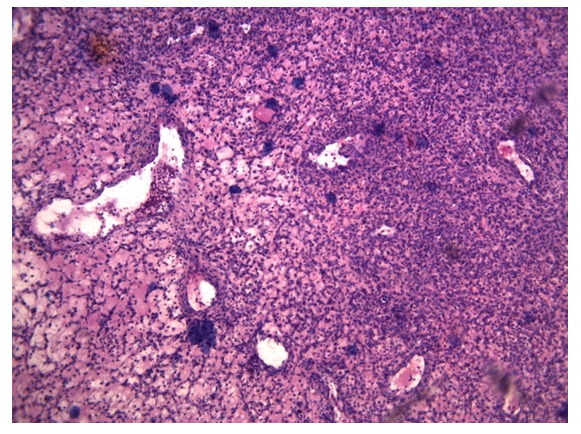 | Figure 1. Capillary hemangioma, stained with hematoxylin-eosin |
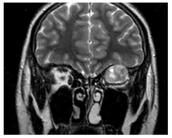 | Figure 2. In the MRI image, the hemangioma of the nasal cavity on the left |