-
Paper Information
- Next Paper
- Paper Submission
-
Journal Information
- About This Journal
- Editorial Board
- Current Issue
- Archive
- Author Guidelines
- Contact Us
American Journal of Medicine and Medical Sciences
p-ISSN: 2165-901X e-ISSN: 2165-9036
2022; 12(9): 859-862
doi:10.5923/j.ajmms.20221209.01
Received: Aug. 20, 2022; Accepted: Sep. 7, 2022; Published: Sep. 9, 2022

Morphological Characteristics of the Morphometric Parameters of the Gastric Mucous Layer in Polypragmasy with Anti-Inflammatory Drugs
 Abstract
 Reference
 Full-Text PDF
 Full-text HTML
Tashmamatov B. N. 1, Teshaev Sh. J. 2
1Department of Human Anatomy, Samarkand State Medical University, Samarkand, Uzbekistan
2Bukhara State Medical Institute, Bukhara, Uzbekistan
Copyright © 2022 The Author(s). Published by Scientific & Academic Publishing.
This work is licensed under the Creative Commons Attribution International License (CC BY).
http://creativecommons.org/licenses/by/4.0/

Polypharmacy is becoming more common due to an aging population, an increase in the number of people with multiple diseases, and more disease recommendations. Although treating patients with complex medical problems with the right medications can improve clinical outcomes, quality of life, and life expectancy, polypharmacy is also associated with an increased risk of adverse drug reactions, some of which are serious enough to lead to hospitalization and even death. Therefore, it is critical to have systems in place to ensure that medications are only started when indicated, that patients are fully informed of the benefits and potential complications of treatment, and that patients are regularly reviewed to ensure compliance with their medication regimen.
Keywords: Polypharmacy, Multiple diseases, Clinical outcomes, Digestive system, Lymphoid tissues
Cite this paper: Tashmamatov B. N. , Teshaev Sh. J. , Morphological Characteristics of the Morphometric Parameters of the Gastric Mucous Layer in Polypragmasy with Anti-Inflammatory Drugs, American Journal of Medicine and Medical Sciences, Vol. 12 No. 9, 2022, pp. 859-862. doi: 10.5923/j.ajmms.20221209.01.
Article Outline
1. Introduction
- The digestive system plays an important role in the interaction of the body with the external environment. Since the mucous membrane of the alimentary canal is affected by various substances contained in food, it is understandable that the mucous membrane and submucosal base, which are immune organs, have their own lymphoid structures.The mucous membrane of the organs of the digestive system is considered a barrier system that blocks the entry of various agents into the body from the outside, and on the other hand, it participates in the exchange processes of the external and internal environment of the body. Due to its direct proximity to microbiota and food, it is constantly exposed to normal as well as potentially dangerous antigens.Diseases of the gastrointestinal system occupy one of the highest places in terms of morbidity of the population. Epidemiological studies conducted on gastroscopy and morphological evaluation of the gastric mucosa revealed that more than half of the population suffers from chronic gastritis.Immunocompetent tissues of the digestive system are called "lymphoid tissues". Lymphocyte recirculation and the immune response cover the mucosa of the entire gastrointestinal tract.Circulating lymphocytes are involved in the recovery of the cellular composition of irradiated lymph nodes.The arrival of a significant amount of natural flora after birth induces its immunological responses, namely the expansion of the intraepithelial lymphocyte population and the expansion of the distribution of cells in the crypts.The further development of knowledge about the lymphatic system cannot be done without taking into account the processes that occur around the lymphatic capillaries and in the interstitium.The morpho-functional state of the organs of the digestive system changes under the influence of various factors of the external environment. A study of the status of fat cells located in the alimentary canal during space flights showed that the number of fat cells decreased against the background of increased expression of chymase in the stomach in the antigravity state of the organism.Artificial feeding leads to an increase in large neurons of intermuscular ganglia of the stomach and a decrease in neuron ganglions of medium-sized nerve cells (21%).Injuries of the muscle layer of the stomach wall in the form of cutting, crushing and thermocoagulation lead to increased proliferative activity of myocytes near the gastric ulcer.The effect of zirk leaf extract on the surface of gastric ulcer resulted in a significant decrease in ulcer healing times. In patients with arterial hypertension, the healing time of gastric ulcer is prolonged.The purpose of the study. Study of morphological and morphometric parameters of gastric mucosa wall in polypharmacy with anti-inflammatory drugs.Tasks of the research:1. To study changes in the morphological and morphological parameters of the stomach of 5-month-old purebred rats in the norm, as well as when anti-inflammatory drugs are used individually.2. To determine the morphological and morphometric parameters of the gastric wall of 5-month-old white rats when two, three, four and five types of anti-inflammatory drugs are used in different combinations, and to determine the change in the morphometric and morphometric parameters of the gastric wall.3. To study the morphometric and iorphometric changes caused by the simultaneous use of two, three, four and five types of anti-inflammatory drugs in 5-month-old purebred rats.Recent years have also seen the development of smartphone applications such as "My Medicine Passport" to improve communication between patients and health care providers, improve people's understanding of their condition and treatment, and track changes to a patient's medication.Polypharmacy is the simultaneous use of several drugs by one patient. As the population ages in the developed world, particularly the UK, the number of people with chronic diseases is increasing, and there is increasing pressure on doctors to adhere to evidence-based guidelines for the management of chronic diseases.Despite the fact that many studies are conducted in polypragmasy, information about the stomach, which is the central organ of the alimentary canal, is very scarce in the literature.
2. Materials and Methods of Research
- The study was conducted on a total of 180 5-month-old adult white rats. According to the purpose of the study, all laboratory animals were divided into 5 groups. 5 different types of anti-inflammatory drugs were used in polypharmacy in different combinations.The histological material obtained from the cardiac, bottom, body and pyloric parts of the stomach of the experimental white male rats served as the subject of the study.Research methods.- staining micropreparations with hematoxylin-eosin- staining of micropreparations by Van-Gieson methods- Determination by variational statistics method using Strelkov tables
3. Research Results and Discussion
- The stomach of non-white rats lies mainly under the liver. A large curvature of the stomach emerges from under its sharp caudal edge. It is on the left side and slightly caudal to the lesser curvature, dorsal to the gastric fundus and slightly cranial to the pyloric portion. Thus, the stomach of the white rat is located almost transversely (between the sagittal and transverse planes).The entry of the stomach into the duodenum (pyloric) and the cranial part of the duodenum join immediately to the right of the midline, at an open ventrocaudal angle. Below it (caudal) are 12 loops of the duodenum and (ventral to these loops) the terminal part of the acute angle of the ileum, and even more ventrocaudal - the cecum. Dorsocaudal (behind) the pyloric part and body of the stomach are the transverse colon, the body and tail of the pancreas. From the large curvature of the body of the organ and from the bottom of the stomach to the left and dorsally, the spleen is located.The following indicators were determined when the topographic-anatomical and skeletoscopic data of the white rat's stomach were studied. In non-laboratory white rats, the upper or upper back wall of the stomach touches the jejunum and ileal loops on the right side, and the left adrenal gland and the left kidney on the left side. Stomach covers the upper 2/3 of the lower surface of the adrenal gland of the left kidney and closes the upper end of the left kidney, that is, the front end. The left side of the stomach is rounded, it is located mainly under the diaphragm, and on the left side it is located touching the spleen. The right side of the stomach narrows and joins from the last part to the beginning of the duodenum. For the first time, it was found that polypharmacy with anti-inflammatory drugs in 5 combinations had different negative effects on the mucosa and submucosa, which is the structural structure of the stomach wall of white male rats;Structural changes in the organs of the digestive system have been proven to cause profound disturbances in the processes of growth and formation of the mucous membrane of the stomach and its glandular tissue, submucosa base, which is found to be accompanied by a decrease in the total thickness of the stomach wall;When using more than 3 types of anti-inflammatory drugs at the same time, the negative effects of polypharmacy increase significantly. The rate of formation of the structural and functional zones of the stomach wall, the activity of cells in the mucous membrane, the change in the shape of the lymphocytes located at the base of the mucosa, and the decrease in the morphological parameters were analyzed;Using the methods of modern morphological research (organometric, histological, histomorphometric, statistical), new information was obtained directly about the morphological and morphometric parameters of the stomach wall. Changes identified at the tissue, cellular and intercellular levels are characterized by hypotrophic and hypoplastic changes in the structures of the gastric wall of the white male rat.There are several lines from the abdominal organs of white rats to the stomach, and they are as follows: Stomach-splenic line: - from the spleen to the great curvature of the stomach; Diaphragm-stomach line: - from the diaphragm to the left half of the greater curvature of the stomach; Liver-stomach section: - from the area of the liver gate to the small curvature of the stomach; Gastrointestinal tract: - starts from the great curvature of the stomach and continues to the transverse colon.Five-month-old purebred laboratory rats have fully formed stomachs. When the five-month-old rats in the experiment were examined macroscopically, the following data were obtained:The body weight of five-month-old laboratory animals varied from 189-258 g, and the average was 244.6±6.3 g. The total length of the white rat stomach in the control group was 33-35 mm, with an average of 34.62±0.18 mm. The width of the member varied from 13 to 15 mm and averaged 13.81±0.18 mm. The thickness of the studied organ varied from 12 to 15 mm, and the average was 13.69±0.32 mm. The length of the large curvature is around 37 - 38 mm, on average -37.43±0.10 mm. The length of the small curvature was 14 - 15 mm, the average was equal to -14.65±0.10 mm.The following data were collected when examining the microscopic findings of the control group. According to this, the height of the mucous layer of the gastric wall of white rats in the period of five months varies from 433.9 to 524.3 μm in the area of the esophagus to the stomach (cardial part), and the average is 473.9±8.32 μm;(Figure 1). The height of the mucosa at the bottom of the organ varied from 442.1 to 529.3 μm, and the average was 509.4±8.02 μm. The height of this layer in the body area of the stomach is 448.1-546.8 μm, on average 511.8±9.08 μm; the height of the mucous layer in the pyloric part was 476.5 μm from 381.4 μm, and the average index was 427.4±8.75 μm (Fig. 2).
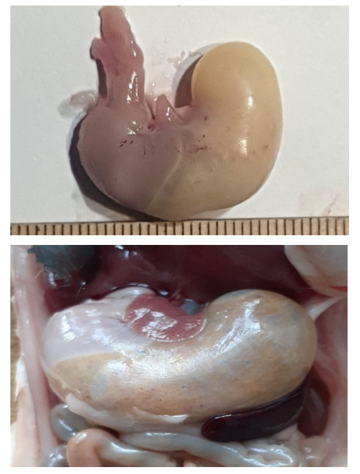 | Figure 1 and 2. Anatomy of the stomach of a 5-month-old purebred rat |
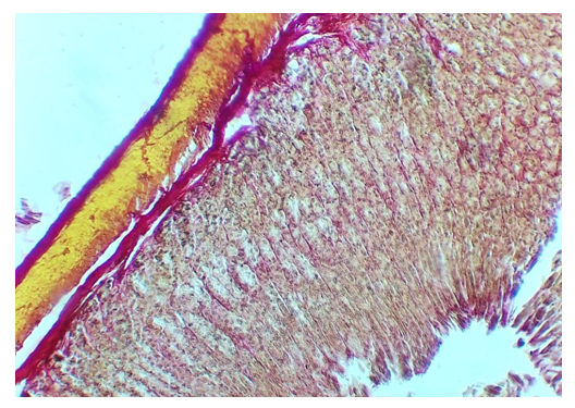 | Figure 3. The structure of the cardiac part of the stomach of 5-month-old white rats. 1 – mucosal layer, 2 – submucosa base, 3 – muscle layer, 4 – bundle of collagen fibers, 5 – cavity between folds. Van – painted according to Gizon.Ok.10hob.4.0 |
4. Conclusions
- 1. Different levels of morphological changes occur under the influence of different amounts of drugs. According to the obtained data, the overall thickness of the stomach wall significantly decreased in groups IV-V due to the decrease in the size of the gastric mucosa and the mucosal base after the effect of drugs. These changes were 1.60% in the cardiac part, 3.27% in the gastric fundus, 3.33% in the body, and 3.65% in the pyloric part of laboratory animals of group IV, and 2.21% in the cardiac part of organ in laboratory animals of group V. %, changed to 3.89% in the base, 3.0% in the body of the stomach and 5.2% in the pyloric part.2. When the adverse effects of polypharmacy with anti-inflammatory drugs were compared between the rats of the experimental control group and the rest of the groups, when the measurements of all the morphometric parameters obtained were viewed in the increasing order of the group, it was observed that the negative effects significantly increased in accordance with it and parallel to it.3. The adverse effect of polypharmacy of anti-inflammatory drugs in the experimental group of rats in the stomach wall, mucosal base and glandular tissue was corrected in the V-group compared to the I-control group. In this case, in the cardiac part of the organ wall of group V, the height of the mucous layer of the gastric wall is 8.4%, the mucous base is 10.5%, and the glandular tissue is 37.0%, the mucous membrane is 7.60%, the mucosa is 17.8% in the bottom of the stomach, and in the gland tissue by 29.7%, the height of the mucous membrane in the organ body by 6.52%, the base of the mucosa by 16.7% and in the gland tissue by 34.4%, the height of the mucous membrane in the pyloric area of the stomach by 6.2%, the mucosa it was found that it did not decrease by 15.9% in the base and by 32.2% in the gland tissue.