-
Paper Information
- Next Paper
- Previous Paper
- Paper Submission
-
Journal Information
- About This Journal
- Editorial Board
- Current Issue
- Archive
- Author Guidelines
- Contact Us
American Journal of Medicine and Medical Sciences
p-ISSN: 2165-901X e-ISSN: 2165-9036
2022; 12(8): 811-814
doi:10.5923/j.ajmms.20221208.09
Received: Jul. 7, 2022; Accepted: Aug. 8, 2022; Published: Aug. 15, 2022

Morphological and Morphometric Indications of Trachea and Bronchial Walls in One-Month-Old Babies
 Abstract
 Reference
 Full-Text PDF
 Full-text HTML
Sultanov Ravshan1, Sodikova Zumrat1, Arsenova Mukhabbat1, Boboyorov Sardor2
1Anatomy, Department of Clinical Anatomy, Tashkent Medical Academy, Uzbekistan
2Student, Termiz branch of the Tashkent Medical Academy, Uzbekistan
Copyright © 2022 The Author(s). Published by Scientific & Academic Publishing.
This work is licensed under the Creative Commons Attribution International License (CC BY).
http://creativecommons.org/licenses/by/4.0/

In this article, the morphological, morphometric and general histological analysis of the trachea and external bronchi of the lungs of babies up to one month old was carried out. Worldwide, early neonatal mortality is a global problem, especially in infants under one year of age, with respiratory diseases being the most common cause of death. Therefore, the study of the morphological, morphometric and general histological structures of the walls of the trachea and lungs of babies up to one month old will effectively help not only pathologists, but also treating doctors to diagnose and prevent the disease.
Keywords: Baby, Trachea, Bronchial tree, Morphometry, Diameter, Lungs, Postnatal ontogeny
Cite this paper: Sultanov Ravshan, Sodikova Zumrat, Arsenova Mukhabbat, Boboyorov Sardor, Morphological and Morphometric Indications of Trachea and Bronchial Walls in One-Month-Old Babies, American Journal of Medicine and Medical Sciences, Vol. 12 No. 8, 2022, pp. 811-814. doi: 10.5923/j.ajmms.20221208.09.
1. Introduction
- According to the World Health Organization, child mortality is one of the main problems in the world medical field. Today, the child mortality rate is 15.6% per 1,000 live births, but the birth rate is very low at 9.1%. [1]. At present, modern endoscopy is used as the most reliable method for damage to the tracheabronchial tree. [2-3] But the morphological structure has not been fully studied. The level of air pollution has increased significantly in recent years, and there is a lot of evidence that exposure to small harmful particles can have adverse effects on breathing. Health effects of environmental exposure to air pollution during the prenatal period may affect lung bronchial organogenesis in particular. [4-5-6].In postnatal ontogenetic development, a study was conducted by Russian scientists to compare the quantitative structure of ciliated and bacillary-shaped epithelial tissue cells of the trachea and main bronchi of rats. (Pavlov A.V., Esev L.I. 2017). [7] However, the morphometric structure of the trachea and bronchial tree in children has not been fully studied.The issues of studying the distribution, structure and quantitative parameters of the glands in the bronchi of the head of a person are presented in the scientific articles of the group of scientists (S. V. Klochkova, T. A. Akmatov, N. T. Alekseeva, D. B. Nikityuk 2021). [8]The level of air pollution has increased significantly in recent years, and there is a lot of evidence that exposure to small harmful particles can have adverse effects on breathing. Health effects of environmental exposure to air pollution during the prenatal period may affect lung bronchial organogenesis in particular. [6-9-10].The purpose of the study: Study of morphological and morphometric analysis of trachea and bronchial wall in one-month-old babies.
2. Research Materials and Methods
- The examination was carried out at the Republican Pathological Anatomy Center on the corpses of babies up to one month of arrival in 2021-2022. In dead children, the lungs were studied in the cadavers of children who died mainly from congenital heart defects and other causes without diseases of the bronchial tract. The causes of death and the main disease were determined in the conclusions of forensic medicine and pathological anatomy. Examination materials were taken in the following parts of the lungs: trachea, right and left lungs were studied by opening the external and internal bronchi. In our study, instrumental (with the help of a Stangen circle), general histological and histochemical methods were used. The obtained materials were fixed in formalin and then 3-5 µm sections were prepared. They were stained with hemotoxylin-eosin, Shik, and Van-gizon methods.
3. Research Results
- Babies up to 1 month without any pathology in the respiratory system, who died mainly from heart attack and various injuries, were taken for the study, and the pathologoanatomical materials taken from the parts of the intrapulmonary bronchus from all corpses were subjected to instrumental, histochemical, and morphological examinations. Trachea and bronchus were taken from all babies for examination.In newborns, the average length of the trachea is 3.8±0.14 cm, and the width is 0.8±0.02 cm. The upper rings of the trachea are weakly developed, thin and small, and the membranous structures of the soft tissue between them are wide. For 6 months after the birth of the baby, the throat wall grows and develops rapidly, after 6 months, the growth slows down, when the baby reaches the age of 1, the throat is almost twice as long, that is, it reaches an average of 6.2 ± 0.42 cm.In babies, the mucous membrane of the trachea is thin, delicate, the glands are poorly developed, the covering epithelium contains a large number of epithelia with a smooth surface, 1.5 times more than the ciliated epithelium. The size of both epithelial cells is relatively small, prismatic in shape, the nuclei are arranged more irregularly, most of the nuclei do not touch the basal layer, they are round and oval in shape.In babies, their tracheas are relatively thin, and their mucous membrane is rich in blood and lymph vessels, so their inflammation is common. Because the wall of the trachea is soft, it is mobile, it narrows during exhalation, it expands during inhalation, it causes a rough wheezing breath and it is called congenital stridor, which disappears at the age of 2 years. In babies, the trachea is wide, short, sometimes long and thin. In children under one year of age, it becomes funnel-shaped, and at the end of 1 year, it becomes cylindrical. In infants, the trachea is poorly developed relative to the connective tissue membrane in the posterior part of the trachea.
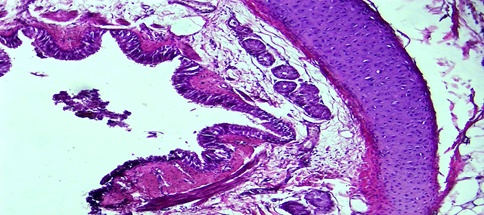 | Figure 1. Bronchi of lung lobes, period of 1 month. The folds of the mucous membrane are relatively small, the submucous layer is thin, there are few tissue structures, and the mucous membrane is multicellular. Paint: G-E. Floor: 10x40 |
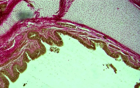 | Figure 2. Infant trachea, 1 month period. Fibrous structures with picrofuchsin are located on the inner and outer surfaces of the submucosa and the mucous membrane. Paint: van Gieson. Floor: 10x40 |
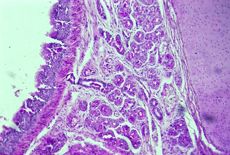 | Figure 3. Infant trachea, 1 month period. Lack of SHIK-positive substance in the covering epithelium, glands and interstitial tissue on the wall of the trachea. Paint: SHIK-reaction. Floor: 10x40 |
|
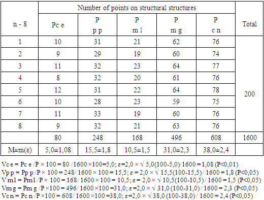
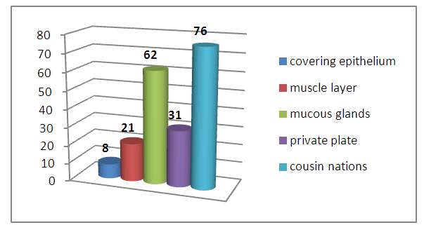 | Diagram 1. Morphometric indicators of tissue structures of the bronchial wall in the early postnatal period in one-month-old infants |
4. Conclusions
- Based on the determined morphometric parameters and histological structures, we conclude that the tracheal mucous membrane is thin, delicate, glands are poorly developed in the babies under 1 month of the research results, the covering epithelium has a large number of smooth surface epithelia compared to the ciliated epithelium, 1.5 times more in number. This causes a lot of acute respiratory diseases among children.In infants up to 1 month, the piece of bronchus occupies a relatively large area, and when examined by histology, the chondrocyte cells in its composition are young and not well developed, as a result of which it is relatively large, the chondroid material in between is relatively pale and the swelling process prevails, so the area of the bronchi is wider and occupies more space. is determined.