-
Paper Information
- Next Paper
- Previous Paper
- Paper Submission
-
Journal Information
- About This Journal
- Editorial Board
- Current Issue
- Archive
- Author Guidelines
- Contact Us
American Journal of Medicine and Medical Sciences
p-ISSN: 2165-901X e-ISSN: 2165-9036
2022; 12(8): 809-810
doi:10.5923/j.ajmms.20221208.08
Received: Jul. 13, 2022; Accepted: Aug. 2, 2022; Published: Aug. 15, 2022

Scientific Reason for the Change of TNF-α and IL-4 in the Development of Silicosis
 Abstract
 Reference
 Full-Text PDF
 Full-text HTML
Dilafruz Bahadirovna Akhmedova
Tashkent Medical Academy, Uzbekistan
Correspondence to: Dilafruz Bahadirovna Akhmedova, Tashkent Medical Academy, Uzbekistan.
Copyright © 2022 The Author(s). Published by Scientific & Academic Publishing.
This work is licensed under the Creative Commons Attribution International License (CC BY).
http://creativecommons.org/licenses/by/4.0/

Symptoms of the course of pulmonary dust diseases at early stages are studied to a lesser degree, functional and biological markers - criteria of individual prognosis of a patient's condition at this pathology are not found. For this reason the question of studying of immunological indexes at pneumoconiosis is actual. The aim of this study is to substantiate the role of cytokines IL-4 and TNF-α in silicosis development caused by silica dust exposure at different stages of the disease. The material and method of the present research were patients who were treated in the clinics of the Research Institute of Sanitary, Hygiene and Occupational Diseases of the Ministry of Health of the Republic of Uzbekistan (n=180). For IL-4 and TNF-α determination we used Vector-Best reagent kit, interleukin concentration was measured by ELISA MR 96A Mindray. The results of the study scientifically substantiated the immunological individuality of the course of silicosis caused by silica-containing dust. The role of TNF-α and IL-4 in the formation, course and progression of silicosis was proved. These studies confirm the necessity of immune status research as an early diagnosis, prediction of the course of the disease and its prevention.
Keywords: Silicosis, Pneumoconiosis, Interleukins, Cytokines
Cite this paper: Dilafruz Bahadirovna Akhmedova, Scientific Reason for the Change of TNF-α and IL-4 in the Development of Silicosis, American Journal of Medicine and Medical Sciences, Vol. 12 No. 8, 2022, pp. 809-810. doi: 10.5923/j.ajmms.20221208.08.
1. Introduction
- Pneumoconiosis is an occupational lung disease often seen in general medical practice. Differential diagnosis of different forms of pneumoconiosis in some cases is aggravated based on the vicinity of their clinical symptomatology [1]. In occupational exposure to silica primarily group of inflammatory and fibrotic mediators are involved in the development of fibrotic lung damage. Free radicals, AFK, lipid peroxidase, TNF, IL-1, IL-6, IL-8 and IFN are all subjectively involved in the inflammatory response, whereas TNF, TGF and IL-4 are more likely to be involved in fibrotic processes. Among cytokines, TNF plays an important role in inflammatory processes, controlling cytokines such as IL-1, IL-4, IL-6 and IL-8, and IL-6 in turn regulates fibrosis [2]. Signs of the course of dust-induced lung diseases at early stages have not been studied to a great extent, functional and biological markers - criteria of individual prognosis of a patient's condition in this pathology - have not been found [3]. For this reason the issue of studying immunological indices in pneumoconiosis from the influence of quartz-containing dust is topical. Objective of the study: To substantiate the role of interleukin-4 (IL-4) and tumour necrosis factor-α (TNF-α) cytokines in the development of silicosis induced by exposure to silica dust at different stages of the disease.
2. Material and Methods
- In order to substantiate a systematic approach to early diagnosis and prediction of the course of the disease under study, we conducted research at the Research Institute of Sanitation, Hygiene and Occupational Diseases of the Ministry of Health of the RUz, the participants were patients (n=180, gender - male) who were treated at the Institute clinic between 2016 and 2018 with the diagnosis of occupational lung pathology "silicosis". To establish the diagnosis of the disease in question, cytokine biomarkers (interleukin-4 (IL-4), tumour necrosis factor-α (TNF-α),) whose concentration was studied on a semiautomatic plate photometer MR 96A Mindray with automatic plate feeding using Vector-Best reagent kit for immunoassay of interleukin concentration in blood serum were used.
3. Results and Discussion
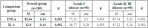
- Silicosis is a chronic irreversible interstitial lung disease due to recurrent aspiration of crystalline silica dust [4,5]. However, the molecular mechanisms of silicosis pathogenesis are unknown until now in all respects, crystalline silica phagocytosis involves active phagosome inflammation followed by phagosome destabilisation, releasing contents into the cytosol, with corresponding activation of the active enzyme complex formed. This complex activates cytokines [6], those engage fibroblasts to build up layers of collagen fibres and trigger an inflammatory cascade, including pro-inflammatory cytokines such as tumour necrosis factor (TNF-α) and others [7]. TNF-α is a pro-inflammatory mediator that is released early in response to exposure to silica aerosol. TNF-α initiates an immune response and activates other pro-inflammatory genes. Persistent inflammatory responses in the lungs can lead to the development of chronic inflammation, which can lead to tissue remodelling. TNF-α is involved in the deposition of collagen during incomplete lung tissue repair, which in turn can lead to fibrosis. Normal fibrosis activity can be characterised as a process of salvage of the damaged organ and is initiated early in the inflammatory process by the release of growth factors that regulate fibroblast activity [8].The laboratory data indicate an increase in serum TNF-α cytokine concentration compared to normal values (0.5 pg/ml) and was 82.64 pg/ml, which is 165.28 times higher, respectively. In turn, it is not unimportant to consider the difference in the serum concentration of the interleukin studied according to the degree of bronchopulmonary system damage. The results of our studies similarly showed higher levels of TNF-α in patients with the diagnosis of silicosis stages II and III, as compared to those with stage I of the disease under study. The findings showed a 155,1-fold increase in the values in those diagnosed with stage I silicosis and 175,48% in the group with stage II and III disease, and there was a significant difference (p<0,001) between groups I and II, stage III silicosis (Table 1).
|
4. Conclusions
- The immunological course of silicosis caused by silica-containing dust has been scientifically proved by the results of investigation. We have proved the role of TNF-α and IL-4 in formation, course and progression of silicosis. The results confirm the necessity of immune status research as an early diagnosis, prediction of the course of the disease and its prevention.