-
Paper Information
- Next Paper
- Previous Paper
- Paper Submission
-
Journal Information
- About This Journal
- Editorial Board
- Current Issue
- Archive
- Author Guidelines
- Contact Us
American Journal of Medicine and Medical Sciences
p-ISSN: 2165-901X e-ISSN: 2165-9036
2022; 12(8): 797-801
doi:10.5923/j.ajmms.20221208.05
Received: Jul. 27, 2022; Accepted: Aug. 5, 2022; Published: Aug. 15, 2022

The Role of Morphometric Study of Breast Epithelium at Different Stages of Carcinogenesis
 Abstract
 Reference
 Full-Text PDF
 Full-text HTML
L. T. Alimkhodjayeva, L. M. Bozarova
Republican Specialized Scientific and Practical Medical Center of Oncology and Radiology, Tashkent, Uzbekistan
Copyright © 2022 The Author(s). Published by Scientific & Academic Publishing.
This work is licensed under the Creative Commons Attribution International License (CC BY).
http://creativecommons.org/licenses/by/4.0/

Aim of the study was to improve the morphological diagnostics of the carcinogenesis stages in the mammary gland based on the data of morphometric and ploidometric studies. The problem of breast neoplasms contains a number of unresolved issues related to the early diagnosis of carcinomas and precancerous changes in the mammary gland. There are often difficulties in interpreting histogenesis, the degree of differentiation of neoplasms, determining the stages of the initial forms of the neoplastic process. This is due to the fact that many aspects of the morphogenesis of hyperplastic and neoplastic processes in the breast epithelium remain poorly understood, there is no unified and generally accepted classification of the stages of the neoplastic process in the breast epithelium and histological diagnosis of this condition is associated with a large share of subjectivity. In order to achieve greater objectivity in the diagnosis of such pathological changes, not only a detailed description of morphological changes in the histological structures of organs, but also the use of a set of additional morphological research methods clarifying the possible rate of proliferative activity of neoplasm cells and changes in their biological properties are required. The use of morphometric and ploidometric research methods in studying the process of carcinogenesis in the breast epithelium may open up new opportunities for differential diagnostics of breast neoplasms.
Keywords: Breast cancer, Carcinogenesis, Diagnostics, Prognosis factors
Cite this paper: L. T. Alimkhodjayeva, L. M. Bozarova, The Role of Morphometric Study of Breast Epithelium at Different Stages of Carcinogenesis, American Journal of Medicine and Medical Sciences, Vol. 12 No. 8, 2022, pp. 797-801. doi: 10.5923/j.ajmms.20221208.05.
1. Introduction
- 25,578 cases of malignant neoplasms were detected in the Republic of Uzbekistan in 2021 (in 2020 – 21, 976). The growth rate of this indicator in compare with 2020 amounted to 16.4%. The highest incidence rates per 100,000 population were found in breast cancer (12.0). 19.8% of women were registered with breast cancer in 2021. The neglect rate was 32.8%. The mortality rate was 5.2 [1]. At the present stage it has not been possible to achieve great success in reducing the incidence of breast carcinoma despite the availability of self-examination in women, the introduction of screening programs for the female population, the use of new diagnostic methods. At the 1st stage the disease is detected in only 13-16% of cases, at the 2nd stage - in 43-47%, at the 3rd - in 33% of cases [1]. Timely treatment of precancerous processes with the potential for malignancy has great practical value [2-4]. These data indicate the need for research to improve the accuracy of cytological and histological diagnostics of breast neoplasms, especially their borderline forms. The same reproducibility of pathohistological stages of neoplastic processes in the mammary gland is one of the important practical problems and needs maximum objectification of the morphological diagnostics process of neoplasms. Therefore, it is necessary to search for morphological signs that could serve to improve the methods of differential diagnostics and earlier detection of carcinomas. Modern methods of studying the proliferative activity of cells are carried out using methods that require expensive consumables (electron microscopy, immunohisto- and immunocytochemical research, molecular biological methods). At the same time, the study of the content of genetic material in the nuclei of epithelial cells is not widely used for this purpose, although histophysical methods of DNA research do not require consumables [6]. However, if microspectrophotometric examination of cytological preparations is associated with the study of whole cells and their nuclei, then in histological sections, measurement data are always associated with random sections of cellular elements and require special methods of mathematical analysis of the data obtained. The most accessible of them is the method of comparative microspectorphotometry. Microspectrophotometry is a method of qualitative and quantitative analysis of microscopic objects using measurements of light energy during its emission, propagation, absorption and scattering [7-9]. The energy distribution is investigated along the spectrum of the radiation source and the scattering of light beams through the thickness of a biological object (tissues - histophotometry, cells - cytophotometry). Ploidometry is a measurement of the ploidy of cells, determined by the content of genetic material in them. A single set of chromosomes in the interphase nucleus is taken as a unit of measurement. As microspectrophotometry of histological preparations has its own characteristics (sections of nuclei are at different levels), it is necessary to use the comparative microspectrophotometry method which is characterized by obtaining relative indicators and reflects the dynamics of pathological changes in cells and nuclei in histological sections [6]. Benign tumors have a modal class of stem line nuclei with a diploid set of chromosomes. The proliferative activity of the tissue is indicated by the presence of single cells with tetraploid nuclei. An increase in the heterogeneity of cells with an increased DNA content is one of the diagnostic signs of malignancy [5]. Significant genetic heterogeneity in malignancy is a consequence of aneuploid DNA content in the nuclei, along with an increase of polyploidy [9, 6-8]. For a pathologist, a typical sample of cells that characterizes a tumor is more informative, that is, measuring the amount of DNA only in the nuclei of cells in the growth zones that determine tissue growth. Tumor cells, unlike normal somatic cells, are characterized by a wide variability of the DNA content and an increase in the average amount of DNA per cell population, which can be used in the differential diagnostics of benign and malignant neoplasms [5]. Morphometric analysis makes allows to clarify the dependence of the nuclei size on the histological shape of the tumor, as well as the possibility of using morphometric and immunohistochemical characteristics of tumor microvessels and the perifocal field in the prognosis of breast carcinomas.Aim of the study was to improve the morphological diagnostics of the carcinogenesis stages in the mammary gland based on the data of morphometric and ploidometric studies.
2. Material and Method
- The study is based on a retrospective study of biopsy and sectional material of the pathology department of the Republican Specialized Scientific and Practical Medical Center of Oncology and Radiology and its branches. Histological diagnostics of tumors and tumor-like formations of the breast was carried out in accordance with the International Histological Classification of Breast Tumors. Biopsies obtained during breast surgery from 92 patients who were diagnosed with the following histological diagnoses were examined: fibroadenoma (D-24 М9010/0) – (n=16), fibrocystic breast disease (N-60 М74320) – (n=31), infiltrating lobular (С-50 М8520/3) – (n=23) ductal breast cancer (С-50 М8500/3) – (n=22). By age groups, the material was distributed as follows: from 30 to 39 years – (n=7), from 40 to 49 years – (n=20), from 50 to 59 years – (n=19), 60 - 69 years – (n=31), from 70 to 79 years – (n=15) biopsies. The comparison group included sectional material (10 observations) of mammary glands of women aged 30-40 years who died from diseases unrelated to the pathology of the reproductive system. The material after sampling was fixed in a 10% solution of neutral formalin. Fixation took place in the thermostat at a temperature of 37°-40°. After fixation and dehydration, the pieces of tissue were poured into paraffin, followed by the preparation of histological sections with a thickness of 8 µm. The sections were stained with hematoxylin and eosin for histological examination and according to Feulgen stain - for microspectrophotometric examination. The study was carried out using a light microscope using lenses x10, x40. The peculiarity of the Feulgen stain is that the dye molecule (leucofuxin) binds to DNA strictly quantitatively, which allows not only to detect the localization of DNA, but also to measure its quantity. A system of tissue sections in three-plane orientation (frontal, sagittal and transverse) was used to study the histoarchitectonics of the normal breast on the sectional material. 6 pieces were cut using a knife from each breast strictly perpendicular to each other in the form of strips of standard width (5 mm). Avtandilov's ocular stereometric grid containing 100 test points was used in the study of histoarchitectonics of normal breast preparations, as well as areas of fibroadenoma, fibrocystic breast disease, ductal and lobular infiltrative breast carcinomas. An x7 eyepiece and an x10 lens were used. 20 visual fields were analyzed in each preparation stained with hematoxylin and eosin. The percentage ratio of the studied objects (epithelium, connective and adipose tissue) was determined by simply counting the coinciding points of the ocular grid with the studied objects. Determination of the percentage ratio of the volume fractions of epithelial (EV), connective (CV) and adipose (AV) tissue in the studied samples allowed to obtain information about the "tissue volume profile" (TVP) in normal mammary gland, fibrocystic breast disease and fibroadenomas, invasive ductal and lobular breast carcinomas.After histological diagnostics, the preparations stained with hematoxylin and eosin and according to the Feulgen stain were divided into the following groups: 1. Areas with a normal histological structure of the lobular and ductal epithelium of the mammary gland; 2. Areas with signs of hyperplasia of the lobular and ductal epithelium of the mammary gland; 3. Areas with a pattern of: a). mild, b). moderate, and c). severe dysplasia and carcinoma "in situ" of the lobular and ductal epithelium of the mammary gland;4. Preparations with typical areas of invasive lobular and ductal breast carcinomas. It was found after a ploidometric and morphometric study of the preparations, that hyperplasia and moderate dysplasia of the mammary epithelium have no statistically significant differences in ploidy and area with neighboring diagnostic groups by stages of carcinogenesis. Therefore, hyperplasia was combined with the "norm" group, and moderate dysplasia was included in the "mild dysplasia" group.
3. Results
- The histological picture of the mammary epithelium hyperplasia is characterized by the presence of a slightly hyperchromic epithelium lining the lobules and ducts of the mammary gland, the number of epithelial cells is increased, they can be layered on top of each other, but always arranged in a row. No signs of cellular atypia were observed. When examining breast biopsies with fibrocystic breast disease and fibroadenoma of the breast, the histological picture of mild, moderate and severe degrees of dysplasia of the lobular and ductal epithelium of the breast was studied. Epithelial dysplasia of varying severity was found in 42% of the biopsies studied with data of benign changes in the mammary gland. A mild degree of epithelial dysplasia is characterized by the presence of an epithelium in one or two rows lining lobules and ducts. Individual nuclei are hyperchromic, with uneven chromatin distribution. A weakly pronounced violation of polarity in the location of epithelial cells was observed. At a moderate degree of dysplasia, part of the epithelial cells acquires signs of atypia — the size of the nuclei increases, hyperchromia and polymorphism of the nuclei were more pronounced, single atypical mitoses were observed. With severe dysplasia, there is a sharp increase in the size of lobules and ducts filled with hyperchromic atypical cells that build various structures (in this study, cribrous and papillary structures were observed more often in the ducts, solid ones were revealed in the lobules). Atypical mitotic figures were multiple. The histological picture of intraepithelial lobular and ductal breast carcinomas is very similar to the histological picture of severe epithelial dysplasia of the same localization. The tumor is represented by sharply polymorphic atypical cells with a large number of atypical mitoses. The cells lose their correct orientation relative to the basement membrane. Foci of intraepithelial carcinoma in the studied material were found against the background of fibrocystic breast disease. This pathology was not detected in fibroadenomas preparations. Noninvasive ductal carcinoma in all cases had a cribrous type of structure, noninvasive lobular had solid structure. Invasive lobular carcinoma in most cases had a classic version of the structure in the form of tumor cells growth with light cytoplasm in the stroma chains. Ductal carcinoma in typical preparations was characterized by the growth of atypical hyperchromic polymorphic cells in the form of strands.Determination of the percentage ratio of epithelial, connective and adipose tissue within the terminal ductal-lobular unit in breast biopsies in normal, fibroadenomas, fibrocystic breast disease and invasive ductal and lobular carcinomas allowed us to trace the change in their volume ratios as the tumor process was developing. The data obtained indicate a change in the histoarchitectonics of biopsies stained with hematoxylin and eosin towards an increase in the volume of epithelial tissue and a decrease in the volume fraction of connective and adipose tissues during the development of carcinomas (Tab. 1., Fig. 1).
|
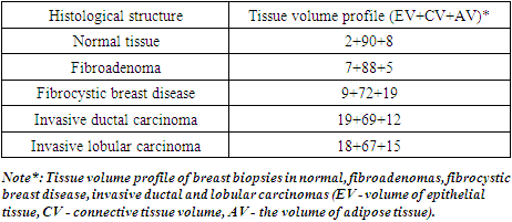
 | Figure 1. Volume ratios of tissues in breast biopsies |
|
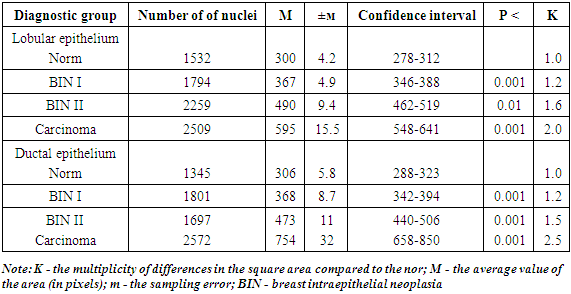
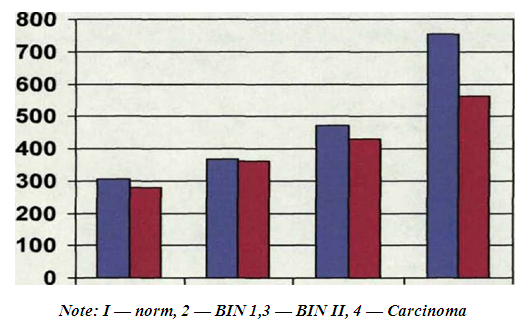
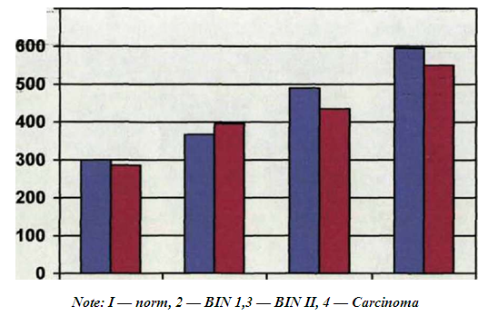 | Figure 2. Lobular epithelium. Average values of the area of nuclei (in pixels) stained with hematoxylin and eosin and according to the Feulgen’s technique |
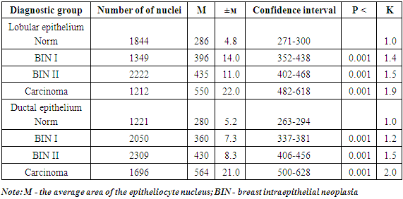
 | Figure 3. Ductal epithelium. Average values of the area of nuclei (in pixels) stained with hematoxylin and eosin and according to the Feulgen’s technique |
4. Conclusions
- The data obtained by morphometric examination of mammary epithelial cells will help to clarify the histological diagnosis, especially in the transitional stages of carcinogenesis. An increase in the area of epithelial cell nuclei compared to the norm by 1.5 times allows to diagnose mammary intraepithelial neoplasia of the 2nd degree, infiltrating carcinoma - by 2 times. The results of the study obtained by us can be used as additional differential diagnostic criteria for the morphological diagnosis of the carcinogenesis stages in the mammary gland.Therefore, this research method allows to clarify the diagnostics of the carcinogenesis stages in the mammary gland and to make informed decisions regarding benign processes, borderline states and malignant neoplasms.The authors declare no conflict of interest. This study does not include the involvement of any budgetary, grant or other funds. The article is published for the first time and is part of a scientific work.
References
| [1] | The state of oncological care to the population of the Republic of Uzbekistan in 2021. In Russian\\ -Publishing house "Hulk" 2022. P. 4-7. |
| [2] | Semiglazov V. F., Chernomordikova M. F., Orlov A. A., etc. Methods of detection and treatment of minimal forms of breast cancer. In Russian. // Vopr. oncol. - 2019. - No. 4. - P. 117. |
| [3] | Pletnev S. D., Mazurin V. G. Modern possibilities of breast cancer recognition at the stage of morphological formation. In Russian. // Diagnosis and treatment of breast cancer. - Moscow: 2021. - P. 57 - 62. |
| [4] | Yakovleva I. A., Cherny A. P., Bondar E. R. Cervical epithelium in the process of malignancy. - Chisinau, 2021. - P.128. |
| [5] | Standards for the diagnosis and treatment of malignant neoplasms. Complex “Print Publishing” House2022. P. 6-9. |
| [6] | Evaluation of proliferative activity of cells in histopathological diagnostics. In Russian. // Pacific Medical Journal. — 2000. - No. 4.- P. 9-11 (co-author. Avtandilov G. G., Loranskaya I. D., Petrenko N. V.) |
| [7] | Pathohistological differential diagnosis of intraepithelial neoplasia and breast carcinomas. In Russian. // Russian Medical News. - 2001. - Volume VI. - No. 2. - pp. 54-57. |
| [8] | Ploidometry in clarifying the diagnosis of the stages of the tumor process in the mammary gland //Diagnostic medical' morphometry. In Russian. /Collection dedicated to the 80th anniversary of Georgy Gerasimovich Avtandilov.- Moscow.: RMAPO, 2002.- pp. 175 - 179. |
| [9] | "Tissue volume profile" of breast biopsy material. In Russian. // Successes of theoretical and clinical medicine. - M., RMAPO, 1999. - Issue 3. - P. 71. |