-
Paper Information
- Next Paper
- Paper Submission
-
Journal Information
- About This Journal
- Editorial Board
- Current Issue
- Archive
- Author Guidelines
- Contact Us
American Journal of Medicine and Medical Sciences
p-ISSN: 2165-901X e-ISSN: 2165-9036
2022; 12(8): 777-780
doi:10.5923/j.ajmms.20221208.01
Received: Jul. 4, 2022; Accepted: Aug. 3, 2022; Published: Aug. 15, 2022

Morphological Features of the Structure of the Nervous Structures of the Muscular Membrane of Various Parts of the Esophagus
 Abstract
 Reference
 Full-Text PDF
 Full-text HTML
Khamraev Akbar Khairulloevich
Samarkand State Medical University, Uzbekistan
Correspondence to: Khamraev Akbar Khairulloevich, Samarkand State Medical University, Uzbekistan.
| Email: |  |
Copyright © 2022 The Author(s). Published by Scientific & Academic Publishing.
This work is licensed under the Creative Commons Attribution International License (CC BY).
http://creativecommons.org/licenses/by/4.0/

The esophagus has its own structural features as the initial part of the digestive tract, especially the structure of the muscular membrane of its various departments and the distinctive signs of innervation differ from other departments of the digestive canal. Material and methods. The esophagus of adult rabbits served as the material for the study. After killing animals according to the rules of bioethics, the taken organ was divided into three parts: cervical, thoracic and abdominal. Each department was divided into two more parts. The first part is placed in a cryostat for taking frozen sections, in order to study adrenergic nerve structures by treating the material with 2% glyoxylic acid. The second part was fixed in a 12% solution of neutral formalin. The results of the study showed that in the intermuscular and submucosal nerve nodes there are mainly long-axon and equidistant Dogel neurons and in a small number of neurons of the third type. In all cases, the number of long-axon neurons in the intermuscular nerve nodes is relatively greater. All nerve fibers and bundles of the esophageal wall are strongly writhing. Conclusions. This structure of nerve fibers and bundles creates a reserve length and prevents strong stretching of the fibers during the expansion of the esophageal wall during the passage of the food lump.
Keywords: Esophagus, Intramural nerve structures, Neurocytes, Muscle fibers
Cite this paper: Khamraev Akbar Khairulloevich, Morphological Features of the Structure of the Nervous Structures of the Muscular Membrane of Various Parts of the Esophagus, American Journal of Medicine and Medical Sciences, Vol. 12 No. 8, 2022, pp. 777-780. doi: 10.5923/j.ajmms.20221208.01.
Article Outline
1. Relevance
- The esophagus is the initial part of the digestive canal, where striated muscle tissue turns into smooth, there is a joint arrangement and gradual combination of somatic nerve structures with vegetative ones. In recent years, the work devoted to the general innervation of the esophagus, especially the nervous structures of the muscular membrane of the organ has a special place. The reason for this is, firstly, the features of the structure of the muscular membrane of various parts of the esophagus, and secondly, the distinctive signs of innervation of the striated muscle tissue of the proximal esophagus from the innervation of the striated muscle tissue of other parts of the digestive canal. There are problems concerning the study of the structure of the esophageal wall, especially its muscular membrane and its innervation. Solving these problems will make it possible to determine the mechanism of pathogenesis of diseases of this part of the digestive canal, their prevention, as well as treatment. In this regard, we have set ourselves the task of studying the morphology of nerve fibers and nerve endings involved in the innervation of smooth and striated muscle tissue of the upper, middle and lower esophagus of rabbits, which has both theoretical and practical significance.
2. The Purpose of the Study
- Study of the nervous structures of various parts of the esophagus in rabbits.
3. Material and Methods
- The esophagus of adult rabbits served as the material for the study. After killing animals according to the rules of bioethics, the taken organ (esophagus) was divided into three parts: cervical, thoracic and abdominal. Each department was divided into two more parts. The first part is placed in a cryostat for taking frozen sections, in order to study adrenergic nerve structures by treating the material with 2% glyoxylic acid. The second part was fixed in a 12% solution of neutral formalin. Formalin was neutralized with sodium tetraborate salts and the PH of formalin was controlled by an RCS indicator. When the medium changed to the acidic side, sections were taken from the fixed material on the cryostat and impregnated with nitric acid silver by the Bilshovsky-Gross and Campos methods. Another part of the material fixed in formalin was filled with paraffin and sections were obtained using a microtome. These sections were stained with hematoxylin-eosin and according to Van Gieson. Preparations treated with glyoxylic acid were studied using a luminescent microscope LUMAM 1-2 (Specialized microscope) using filters FS-1-4 and FS-1-6.
4. The Results of the Study
- Nerve nodes consisting of neurons are located in the muscle membrane. Between the nerve cells are located the nuclei of many gliocytes and connective tissue cells. The shape of the nerve nodes and the number of neurons in it to a certain extent depends on the thickness of the micropreparation, and the morphometric parameters of the nerve nodes depend on the caliber of the overlapping nerve fibers. As can be seen in the figure, the degree of impregnation of neurons located in the node is not the same. Because the intake of silver nitrate salts is associated with the functional state of neurons at the time of exposure to the drug. The size of the intermuscular nerve nodes and the number of neurons in them are diverse and the density of the location is high compared to submucosal nerve nodes (Figure 1).
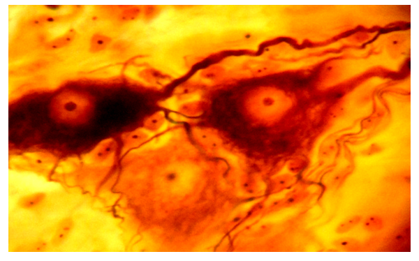 | Figure 1. Long-axon neurocytes in the intermuscular nerve plexuses of the rabbit esophagus. Coloring according to Bilshovsky–Gross. Vol. 40, approx.7 |
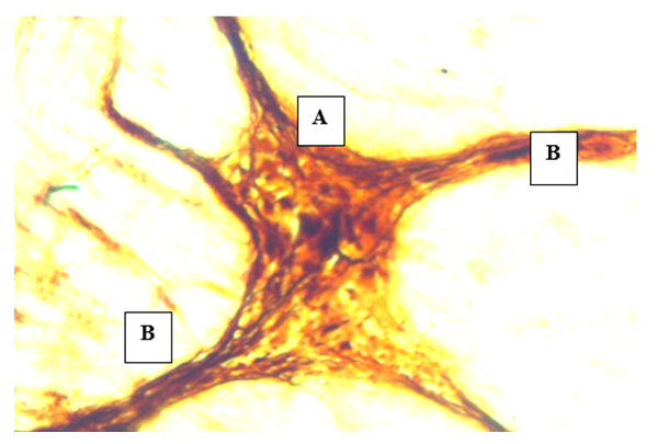 | Figure 2. The nerve node (A) of the intermuscular nerve plexus and bundles of nerve fibers (B) of the rabbit esophagus. The Bilshovsky method –Gross. Vol.20, approx 10 |
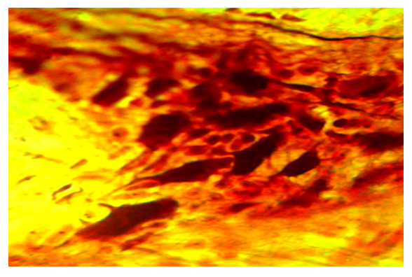 | Figure 3. An intramural nerve node located between the muscular and submucosal membranes of the rabbit esophagus. Dark-impregnated long-axon neurocytes. The Bilshovsky method-Gross Volume 20, approx. 10 |
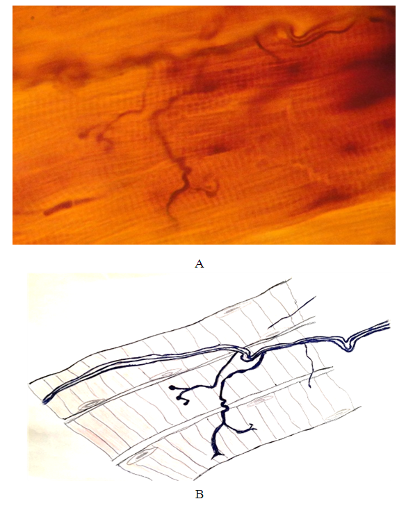 | Figure 4. Motor nerve ending in the wall of the rabbit esophagus (A). Bilshovsky method – Gross. Volume 40, approx.7. Drawing of the cluster-shaped nerve ending sketched using the drawing apparatus DA-1 (B) |
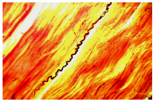 | Figure 5. Spiral nerve bundle of the longitudinal layer of the muscular membrane of the middle part of the rabbit esophagus. The Bilshovsky-Gros method. Pre-coloring with Carmine. Vol.10, ok 7 |
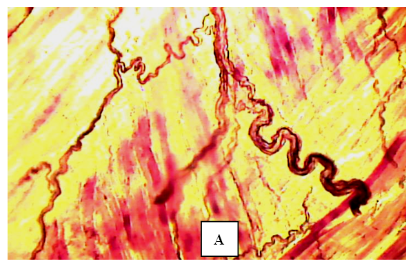 | Figure 6. Spiral-shaped nerve bundles of various calibers and fibers separated from them at the border of the submucosal and muscular membranes of the rabbit esophagus. The Bilshovsky method-Gross Volume 10, approx.7. A – fibers of muscle tissue |
5. Conclusions
- The activity of muscle tissue is regulated by nerve structures, in particular by long-axon neurocytes (Dogel type 1) of the intramural nervous apparatus, as well as by the vagus nerve and the somatic nervous system. Submucosal (Meissner) and intermuscular (Auerbach) nerve plexuses were found in the wall of the rabbit esophagus and between the layers of muscle fibers. These plexuses consist of bundles of nerve fibers and there are intramural nerve nodes at the intersection of the bundles. The nodes contain all three types of Dogel nerve cells (long-axon, equi-process and interneurons) and the quantitative ratio of these neurons varies in the direction of the distal part of the digestive tube. Our studies have shown that submucosal intramural nerve nodes are larger than intermuscular ones and there are more nerve cells in their composition. Naturally, there are more long-axon neurocytes (Dogel type 1) in the nerve nodes of the intermuscular plexuses. For the nervous apparatus of the esophagus, it is characteristic that all nerve fibers and their bundles located in the nerve plexuses strongly twist and they are located in all layers of the esophageal wall. In our opinion, such a structure of nerve fibers and bundles creates a reserve length to prevent strong stretching of the fibers during the expansion of the esophageal wall during the passage of a food lump.Information about the source of support in the form of grants, equipment, and drugs. The authors did not receive financial support from manufacturers of medicines and medical equipment.Conflicts of interest: The authors have no conflicts of interest.