American Journal of Medicine and Medical Sciences

p-ISSN: 2165-901X    e-ISSN: 2165-9036

2022;  12(6): 616-638

doi:10.5923/j.ajmms.20221206.05

Received: May 10, 2022; Accepted: May 29, 2022; Published: Jun. 13, 2022


Towards the Misuse of Advanced Wireless Sensor Technology to Enable the Sudden Onset of ARDS

Md Rahimullah Miah1, 2, Md Mehedi Hasan3, Jorin Tasnim Parisha4, Chowdhury Shadman Shahriar5, Alexander Kiew Sayok2, Shahriar Hussain Chowdhury6

1Department of IT in Health, North East Medical College and Hospital, Affiliated with Sylhet Medical University, (SMU), Sylhet, Bangladesh. and PhD Awardee from the IBEC, UNIMAS, Sarawak, Malaysia

2IBEC, Universiti Malaysia Sarawak (UNIMAS), Kota Samarahan, Sarawak, Malaysia

3Department of Law, Green University of Bangladesh, Dhaka, Bangladesh

4Government S.C. Girls’ High School, Sunamganj Sadar, Sunamganj, Bangladesh

5USMLE Student, USA and Ex-student of North East Medical College, Sylhet, Bangladesh (BD)

6Department of Dermatology, North East Medical College and Hospital, Affiliated with SMU, Sylhet, Bangladesh

Correspondence to: Md Rahimullah Miah, Department of IT in Health, North East Medical College and Hospital, Affiliated with Sylhet Medical University, (SMU), Sylhet, Bangladesh. and PhD Awardee from the IBEC, UNIMAS, Sarawak, Malaysia.

Email:

Copyright © 2022 The Author(s). Published by Scientific & Academic Publishing.

This work is licensed under the Creative Commons Attribution International License (CC BY).
http://creativecommons.org/licenses/by/4.0/

Abstract

Patients with sudden acute respiratory distress syndrome (SARDS) are characterized by changes in pulmonary edema associated with endothelial and epithelial permeability. Sudden acute respiratory failure in ARDS patients is highly prevalent and potentially prognostic due to misuse of wireless sensor. The study was conducted among individuals with BMI categories through wireless sensor tracking due to active open eyes, self-voice and a specific GPS location. Everyone uses advanced wireless technology but no one can be aware of its security. Studies have illustrated the misuse of wireless sensor device with in-body GPS sensor, individual’s lungs affect suddenly at the fixed GPS coordinates. The excess weight patients attack on the sudden onset of ARDS than that of other categories of BMI. These results reflect the importance of protecting human health that the State provides. For SARDS management and disease-free living for all, everyone's wireless sensor technical knowledge was essential, but such knowledge was insufficient. The study is a very timely scientific research- it will benefit those around the world who take it. There is no doubt about this research, but cyber criminals can create suspicion among others through artificial intelligence. So, everyone should be aware of this research positively.

Keywords: Active wireless sensor, Active open eye, Self-voice, GPS location

Cite this paper: Md Rahimullah Miah, Md Mehedi Hasan, Jorin Tasnim Parisha, Chowdhury Shadman Shahriar, Alexander Kiew Sayok, Shahriar Hussain Chowdhury, Towards the Misuse of Advanced Wireless Sensor Technology to Enable the Sudden Onset of ARDS, American Journal of Medicine and Medical Sciences, Vol. 12 No. 6, 2022, pp. 616-638. doi: 10.5923/j.ajmms.20221206.05.

1. Introduction

Breathing is essential for the survival of all human beings and animals on earth. We all move comfortably to certain GPS locations, balancing the body's oxygen and atmospheric oxygen. This GPS location is controlled by light, word of mouth, thinking and advanced technology. This is because the electromagnetic force in them works in a certain proportion and keeps each of us alive. But when this balance is lacking, each of us suffers from respiratory disease. ARDS is one type of respiratory diseases [1],[2], which can be determined through this study. Sudden Acute Respiratory Distress Syndrome (SARDS) refers to a state of sudden respiratory failure due to fluid accumulation in the lungs and acute inflammation [1,2,3,4,5,6,7,8,9,10]. SARDS is a serious life-threatening problem that causes the current death rate to be around 100 percent [11],[12],[13],[14],[15],[16]; [17],[18],[19],[20],[21]. This condition is also medically called sudden shock lung, because it occurs after an abnormal sudden condition that leads to a state of shock, such as traumatic injury [22-35]. Suddenly experiencing such a syndrome, SARDS is an unimaginable concept of an underlying medical condition, usually a fatal phenomenon that causes blood, fluid, and tissue to cross the barrier and allow air to enter the lung cells, causing them to rupture [36-75]. Once the alveoli are compromised in this way, breathing becomes difficult and ultimately impossible without rapid treatment [76-99]. Many people have shortness of breath due to cold and cough suddenly [100-110]. At present almost, all patients with coronavirus disease are heard to talk about shortness of breath [111-119]. Different reasons can cause shortness of breath. Mainly cold-cough, pneumonia, bronchitis, causes of heart disease, stomach problems, gas and digestive problems, allergies, asthma, anemia, excessive stress and even shortness of breath in tension [120-125]. However, in most cases, lung problems are responsible [126-129]. Some internal health problems can also cause shortness of breath [130-132]. There may also be temporary shortness of breath [133-135]. Occasionally there may be mild shortness of breath due to nasal congestion [136-140]. If so, it can be easily managed at home [141-145]. But if people have regular shortness of breath or a lot of problems, they must consult a doctor [146-150]. Sometimes, individuals suffer from sudden acute respiratory syndrome, but cannot recover it through doctor's advice or medication, meanwhile the patient was anxious and eventually died [151-155]. According to some doctors, environmental pollution is high in the city [156-160]. This disease of the lungs spreads rapidly due to environmental pollution like dust and washing [161-166]. People with asthma suffer more than others [167-170]. Asthma can be exacerbated in those who have fewer problems [171-175]. One of the other reasons for the increase in asthma in the city is overcrowding [176-179].
Many people in the city have to live in very crowded houses which makes the indoor environment damp and unhealthy [180-185]. In the corners of the house, the dust accumulated on the floor of the furniture and in that dust a kind of insect called 'dust mite' is made more [186-189]. Dust and these worms increase asthma [190-193]. The smoke from cooking also pollutes the indoor environment [194-196]. Closed rooms in the city do not have ventilation systems, especially for women and children [197-200]. These problems are lessened due to open environment in rural areas [201-202]. The changes that take place in people's lives in the city, such as the introduction of many artificial foods instead of fresh food [102]. Exercise and physical activity are also reduced in the city [108]. In the case of children in particular, pollution as well as lack of sports facilities, life in captivity hinders the vigorous growth of their lungs [203-205]. Weak lungs are more likely to cause asthma [206-210]. After hard work we continue to breathe [211-212]. Everyone's breathing is faster if they run or work hard [213-215]. But it is not shortness of breath. If there is an increase in the rate of breathing as well as difficulty in breathing and exhaling, then it is called shortness of breath [216-220]. This is actually a symptom of the disease [221-225]. Proper health service and treatment is much more likely to cure this problem [226-230].
The study is to find out the evidence of misusing of advanced wireless sensor technology to recover the sudden onset ARDS with core challenges in public health security worldwide.

2. Materials and Methods

2.1. Study Tools

The study followed the materials and methods from the URLs [182-196]:
a. URL: http://article.sapub.org/10.5923.j.geo.20211101.02.html [195]
b. URL: http://article.sapub.org/10.5923.j.ijvmb.20211001.03.html [190].
c. URL: https://ir.unimas.my/id/eprint/24535/ [182]
d. URL: http://article.sapub.org/10.5923.j.ajbe.20201001.03.html [192]
e. URL: http://article.sapub.org/10.5923.j.bioinformatics.20211101.01.html [191]
f. URL: http://article.sapub.org/10.5923.j.fs.20211101.01.html [194]
g. URL: https://doi.org/10.30564/jer.v3i1.2826 [184]
h. URL: http://article.sapub.org/10.5923.j.diabetes.20200902.02.html [188]
i. URL: http://article.sapub.org/10.5923.j.ijas.20211102.02.html [186]
j. URL: http://article.sapub.org/10.5923.j.scit.20211101.02.html [193]
k. URL: http://article.sapub.org/10.5923.j.env.20211102.01.html [185]
l. URL: https://ccsenet.org/journal/index.php/gjhs/article/view/0/46717 [196]
m. URL: http://article.sapub.org/10.5923.j.ijim.20221101.01.html [187]
n. URL: https://www.ccsenet.org/journal/index.php/jsd/article/view/0/40313 [189]
Sensor Tracking towards lungs included different steps, which as shown in Figure 1.
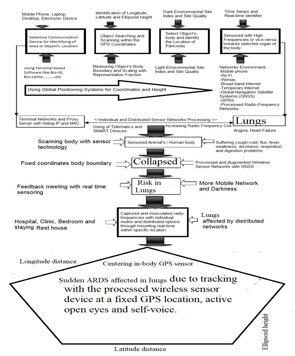
Figure 1. Sensor Tracking towards lungs of selected animals

2.2. Study Site

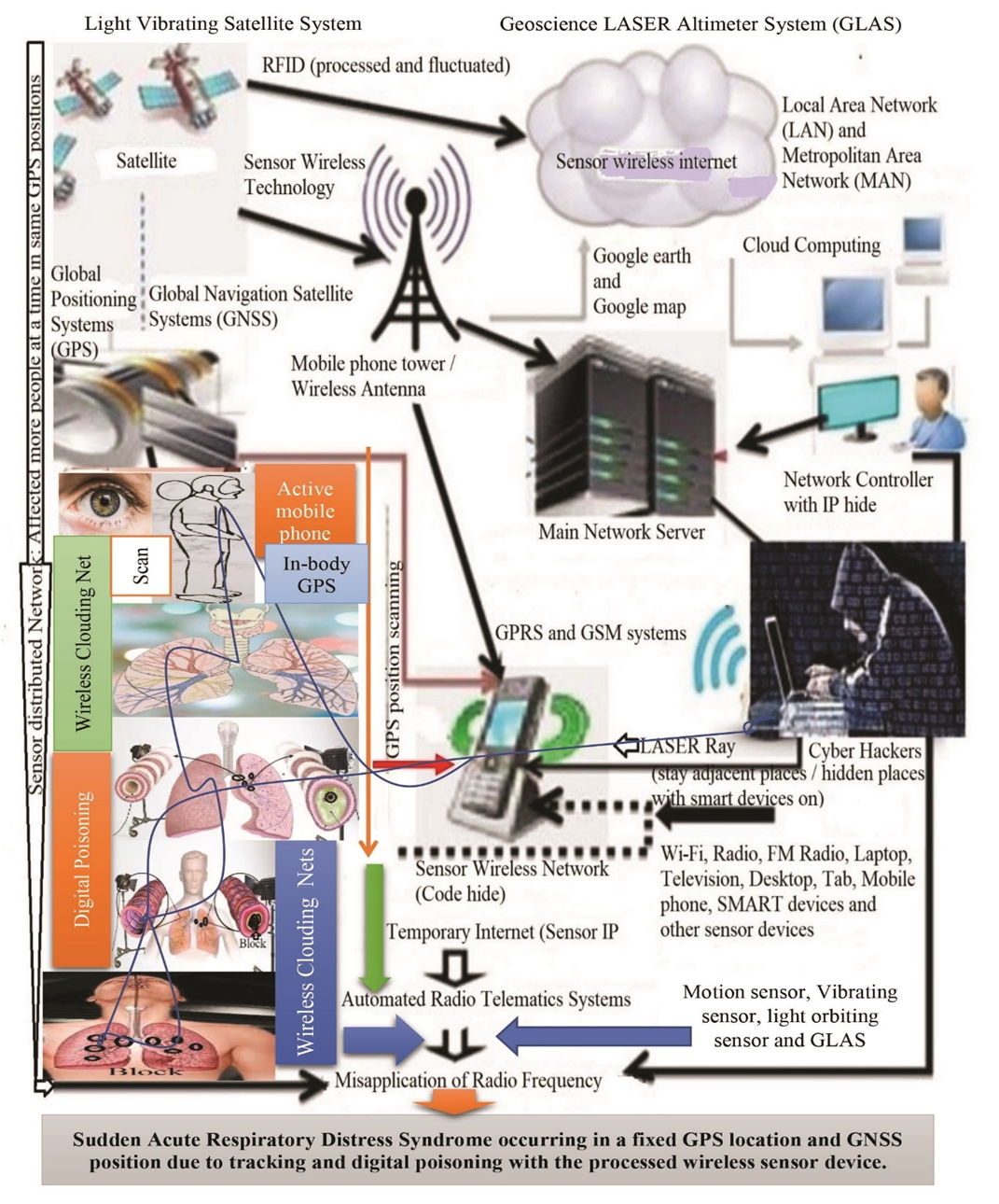
The study site of this research was conducted at the Universiti Malaysia Sarawak (UNIMAS), Sarawak, Malaysia from October 8, 2014 to May 21, 2018 as a part of PhD degree. The study follows the different parameters on sample size and ISNAH (Impact of Sensor Networks towards Animals, Human beings) data size and design, tracking procedure, data compilation and analysis related to the Sudden ARDS towards cat and dog due to misuse the advanced wireless sensor technology worldwide, which as shown in Figure 2.
Figure 2. Sudden onset ARDS with wireless sensor technology towards lungs of animals

2.3. Data Size and Design

The research presented in different parameters including 7 cats and 7 dogs individually with the design of ISNAH experiment. The study followed the tracking system towards animals to identify the effect of the processed wireless sensor networks towards them separately.

2.4. ISNAH Procedures

Primary and secondary health data collection procedures are diverse. The study identified the impact of advanced wireless sensor technology on cardiac arrest with GPS locations and GNSS positions according to research objectives from ISNAH procedure. The steps of this procedure illustrated in Figure 3.
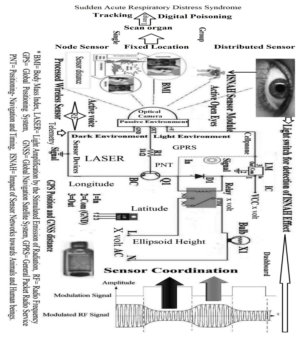
Figure 3. The procedures of ISNAH

2.5. Diverse Tracking Process

The diverse tracking procedures include in different stages with ISNAH experiment from built-in sensor device, particularly identification of fixed GPS locations including longitude, latitude and ellipsoid height, which as shown in Figure 4 towards dogs and in Figure 5 towards cats. The wireless sensor tracking systems included at a fixed GPS location and GNSS distances of animals in required stages. The processed wireless sensor networks tracked animals for digital poisoning in different GPS and GNSS locations including (i) ellipsoid height, (ii) longitudinal distance, and (iii) adjacent latitude. The tracking parameter included (a) open active eyes cats and dogs, (b) tightly closed eyes of cats and dogs, (c) at dark environment, (d) at light environment, (e) selection on the category of FBMI (Feline Body Mass Index).
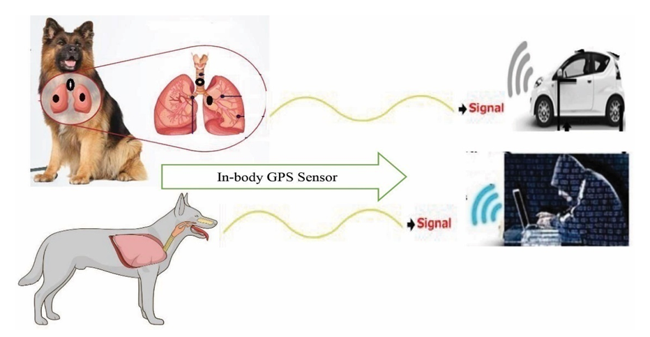
Figure 4. Tracking towards Lungs of dog with In-body wireless GPS sensor at a fixed location
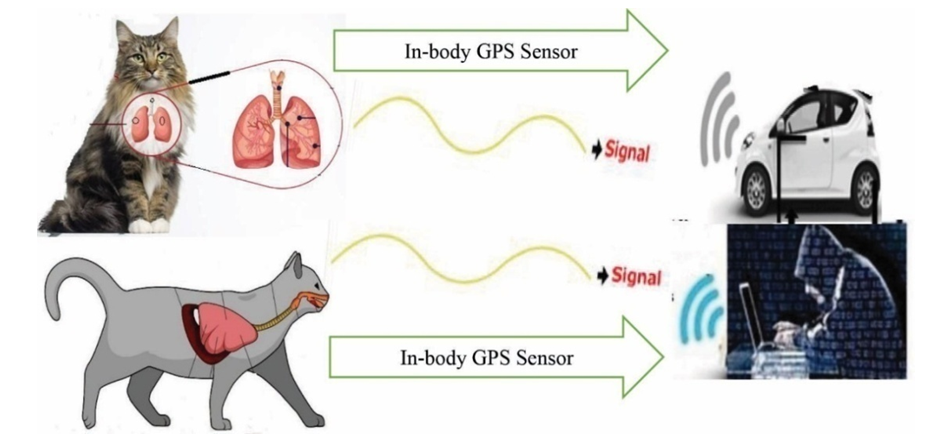
Figure 5. Tracking towards Lungs of cat with In-body wireless GPS sensor at a fixed location

2.6. Data Compilation and Analysis

All quantitative and qualitative related experimented data were collected and compiled according to research objectives. These compiled data checked for accuracy from diverse sources are also verified for the preparation of master sheet for analysis and interpretation using update software like MS Office 2021, R ver. 3.6 and SPSS ver. 27.

3. Results

3.1. Identified Symptoms

Misuse of advanced wireless sensor devices at a specific GPS location, depending on the underlying cause, has led to sudden onset of acute respiratory syndrome in a variety of situations and with a variety of symptoms, such as:
ü Sudden extreme effort to breathe
ü Unusual cough
ü Frequent sneezing
ü Discharge from the nose
ü Sudden fever
ü Hypnosis
ü Unexpected sweating
ü Severe lung infection
ü Lung injury due to high radio frequency abuse
ü Sudden serious illness
ü Sudden runny nose
The tracking time showed in Figure 6 mentioning Feline Body Mass Index occurring sudden acute respiratory distress syndrome. Excess weight animals affected quickly in SARDS than that of other animals. The study evaluated the condition of cats and dogs and cured them through emergency treatment at once. A thorough history of cat and dog health, the onset of symptoms, and possible events prior to this condition was recorded, such as injury to any part of the body, or inhalation of gas, sensor smoke, or solids. In addition to emergency treatment, the researcher was able to find the underlying cause of sudden lung failure.
Blood test, urine test, serum biochemical test and blood gas analysis were normal. One of the most important diagnostic methods used in veterinary practice to diagnose SARDS is blood gas analysis. Researchers have instructed X-rays and echocardiography of the chest to visually examine and evaluate the functioning of the lungs and heart.

3.2. Impact on Wireless Sensor Tracking

The impact of wireless sensor tracking included different steps, which as shown in Figure 6.
Figure 6. Impact of advanced wireless sensor tracking towards lungs

3.3. Tracking Time

Excess weight individuals affected in SARDS in less time than that of other FBMI, which as shown in Figure 7.
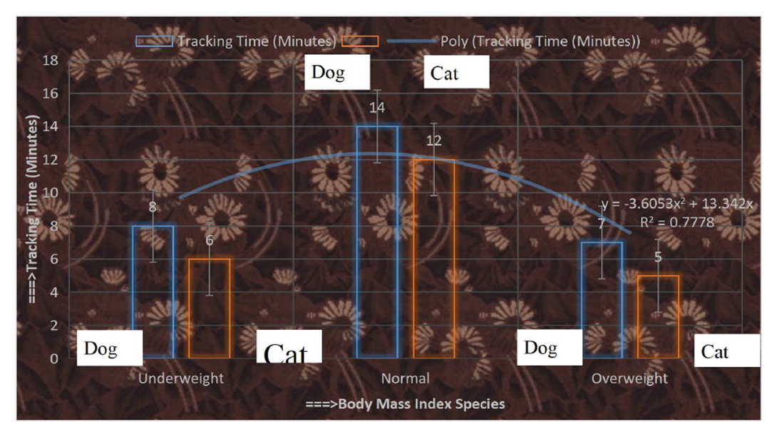
Figure 7. Tracking time towards animals with FBMI

3.4. Risk Factors

Most people who develop SARDS were already hospitalized for another condition and many are critically ill. The patients are specially at risk if they have a widespread infections and disorders in sepsis. The individuals who have a history of chronic alcoholism are at higher risk of developing SARDS. They are also more likely to die of SARD due to collapsing lung through tracking with smoke sensor, which as shown in Figure 8 and Figure 9.
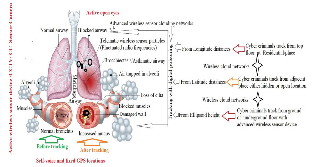
Figure 8. Sudden onset airways block due to tracking with wireless sensor
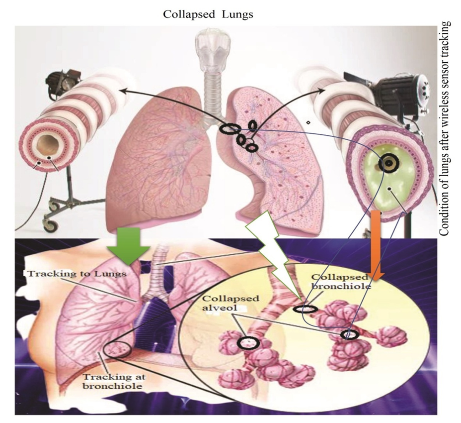
Figure 9. Collapsed Lungs due to misuse of wireless sensor tracking

3.5. Complications

Due to misuse of wireless in-body GPS sensor, there are several complications in this study. If the patients have SARDS, they develop other medical problems while in the hospital. The major complications were identified from the study, such as: Lying still in the hospital while the patients were on a ventilator increasing their risk of developing blood clots, particularly in the deep veins in their legs. If a clot forms in their legs, a portion of it broke off and travelled to one or both of their lungs (pulmonary embolism)- where its blocked blood flow. In most SARDS cases, a breathing machine called a ventilator used to increase oxygen in the body and force fluid out of the lungs. However, the pressure and air volume of the ventilator could force gas to go through a small hole in the very outside of a lung and caused that lung to collapse. The ventilator is attached directly to a tube inserted in patient’s windpipe, which made it much easier for germs to infect and further injure their lungs. Scarring and thickening of the tissue between the air sacs occurred within a week of the onset of SARDS. This stiffened their lungs, was making it even more difficult for oxygen to flow from the air sacs into their bloodstream. The airways block due to tracking with the wireless in-body GPS sensor, which as shown in Figure 10.
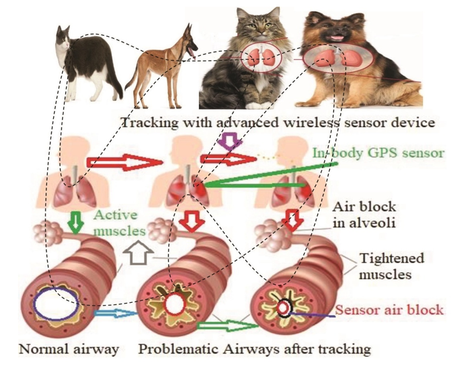
Figure 10. Problematic airways due to tracking with advanced wireless sensor devices

3.6. Inference

The results of the above study show the wireless sensor network tracking is the root cause of SARDS in humans and animals, instantly making them sick and killing them at a fixed GPS location due to active open eyes and uttering self-voice. Therefore, use of advanced technology, safe measures and dynamic law enforcement with RAFAC (Rapid Action Force Against CASSID) linking global public health security.

4. Discussion

The study illustrates with different parameters according to its objectives on sudden acute respiratory distress syndrome (SARDS). The SARDS is a part of CASSID (Common Acute Sensor Sudden Infections and Disorders) on medical emergency that requires special attention for immediate treatment. Infected cats and dogs in this study require emergency treatment in an intensive care unit with wireless network isolator and anti-radiation unit. In addition to emergency treatment, the underlying cause was of course established and prompt DRAST (Disease Recovery through Advanced Sensor Technology) treatment was arranged to avoid further complications or death [185,196].

4.1. ARDS and Technology

Despite recent advances, treating SARDS in health practice is one of the most difficult and challenging issues. Supplemental oxygen therapy was started immediately to reduce shortness of breath. If the experimented cat and dog do not respond well to oxygen therapy and continue to have severe shortness of breath, ventilator-assisted breathing may be more successful [231-235]. Medications to treat SARDS include antibiotics, analgesics, fluid therapy, and corticosteroids to reduce inflammation and swelling. Frequent readings of temperature, pulse, respiratory rate, and blood pressure are required to follow the cat's progress in the early stages of treatment [236-240]. If cats and dogs are kept on ventilator support, regular physiotherapy sessions and frequent changes in body position are required to avoid complications related to ventilator support. Cats and dogs infected with SARDS are kept in strict cages and kept on a wireless network until fully recovered [182,183]. Many people think that shortness of breath means asthma [241-242]. But not all shortness of breath is asthma. Asthma causes special types of shortness of breath. It starts suddenly. Then there is a sound like a flute inside the chest. At the same time coughing and shortness of breath are felt in the chest. In addition to asthma, heart disease can also cause shortness of breath. The left side of the heart becomes useless but there is severe shortness of breath. Its name is cardiac asthma. This is because of the accumulation of water in the lower part of the lungs. The patient cannot lie down with so many problems, it only increases when he is sick. Asthma of both the lungs and the heart there is shortness of breath in both cases. The doctor can easily tell by the patient's age, symptoms and chest examination what kind of asthma the patient is suffering from. In addition, kidney failure can also cause shortness of breath [243-245]. Difficulties in various organs also cause shortness of breath [246-250]. Because the causes of these shortness of breath are different, the treatment system is different [251-261].

4.2. Modified Definition of SARDS

Any lung problem or disease can cause shortness of breath. The main symptom of pneumonia is shortness of breath. The more parts of the lungs are affected, the more obvious the shortness of breath will be. Chronic bronchitis also causes shortness of breath. Among the various causes of this disease are excessive smoking, dusty and smoky environment and in some cases hereditary causes. Lung asthma has many similarities with chronic bronchitis. Although these two diseases are of completely different types and natures [262-272]. Shortness of breath in chronic bronchitis continues to increase day by day. The earlier definition of ARDS as: ARDS is an acute diffuse, inflammatory lung injury, leading to increased pulmonary vascular permeability, increased lung weight, and loss of aerated lung tissue…[with] hypoxemia and bilateral radiographic opacities, associated with increased venous admixture, increased physiological dead space and decreased lung compliance [229]. There is disagreement among doctors, nurses and others about the current definition of SARDS. The disease is a component of CASSID (Common Acute Sensor Sudden Infections and Disorders), which is caused by sudden sensor poisoning. Cyber trackers can kill selected people and animals via instantaneous wireless sensor tracking in a GPS designated area, where health professionals can easily prevent disease through DRAST (disease recovery through advanced sensor technology) systems. But many physicians, scientists and researchers do not know yet. The current definition of ARDS is very old. Depending on the long time and the discovery of advanced technology, there is no similarity in definition with the symptoms of this disease. Therefore, the World Health Organization (WHO) can redefine the disease as a leading body with the participation of medical experts, sensor technologists and relevant authorities from around the world. However, in this case, this study may serve as an aid.

4.3. SARDS and Chronic Conditions

Sudden head, chest, other major injuries, falls or car accidents, etc., can directly damage the lungs or parts of the brain that control breathing [186-188]. People with severe COVID-19 may have SARDS [190-196]. It also develops pancreatitis (inflammation of the pancreas), extensive blood circulation and burns. The most common cause of SARDS is sepsis, which is a serious and widespread infection of the bloodstream [273-275]. Inhalation of harmful substances, inhalation of smoke or high concentrations of chemical fumes can result in SARDS, such as inhaling (desirable) vomiting or almost drowning. SARDS occurs when fluid accumulates in a tiny and elastic air sac (alveoli) in the person's lungs. The fluid prevents their lungs from filling with enough air, which means less oxygen reaches their bloodstream [276-279]. This deprives the individual's organs of the oxygen they need to function. SARDS usually occurs in people who are already seriously ill or have significant injuries. Acute shortness of breath - the main symptom of SARDS - usually creates within hours to days of the injury or infection [280]. Many people who evolve SARDS do not survive due to avoid risks [281-282]. The risk of death increases with age and severity of illness. Some survivors of SARDS recover completely while others experience chronic damage to their lungs [283-285].

4.4. ICU Admission and Healthcare Technology

Patients with the acute respiratory distress syndrome (ARDS) are characterized, to different degrees, by an alteration in pulmonary endothelial and epithelial permeability with associated lung edema [59]. Acute circulatory failure is highly prevalent and potentially prognostic in ARDS patients [286-290]. Optimal fluid management in these patients remains challenging and controversial because it should provide an adequate oxygen delivery while avoiding inadvertent increase in lung edema, thus balancing a liberal versus a restrictive fluid strategy approach. Positive fluid balance and low serum albumin concentration have been found to be independent risk factors for ARDS development [291]. Moreover, an increased body weight related to cumulative fluid balance has been associated with a worse outcome [293]. Clinical data from patients with ARDS confirm that fluid overload is deleterious for patient outcomes. Early in the course of critical illness, positive fluid balance prior to the development of ARDS portends its development and a higher risk of dying [189]. Net positive fluid balance occurs in the majority of patients at the onset of ARDS even when closely monitored, and predicts prolonged mechanical ventilation, longer intensive care unit (ICU) and hospital stay, and higher mortality [294],[295].

4.5. Focus on Data Richness

Increased hydrostatic pressure is common in ARDS patients, at least intermittently during the course of the disease, and associated with a higher risk of death [296,297]. Intravenous fluid administration is necessary in many critically ill patients, and the ebb and flow phenomenon across time in these patients is evident in observing the lowest mortality for sepsis and ARDS patients receiving adequate early fluid administration followed by later conservative fluid management, compared to patients who received either inadequate early fluids or more liberal later fluid administration [298,299]. Compared to healthy subjects, ARDS patients present a higher amount of extravascular lung water (EVLW) for a given arterial pulmonary pressure and a linear increase in the fluid shift from capillaries to alveoli consequent to any increase in the pulmonary pressure [300],[301],[302].

4.6. Research Potentiality

Research into wireless sensor technology has proven to be revolutionarily beneficial to mankind over the past few decades, but due to the lack of proper security, its misuse is also increasing day by day and cyber criminals are emerging new pandemics [303],[304].

4.7. Diagnosis and Treatment

Pathological features, advance sensor technology, clinical presentation, long-term prognosis, complications and major risk factors for SARDS vary abruptly between different CASSID diseases as a result of being in a cloud network system, which lead to certain deaths. In this moment all types of medicine can’t recover from SARDS due to switch-on at tracking wireless sensor device. So, PANCU (Personal Area Network Control Unit) and tightly closed eyes are essential for life safety net [309].

4.8. Innovation

The lungs are an important part of the human body used for breathing. People die when this breathing stop. Both natural and technological stoppage can occur at a specific GPS location. But technological stoppage occurs sudden due to tracking with advanced wireless sensor devices. Many people are dying everyday in the world due to sudden cessation of breathing. But many of us do not know the secret of the sudden cessation of breathing? Even many scientists and researchers do not know the exact source of this SARDS. But this researcher does believe all mankind will know it from today’s published research paper through diverse media.

4.9. Legal Aspect in Trust

Sensor technology sometimes creates a lot of problems in CASSID that may be subjected our life to be lost. The victimization of such deed is not only any individual but also national, regional and global levels, which as shown in Figure 11. Some legal aspects in trust are outlined to tackle this sensor health crime, particularly in SARDS, such as:
i. Public Awareness or Social Movement: Social movement should be launched and public awareness should be raised against these crimes through the media of press, TV, newspaper and other audio-visual aid. Seminar, symposium etc. should be arranged to aware the public.
ii. Invention of New Technology: Modern and sophisticated devices should be invented as well as upgraded to identify such incident. Adequate training should be provided to the law enforcing authority.
iii. Amendment of Law providing Vehement Punishment: The Digital Security Act 2018 needs to be amended and should incorporate vehement punishment for the violator who commits crime against human body using sensor technology.
iv. Incorporation of Healthcare Education to Curriculum: Healthcare Education should be incorporated to the curriculum of every disciplines so that the learners can be acquainted and be aware of the bad impact of cyber technology.
v. Monitoring system: Proper monitoring is perquisite to such types of crime. Lack of vigilance paves the criminal a way to commit crime. So, the offenders should always be monitored to curb this crime.
vi. Global Effort: The problems is not confined to the territory of any individual state rather it is a global problem. So, pragmatic approach and effort of the whole world needs to be taken.
Figure 11. Legal aspects in Trust for sustained life

4.10. Challenges

We are still investigating the dynamic security measures of the proposed public health policy to address real-world challenges. This study is a challenging step in image processing, which plays a key role in identifying humans, animals and objects, and their patterns [305,306,307,308]. On the other hand, after the publication of this study, many researchers, scientists, health professionals and others will be misled by cyber criminals in various ways through message bounce, scamming text, phishing voice and false interface. As a result, cybercriminals will infect the readers through CASSIDs by keeping their eyes open, talking through their mouths, and having a specific GPS location. However, the conscious community will be fully aware of this and will not be disappointed, as opportunities for cooperation between the World Health Organization (WHO), INTERPOL and RAFAC (Rapid Action Force against CASSID) will increase the security assistance to victims as citizens of UN member states.

4.11. Paths Forwards

If the SARDS patient is taken to the hospital's IUC (Intensive Care Unit), the patient is at greater risk if the ICU does not have a wireless network control unit. Since the patient's bed is in a specific GPS location, cybercriminals make it even sicker by cloud-tracking around the hospital. This cloud wireless network disrupts the flow of oxygen through digital poisoning and is virtualized by wireless telematic particles in the patient's lungs, and cybercriminals increase the amount of radio frequency to further complicate the patient's condition. When this cloud wireless network applied to the patient is connected, the amount of digital poisoning due to the obstruction of oxygen flow increases, more mucus is produced instantly and the primary alveoli are blocked by digital knots. Eventually, the patient died instantly due to severe shortness of breath. Again, wireless digital gravity fluctuates body-temperature and blood pressure but causes sudden shortness of breath, which is blocked by node tracking for one person and re-tracking is distributed to multiple people to block the respiratory system at selected GPS locations. In this case, the patient cannot be cured by various high quality medicines, treatment by an experienced doctor or advanced care by a nurse. Therefore, the first and foremost task of such patient recovery is to treat the patient through an 'anti-radiation zone' and a private network control unit. People are responsible for SARDS and people know how to cure this disease. This requires participation, cooperation, integration and awareness of all. Global health security can be ensured through timely government effective legislation, administrative support from higher authorities, coordination of development agencies and public participation in the media.

5. Conclusions

In concluding, the ARDS occurs through the misuse of advanced wireless sensor technology due to active open eyes, self-voice and a specific coordinate. For this reason, SARDS is a very serious health problem that requires endless proper awareness for successful treatment, dynamic management and effective healthcare of the condition. The patients must follow the doctor's instructions closely and they consult their doctors if they have any doubts. If SARDS is not finally diagnosed and resolved, another occurrence of shortness of breath may follow. The patients who have survived in SARDS usually need time, rest and good nutrition for a full recovery. Patients should not be confined to crowded or hot places and they have enough time to walk or exercise. Even after the condition has resolved, many patients may have scarring in the lungs, a condition called fibrosis, and the lung tissue may become stiff and unable to hold oxygen. It is therefore very important to follow the diet and direction made by the physician and adhere to the best measures to prevent recurrence to keep the activity to a minimum. Lastly, this is a very timely study- it will benefit those around the world who take it. There is no doubt about this research, but cyber criminals can create suspicion among people. So, everyone should be aware of this research.

6. Declaration

Funding
This research work is a part of PhD Thesis, which was funded by the Zamalah Postgraduate Scholarship of UNIMAS, Malaysia and also sponsored by the Information and Communication Technology Division, Ministry of Posts, Telecommunication and Information Technology, Government of People’s Republic of Bangladesh. The funders had no role in the design of the research, in data collection, analyses or final interpretation of data, in the writings of the manuscript, or in the decision to publish the findings.
Data Availability
The data being used to support the findings of this research work are available from the corresponding author upon request.
Competing Interests
The authors declare no potential conflict of interests in this research work.

ACKNOWLEDGEMENTS

The authors acknowledged the authority of Universiti of Malaysia Sarawak (UNIMAS), Malaysia for providing the Zamalah Postgraduate Scholarship for the completion of PhD degree. The authors are also grateful to the authority of the Information and Communication Technology Division, Ministry of Posts, Telecommunication and Information Technology, Government of People’s Republic of Bangladesh, for PhD Fellowship during the higher study in Malaysia. The authors acknowledged the authority of North East Medical College & Hospital, affiliated with Sylhet Medical University, Sylhet, Bangladesh for kind supports.

References

[1]  Abdulnour, R. E. et al. (2018). Early intravascular events are associated with development of acute respiratory distress syndrome. A substudy of the LIPS-A clinical trial. Am. J. Respir. Crit. Care Med. 197, 1575–1585.
[2]  Afshar, M. et al. (2018). Injury characteristics and von Willebrand Factor for the prediction of acute respiratory distress syndrome in patients with burn injury: development and internal validation. Ann. Surg. https://doi.org/10.1097/SLA.0000000000002795.
[3]  Agrawal, A. et al. (2013). Plasma angiopoietin-2 predicts the onset of acute lung injury in critically ill patients. Am. J. Respir. Crit. Care Med. 187, 736–742.
[4]  Ahmed, A. H. et al. (2014). The role of potentially preventable hospital exposures in the development of acute respiratory distress syndrome: a population-based study. Crit. Care Med. 42, 31–39.
[5]  Albert, R. K. (2012). The role of ventilation-induced surfactant dysfunction and atelectasis in causing acute respiratory distress syndrome. Am. J. Respir. Crit. Care Med. 185, 702–708.
[6]  Albertine, K. H. et al. (2002). Fas and fas ligand are upregulated in pulmonary edema fluid and lung tissue of patients with acute lung injury and the acute respiratory distress syndrome. Am. J. Pathol. 161, 1783–1796.
[7]  Amato, M. B. et al. (2015). Driving pressure and survival in the acute respiratory distress syndrome. N. Engl. J. Med. 372, 747–755.
[8]  Amato, M.B.; Meade, M.O.; Slutsky, A.S.; Brochard, L.; Costa, E.L.; Schoenfeld, D.A.; Stewart, T.E.; Briel, M.; Talmor, D.; Mercat, A.; et al. (2015). Driving pressure and survival in the acute respiratory distress syndrome. N. Engl. J. Med., 372, 747–755. [CrossRef] [PubMed]
[9]  Andrews, P.L.; Sadowitz, B.; Kollisch-Singule, M.; Satalin, J.; Roy, S.; Snyder, K.; Gatto, L.A.; Nieman, G.F.; Habashi, N.M. (2015). Alveolar instability (atelectrauma) is not identifified by arterial oxygenation predisposing the development of an occult ventilator-induced lung injury. Intensive Care Med. Exp., 3, 1–12.
[10]  Angus, D. C. et al. (2001). Quality-adjusted survival in the first year after the acute respiratory distress syndrome. Am. J. Respir. Crit. Care Med. 163, 1389–1394.
[11]  Antonelli, M. et al. (1998). A comparison of noninvasive positive-pressure ventilation and conventional mechanical ventilation in patients with acute respiratory failure. N. Engl. J. Med. 339, 429–435.
[12]  Ashbaugh, D. G., Bigelow, D. B., Petty, T. L. & Levine, B. E. (1967). Acute respiratory distress in adults. Lancet 2, 319–323.
[13]  Azoulay, E. et al. (2017). Recovery after critical illness: putting the puzzle together-a consensus of 29. Crit. Care 21, 296.
[14]  Azoulay, E. et al. (2018). Effect of high-flow nasal oxygen versus standard oxygen on 28-day mortality in immunocompromised patients with acute respiratory failure: the HIGH randomized clinical trial. JAMA 320, 2099–2107.
[15]  Bachofen, M. & Weibel, E. R. (1977). Alterations of the gas exchange apparatus in adult respiratory insufficiency associated with septicemia. Am. Rev. Respir. Dis. 116, 589–615.
[16]  Bachofen, M. & Weibel, E. R. (1982). Structural alterations of lung parenchyma in the adult respiratory distress syndrome. Clin. Chest Med. 3, 35–56.
[17]  Bastarache, J. A., Fremont, R. D., Kropski, J. A., Bossert, F. R. & Ware, L. B. (2009). Procoagulant alveolar microparticles in the lungs of patients with acute respiratory distress syndrome. Am. J. Physiol. Lung Cell. Mol. Physiol. 297, L1035–L1041.
[18]  Bateman, S. T. et al. (2016). Early high-frequency oscillatory ventilation in pediatric acute respiratory failure. A propensity score analysis. Am. J. Respir. Crit. Care Med. 193, 495–503.
[19]  Baydur, A.; Behrakis, P.K.; Zin, W.A.; Jaeger, M.; Milic-Emili, J. (1982). A simple method for assessing the validity of the esophageal balloon technique. Am. Rev. Respir. Dis., 126, 788–791.
[20]  Bein, T. et al. (2013). Lower tidal volume strategy (approximately 3 ml/kg) combined with extracorporeal CO2 removal versus ‘conventional’ protective ventilation (6 ml/kg) in severe ARDS: the prospective randomized Xtravent-study. Intensive Care Med. 39, 847–856.
[21]  Beitler, J. R. et al. (2014). Prone positioning reduces mortality from acute respiratory distress syndrome in the low tidal volume era: a meta-analysis. Intensive Care Med. 40, 332–341.
[22]  Beitler, J. R. et al. (2016). Quantifying unintended exposure to high tidal volumes from breath stacking dyssynchrony in ARDS: the BREATH criteria. Intensive Care Med. 42, 1427–1436.
[23]  Beitler, J. R. et al. (2016). Volume delivered during recruitment maneuver predicts lung stress in acute respiratory distress syndrome. Crit. Care Med. 44, 91–99.
[24]  Beitler, J. R. et al. EPVent-2 Study Group. (2019). Effect of titrating positive end-expiratory pressure (PEEP) with an esophageal pressure-guided strategy versus an empiric high PEEP-FiO2 strategy on death and days free from mechanical ventilation among patients with acute respiratory distress syndrome: a randomized clinical trial. JAMA, 321(9): 846-857. doi:10.1001/jama.2019.0555.
[25]  Beitler, J. R., Owens, R. L. & Malhotra, A. (2016). Unmasking a role for noninvasive ventilation in early acute respiratory distress syndrome. JAMA 315, 2401–2403.
[26]  Bellani, G. et al. (2016). Epidemiology, patterns of care, and mortality for patients with acute respiratory distress syndrome in intensive care units in 50 countries. JAMA 315, 788–800.
[27]  Bellani, G. et al. (2017). Noninvasive ventilation of patients with acute respiratory distress syndrome. Insights from the LUNG SAFE Study. Am. J. Respir. Crit. Care Med. 195, 67–77.
[28]  Bellani, G.; Laffffey, J.G.; Pham, T.; Fan, E.; Brochard, L.; Esteban, A.; Gattinoni, L.; Van Haren, F.; Larsson, A.; McAuley, D.F.; et al. (2016). Epidemiology, Patterns of Care, and Mortality for Patients with Acute Respiratory Distress Syndrome in Intensive Care Units in 50 Countries. JAMA, 315, 788–800.
[29]  Bernard, G. R. et al. (1994). The American-European Consensus Conference on ARDS. Definitions, mechanisms, relevant outcomes, and clinical trial coordination. Am. J. Respir. Crit. Care Med. 149, 818–824.
[30]  Bhattacharya, J. & Matthay, M. A. (2013). Regulation and repair of the alveolar-capillary barrier in acute lung injury. Annu. Rev. Physiol. 75, 593–615.
[31]  Bindl, L. et al. (2003). Gender-based differences in children with sepsis and ARDS: the ESPNIC ARDS Database Group. Intensive Care Med. 29, 1770–1773.
[32]  Bos, L. D. et al. (2019). Understanding heterogeneity in biological phenotypes of ARDS by leukocyte expression profiles. Am. J. Respir. Crit. Care Med., 200(1): 42-50. https://doi.org/10.1164/rccm.201809-1808OC.
[33]  Boyle, A. J. et al. (2018). Extracorporeal carbon dioxide removal for lowering the risk of mechanical ventilation: research questions and clinical potential for the future. Lancet Respir. Med. 6, 874–884.
[34]  Boyle, A. J. et al. (2018). Identifying associations between diabetes and acute respiratory distress syndrome in patients with acute hypoxemic respiratory failure: an analysis of the LUNG SAFE database. Crit. Care 22, 268.
[35]  Brauer, R. et al. (2016). Syndecan-1 attenuates lung injury during influenza infection by potentiating c-Met signaling to suppress epithelial apoptosis. Am. J. Respir. Crit. Care Med. 194, 333–344.
[36]  Briel, M. et al. (2010). Higher versus lower positive endexpiratory pressure in patients with acute lung injury and acute respiratory distress syndrome: systematic review and meta-analysis. JAMA 303, 865–873.
[37]  Brigham, K. L., Woolverton, W. C., Blake, L. H. & Staub, N. C. Increased sheep lung vascular permeability caused by pseudomonas bacteremia. J. Clin. Invest. 54, 792–804 (1974).
[38]  Brochard, L., Slutsky, A. & Pesenti, A. (2017). Mechanical ventilation to minimize progression of lung injury in acute respiratory failure. Am. J. Respir. Crit. Care Med. 195, 438–442.
[39]  Broermann, A. et al. (2011). Dissociation of VE-PTP from VE-cadherin is required for leukocyte extravasation and for VEGF-induced vascular permeability in vivo. J. Exp. Med. 208, 2393–2401.
[40]  Brower, R. G. et al. (2000). Ventilation with lower tidal volumes as compared with traditional tidal volumes for acute lung injury and the acute respiratory distress syndrome. N. Engl. J. Med. 342, 1301–1308.
[41]  Brower, R.G.; Lanken, P.N.; MacIntyre, N.; Matthay, M.A.; Morris, A.; Ancukiewicz, M.; Schoenfeld, D.; Thompson, B.T.; The National Heart, Lung, and Blood Institute ARDS Clinical Trials Network. (2004). Higher versus lower positive end-expiratory pressures in patients with the acute respiratory distress syndrome. N. Engl. J. Med., 351, 327–336.
[42]  Brower, R.G.; Matthay, M.A.; Morris, A.; Schoenfeld, D.; Thompson, B.T.; Wheeler, A. and Acute Respiratory Distress Syndrome Network. (2000). Ventilation with lower tidal volumes as compared with traditional tidal volumes for acute lung injury and the acute respiratory distress syndrome. N. Engl. J. Med., 342, 1301–1308.
[43]  Budinger, G. R. et al. (2011). Epithelial cell death is an important contributor to oxidant-mediated acute lung injury. Am. J. Respir. Crit. Care Med. 183, 1043–1054.
[44]  Caironi, P.; Cressoni, M.; Chiumello, D.; Ranieri, M.; Quintel, M.; Russo, S.G.; Cornejo, R.; Bugedo, G.; Carlesso, E.; Russo, R.; et al. (2010). Lung opening and closing during ventilation of acute respiratory distress syndrome. Am. J. Respir. Crit. Care Med., 181, 578–586. [CrossRef]
[45]  Calfee, C. S. & Matthay, M. A. (2007). Non-ventilatory treatments for acute lung injury and ARDS. Chest 131, 913–920.
[46]  Calfee, C. S. et al. (2008). Plasma receptor for advanced glycation end products and clinical outcomes in acute lung injury. Thorax 63, 1083–1089.
[47]  Calfee, C. S. et al. (2011). Active and passive cigarette smoking and acute lung injury after severe blunt trauma. Am. J. Respir. Crit. Care Med. 183, 1660–1665.
[48]  Calfee, C. S. et al. (2014). Subphenotypes in acute respiratory distress syndrome: latent class analysis of data from two randomised controlled trials. Lancet Respir. Med. 2, 611–620.
[49]  Calfee, C. S. et al. (2015). Cigarette smoke exposure and the acute respiratory distress syndrome. Crit. Care Med. 43, 1790–1797.
[50]  Calfee, C. S. et al. (2015). Distinct molecular phenotypes of direct versus indirect ARDS in single-center and multicenter studies. Chest 147, 1539–1548.
[51]  Calfee, C. S. et al. (2018). Acute respiratory distress syndrome subphenotypes and differential response to simvastatin: secondary analysis of a randomised controlled trial. Lancet Respir. Med. 6, 691–698.
[52]  Cardinal-Fernandez, P. et al. (2016). The presence of diffuse alveolar damage on open lung biopsy is associated with mortality in patients with acute respiratory distress syndrome: a systematic review and metaanalysis. Chest 149, 1155–1164.
[53]  Cardinal-Fernandez, P., Lorente, J. A., Ballen-Barragan, A. & Matute-Bello, G. (2017). Acute respiratory distress syndrome and diffuse alveolar damage. New insights on a complex relationship. Ann. Am. Thorac Soc. 14, 844–850.
[54]  Cavalcanti, A. B. et al. (2017). Effect of lung recruitment and titrated positive end-expiratory pressure (PEEP) vs low PEEP on mortality in patients with acute respiratory distress syndrome: a randomized clinical trial. JAMA 318, 1335–1345.
[55]  Cheng, K. T. et al. Caspase-11-mediated endothelial pyroptosis underlies endotoxemia-induced lung injury. J. Clin. Invest. 127, 4124–4135 (2017).
[56]  Cochi, S. E., Kempker, J. A., Annangi, S., Kramer, M. R. & Martin, G. S. (2016). Mortality trends of acute respiratory distress syndrome in the United States from 1999 to 2013. Ann. Am. Thorac Soc. 13, 1742–1751.
[57]  Combes, A. et al. (2014). Position paper for the organization of extracorporeal membrane oxygenation programs for acute respiratory failure in adult patients. Am. J. Respir. Crit. Care Med. 190, 488–496.
[58]  Combes, A. et al. (2018). Extracorporeal membrane oxygenation for severe acute respiratory distress syndrome. N. Engl. J. Med. 378, 1965–1975.
[59]  Cong, X., Hubmayr, R. D., Li, C. & Zhao, X. (2017). Plasma membrane wounding and repair in pulmonary diseases. Am. J. Physiol. Lung Cell. Mol. Physiol. 312, L371–L391.
[60]  Corada, M. et al. (1999). Vascular endothelial-cadherin is an important determinant of microvascular integrity in vivo. Proc. Natl Acad. Sci. USA 96, 9815–9820.
[61]  Cortegiani, A. et al. (2018). Immunocompromised patients with acute respiratory distress syndrome: secondary analysis of the LUNG SAFE database. Crit. Care 22, 157.
[62]  Costa, E.L.; Borges, J.B.; Melo, A.; Suarez-Sipmann, F.; Toufen, C., Jr.; Bohm, S.H.; Amato, M.B. (2009). Bedside estimation of recruitable alveolar collapse and hyperdistension by electrical impedance tomography. Intensive Care Med., 35, 1132–1137. [CrossRef] [PubMed]
[63]  Cressoni, M.; Cadringher, P.; Chiurazzi, C.; Amini, M.; Gallazzi, E.; Marino, A.; Brioni, M.; Carlesso, E.; Chiumello, D.; Quintel, M.; et al. (2014). Lung inhomogeneity in patients with acute respiratory distress syndrome. Am. J. Respir. Crit. Care Med., 189, 149–158. [CrossRef]
[64]  Davidson, T. A., Caldwell, E. S., Curtis, J. R., Hudson, L. D. & Steinberg, K. P. (1999). Reduced quality of life in survivors of acute respiratory distress syndrome compared with critically ill control patients. JAMA, 281, 354–360.
[65]  Davies, A. et al. (2009). Extracorporeal membrane oxygenation for 2009 influenza A(H1N1) acute respiratory distress syndrome. JAMA 302, 1888–1895.
[66]  De Jong, A.; Cossic, J.; Verzilli, D.; Monet, C.; Carr, J.; Conseil, M.; Monnin, M.; Cisse, M.; Belafifia, F.; Molinari, N.; et al. (2018). Impact of the driving pressure on mortality in obese and non-obese ARDS patients: A retrospective study of 362 cases. Intensive Care Med., 44, 1106–1114. [CrossRef]
[67]  De Luca, D. et al. (2017). The Montreux definition of neonatal ARDS: biological and clinical background behind the description of a new entity. Lancet Respir. Med. 5, 657–666.
[68]  de Roulet, A., Burke, R.V., Lim, J., Papillon, S., David W. Bliss, D.W., Ford, H.R., Upperman, J.S., Inaba, K., Jensen, A.R. (2018). Pediatric trauma-associated acute respiratory distress syndrome: incidence, risk factors, and outcomes. J. Pediatr. Surg., 54(7), 1405-1410. https://doi.org/10.1016/j.jpedsurg.2018.07.005.
[69]  Dellamonica, J.; Lerolle, N.; Sargentini, C.; Beduneau, G.; Di Marco, F.; Mercat, A.; Richard, J.C.; Diehl, J.L.; Mancebo, J.; Rouby, J.J.; et al. (2011). Accuracy and precision of end-expiratory lung-volume measurements by automated nitrogen washout/washin technique in patients with acute respiratory distress syndrome. Crit. Care, 15, R294. [CrossRef] [PubMed]
[70]  Determann, R. M. et al. (2010). Ventilation with lower tidal volumes as compared with conventional tidal volumes for patients without acute lung injury: a preventive randomized controlled trial. Crit. Care 14, R1.
[71]  Dial, C. F., Tune, M. K., Doerschuk, C. M. & Mock, J. R. (2017). Foxp3+regulatory T cell expression of keratinocyte growth factor enhances lung epithelial proliferation. Am. J. Respir. Cell Mol. Biol. 57, 162–173.
[72]  Donnelly, S. C. et al. (1994). Role of selectins in development of adult respiratory distress syndrome. Lancet 344, 215–219.
[73]  Dos Santos, C. et al. (2016). Mechanisms of chronic muscle wasting and dysfunction after an intensive care unit stay. A pilot study. Am. J. Respir. Crit. Care Med. 194, 821–830.
[74]  Dowdy, D. W. et al. (2008). Intensive care unit hypoglycemia predicts depression during early recovery from acute lung injury. Crit. Care Med. 36, 2726–2733.
[75]  Downs, J. B. & Olsen, G. N. (1974). Pulmonary function following adult respiratory distress syndrome. Chest 65, 92–93.
[76]  Dreyfuss, D., Soler, P., Basset, G. & Saumon, G. (1988). High inflation pressure pulmonary edema. Respective effects of high airway pressure, high tidal volume, and positive end-expiratory pressure. Am. Rev. Respir. Dis. 137, 1159–1164.
[77]  Drusano, G. L. (2011). What are the properties that make an antibiotic acceptable for therapy of communityacquired pneumonia? J. Antimicrob. Chemother. 66 (Suppl. 3), 61–67.
[78]  Eisner, M. D. et al. (2003). Plasma surfactant protein levels and clinical outcomes in patients with acute lung injury. Thorax 58, 983–988.
[79]  Ely, E. W. et al. (2004). Delirium as a predictor of mortality in mechanically ventilated patients in the intensive care unit. JAMA 291, 1753–1762.
[80]  Erickson, S. E. et al. (2009). Racial and ethnic disparities in mortality from acute lung injury. Crit. Care Med. 37, 1–6.
[81]  Ewald, H. et al. (2015). Adjunctive corticosteroids for Pneumocystis jiroveci pneumonia in patients with HIV infection. Cochrane Database Syst. Rev. 4, CD006150.
[82]  Famous, K. R. et al. (2017). Acute respiratory distress syndrome subphenotypes respond differently to randomized fluid management strategy. Am. J. Respir. Crit. Care Med. 195, 331–338.
[83]  Fan, E. et al. (2014). Physical complications in acute lung injury survivors: a two-year longitudinal prospective study. Crit. Care Med. 42, 849–859.
[84]  Fan, E. et al. (2017). An Official American Thoracic Society / European Society of Intensive Care Medicine / Society of Critical Care Medicine Clinical Practice Guideline: mechanical ventilation in adult patients with acute respiratory distress syndrome. Am. J. Respir. Crit. Care Med. 195, 1253–1263.
[85]  Fan, E.; Del Sorbo, L.; Goligher, E.C.; Hodgson, C.L.; Munshi, L.; Walkey, A.J.; Adhikari, N.K.J.; Amato, M.B.P.; Branson, R.; Brower, R.G.; et al. (2017). An offiffifficial American Thoracic Society/European Society of Intensive Care Medicine/Society of Critical Care Medicine Clinical Practice Guideline: Mechanical Ventilation in Adult Patients with Acute Respiratory Distress Syndrome. Am. J. Respir. Crit. Care Med., 195, 1253–1263.
[86]  Fanelli, V. et al. (2016). Feasibility and safety of low-flow extracorporeal carbon dioxide removal to facilitate ultra-protective ventilation in patients with moderate acute respiratory distress sindrome. Crit. Care 20, 36.
[87]  Fang, X., Neyrinck, A. P., Matthay, M. A. & Lee, J. W. (2010). Allogeneic human mesenchymal stem cells restore epithelial protein permeability in cultured human alveolar type II cells by secretion of angiopoietin-1. J. Biol. Chem. 285, 26211–26222.
[88]  Fein, A. et al. (1979). The value of edema fluid protein measurement in patients with pulmonary edema. Am. J. Med. 67, 32–38.
[89]  Ferguson ND, Meade MO, Hallett DC, Stewart TE (2002) High values of the pulmonary artery wedge pressure in patients with acute lung injury and acute respiratory distress syndrome. Intensive Care Med., 28:1073–1077.
[90]  Ferguson, N. D. et al. (2007). Clinical risk conditions for acute lung injury in the intensive care unit and hospital ward: a prospective observational study. Crit. Care 11, R96.
[91]  Ferguson, N. D. et al. (2013). High-frequency oscillation in early acute respiratory distress syndrome. N. Engl. J. Med. 368, 795–805.
[92]  Ferrante, L. E. et al. (2015). Functional trajectories among older persons before and after critical illness. JAMA Intern. Med. 175, 523–529.
[93]  Festic, E. et al. (2017). Randomized clinical trial of a combination of an inhaled corticosteroid and beta agonist in patients at risk of developing the acute respiratory distress syndrome. Crit. Care Med. 45, 798–805.
[94]  Fiedler, M.O., Diktanaite, D., Simeliunas, E., Pilz, M., and Kalenka, A. (2020). Prospective Observational Study to Evaluate the Effffect of Difffferent Levels of Positive End-Expiratory Pressure on Lung Mechanics in Patients with and without Acute Respiratory Distress Syndrome. Journal of Clinical Medicine, 9, 2446; doi:10.3390/jcm9082446.
[95]  Fielding-Singh, V., Matthay, M. A. & Calfee, C. S. (2018). Beyond low tidal volume ventilation: treatment adjuncts for severe respiratory failure in acute respiratory distress syndrome. Crit. Care Med. 46, 1820–1831.
[96]  Fischer, N. et al. (2014). Rapid metagenomic diagnostics for suspected outbreak of severe pneumonia. Emerg. Infect. Dis. 20, 1072–1075.
[97]  Florio, G.; Ferrari, M.; Bittner, E.A.; De Santis Santiago, R.; Pirrone, M.; Fumagalli, J.; Droghi, M.T.; Mietto, C.; Pinciroli, R.; Berg, S.; et al. (2020). A lung rescue team improves survival in obesity with acute respiratory distress syndrome. Crit. Care, 24, 1–11. [CrossRef]
[98]  Force, A.D.T.; Ranieri, V.M.; Rubenfeld, G.D.; Thompson, B.T.; Ferguson, N.D.; Caldwell, E.; Fan, E.; Camporota, L.; Slutsky, A.S. (2012). Acute respiratory distress syndrome: The Berlin Defifinition. JAMA, 307, 2526–2533.
[99]  Frank, J. A. et al. (2002). Low tidal volume reduces epithelial and endothelial injury in acid-injured rat lungs. Am. J. Respir. Crit. Care Med. 165, 242–249.
[100]  Frat, J. P. et al. (2015). High-flow oxygen through nasal cannula in acute hypoxemic respiratory failure. N. Engl. J. Med. 372, 2185–2196.
[101]  Frye, M. et al. (2015). Interfering with VE-PTP stabilizes endothelial junctions in vivo via Tie-2 in the absence of VE-cadherin. J. Exp. Med. 212, 2267–2287.
[102]  Fuller, B.M.; Page, D.; Stephens, R.J.; Roberts, B.W.; Drewry, A.M.; Ablordeppey, E.; Mohr, N.M.; Kollef, M.H. (2020). Pulmonary Mechanics and Mortality in Mechanically Ventilated Patients without Acute Respiratory Distress Syndrome: A Cohort Study. Shock, 49, 311–316. [CrossRef] [PubMed]J. Clin. Med., 9, 2446.
[103]  Fumagalli, J.; Berra, L.; Zhang, C.; Pirrone, M.; Santiago, R.R.S.; Gomes, S.; Magni, F.; Dos Santos, G.A.B.; Bennett, D.; Torsani, V.; et al. (2017). Transpulmonary Pressure Describes Lung Morphology during Decremental Positive End-Expiratory Pressure Trials in Obesity. Crit. Care Med., 45, 1374–1381. [CrossRef]
[104]  Fumagalli, J.; Santiago, R.R.S.; Droghi, M.T.; Zhang, C.; Fintelmann, F.J.; Troschel, F.M.; Morais, C.C.A.; Amato, M.B.P.; Kacmarek, R.M.; Berra, L.; et al. (2019). Lung Recruitment in Obese Patients with Acute Respiratory Distress Syndrome. Anesthesiology, 130, 791–803. [CrossRef] [PubMed]
[105]  Gajic, O. et al. (2011). Early identification of patients at risk of acute lung injury: evaluation of lung injury prediction score in a multicenter cohort study. Am. J. Respir. Crit. Care Med. 183, 462–470.
[106]  Gattinoni, L. & Pesenti, A. (2005). The concept of “baby lung”. Intensive Care Med. 31, 776–784.
[107]  Gattinoni, L. et al. (2006). Lung recruitment in patients with the acute respiratory distress syndrome. N. Engl. J. Med. 354, 1775–1786.
[108]  Gattinoni, L., Taccone, P., Carlesso, E. & Marini, J. J. (2013). Prone position in acute respiratory distress syndrome. Rationale, indications, and limits. Am. J. Respir. Crit. Care Med. 188, 1286–1293.
[109]  Gattinoni, L.; Carlesso, E.; Cressoni, M. (2015). Selecting the ‘right’ positive end-expiratory pressure level. Curr. Opin. Crit. Care, 21, 50–57. [CrossRef]
[110]  Giannotta, M., Trani, M. & Dejana, E. (2013). VE-cadherin and endothelial adherens junctions: active guardians of vascular integrity. Dev. Cell 26, 441–454.
[111]  Goligher, E. C. et al. (2017). Lung recruitment maneuvers for adult patients with acute respiratory distress syndrome. A systematic review and meta-analysis. Ann. Am. Thorac Soc. 14, S304–S311.
[112]  Goligher, E.C.; Kavanagh, B.P.; Rubenfeld, G.D.; Adhikari, N.K.; Pinto, R.; Fan, E.; Brochard, L.J.; Granton, J.T.; Mercat, A.; Richard, J.C.M.; et al. (2014). Oxygenation response to positive end-expiratory pressure predicts mortality in acute respiratory distress syndrome. A secondary analysis of the LOVS and ExPress trials. Am. J. Respir. Crit. Care Med., 190, 70–76. [PubMed]
[113]  Gotts, J. E., Abbott, J. & Matthay, M. A. (2014). Influenza causes prolonged disruption of the alveolar-capillary barrier in mice unresponsive to mesenchymal stem cell therapy. Am. J. Physiol. Lung Cell. Mol. Physiol. 307, L395–L406.
[114]  Grieco, D.L.; Chen, L.; Brochard, L. (2017). Transpulmonary pressure: Importance and limits. Ann. Transl. Med., 5, 14.
[115]  Griffiths, M. J. & Evans, T. W. (2005). Inhaled nitric oxide therapy in adults. N. Engl. J. Med. 353, 2683–2695.
[116]  Grune, J.; Tabuchi, A.; Kuebler, W.M. (2019). Alveolar dynamics during mechanical ventilation in the healthy and injured lung. Intensive Care Med. Exp., 7, 1–20. [CrossRef]
[117]  Guerin, C. et al. (2013). Prone positioning in severe acute respiratory distress syndrome. N. Engl. J. Med. 368, 2159–2168.
[118]  Guerin, C.; Papazian, L.; Reignier, J.; Ayzac, L.; Loundou, A.; Forel, J.M. (2016). Investigators of the Acurasys and Proseva Trials. Effffect of driving pressure on mortality in ARDS patients during lung protective mechanical ventilation in two randomized controlled trials. Crit. Care, 20, 384. [CrossRef]
[119]  Gwozdzinska, P. et al. (2017). Hypercapnia impairs ENaC cell surface stability by promoting phosphorylation, polyubiquitination and endocytosis of beta-ENaC in a human alveolar epithelial cell line. Front. Immunol. 8, 591.
[120]  Hasvold, J., Sjoding, M., Pohl, K., Cooke, C. & Hyzy, R. C. (2016). The role of human metapneumovirus in the critically ill adult patient. J. Crit. Care 31, 233–237.
[121]  Herrero, R. et al. (2011). The biological activity of FasL in human and mouse lungs is determined by the structure of its stalk region. J. Clin. Invest. 121, 1174–1190.
[122]  Herridge, M. S. et al. (2003). One-year outcomes in survivors of the acute respiratory distress syndrome. N. Engl. J. Med. 348, 683–693.
[123]  Herridge, M. S. et al. (2011). Functional disability 5 years after acute respiratory distress syndrome. N. Engl. J. Med. 364, 1293–1304.
[124]  Hogan, B. L. et al. (2014). Repair and regeneration of the respiratory system: complexity, plasticity, and mechanisms of lung stem cell function. Cell Stem Cell 15, 123–138.
[125]  Hogner, K. et al. (2013). Macrophage-expressed IFN-beta contributes to apoptotic alveolar epithelial cell injury in severe influenza virus pneumonia. PLOS Pathog. 9, e1003188.
[126]  Hong, D. K. et al. (2018). Liquid biopsy for infectious diseases: sequencing of cell-free plasma to detect pathogen DNA in patients with invasive fungal disease. Diagn. Microbiol. Infect. Dis. 92, 210–213.
[127]  Honiden, S. & Gong, M. N. (2009). Diabetes, insulin, and development of acute lung injury. Crit. Care Med. 37, 2455–2464.
[128]  Hook, J. L. et al. (2018). Disruption of staphylococcal aggregation protects against lethal lung injury. J. Clin. Invest. 128, 1074–1086.
[129]  Hopkins, R. O. et al. (1999). Neuropsychological sequelae and impaired health status in survivors of severe acute respiratory distress syndrome. Am. J. Respir. Crit. Care Med. 160, 50–56.
[130]  Hopkins, R. O. et al. (2005). Two-year cognitive, emotional, and quality-of-life outcomes in acute respiratory distress syndrome. Am. J. Respir. Crit. Care Med. 171, 340–347.
[131]  Howard, B. M. et al. (2015). Differences in degree, differences in kind: characterizing lung injury in trauma. J. Trauma Acute Care Surg. 78, 735–741.
[132]  Huang, D. T. et al. (2017). Design and rationale of the reevaluation of systemic early neuromuscular blockade trial for acute respiratory distress syndrome. Ann. Am. Thorac Soc. 14, 124–133.
[133]  Idell, S. et al. (1987). Angiotensin converting enzyme in bronchoalveolar lavage in ARDS. Chest 91, 52–56.
[134]  Imai, Y. et al. (2003). Injurious mechanical ventilation and end-organ epithelial cell apoptosis and organ dysfunction in an experimental model of acute respiratory distress syndrome. JAMA 289, 2104–2112.
[135]  Jabaudon, M. et al. (2018). Receptor for advanced glycation end-products and ARDS prediction: a multicentre observational study. Sci. Rep. 8, 2603.
[136]  Jansing, N. L. et al. (2017). Unbiased quantitation of alveolar type II to alveolar type I cell transdifferentiation during repair after lung injury in mice. Am. J. Respir. Cell Mol. Biol. 57, 519–526.
[137]  Jia X, Malhotra A, Saeed M, Mark RG, Talmor D. (2008) Risk factors for ARDS in patients receiving mechanical ventilation for > 48 h. Chest 133:853–861.
[138]  Kangelaris, K. N. et al. (2015). Increased expression of neutrophil-related genes in patients with early sepsisinduced ARDS. Am. J. Physiol. Lung Cell. Mol. Physiol. 308, L1102–L1113.
[139]  Kao, K. C. et al. (2017). Coinfection and mortality in pneumonia-related acute respiratory distress syndrome patients with bronchoalveolar lavage: a prospective observational study. Shock 47, 615–620.
[140]  Katzenstein, A. L., Bloor, C. M. & Leibow, A. (1976). A. Diffuse alveolar damage—the role of oxygen, shock, and related factors. A review. Am. J. Pathol. 85, 209–228.
[141]  Khemani, R. G. et al. (2018). Paediatric acute respiratory distress syndrome incidence and epidemiology (PARDIE): an international, observational study. Lancet Respir. Med. 7, 115–128.
[142]  Khemani, R. G., Smith, L. S., Zimmerman, J. J. & Erickson, S. (2015). Pediatric acute respiratory distress syndrome: definition, incidence, and epidemiology: proceedings from the Pediatric Acute Lung Injury Consensus Conference. Pediatr. Crit. Care Med. 16, S23–S40.
[143]  Klein, J. J., van Haeringen, J. R., Sluiter, H. J., Holloway, R. & Peset, R. (1976). Pulmonary function after recovery from the adult respiratory distress syndrome. Chest 69, 350–355.
[144]  Kor, D. J. et al. (2016). Effect of aspirin on development of ARDS in at-risk patients presenting to the emergency department: the LIPS-A randomized clinical trial. JAMA 315, 2406–2414.
[145]  Koval, M. et al. (2010). Extracellular matrix influences alveolar epithelial claudin expression and barrier function. Am. J. Respir. Cell Mol. Biol. 42, 172–180.
[146]  Krebs, J.; Pelosi, P.; Tsagogiorgas, C.; Alb, M.; Luecke, T. (2009). Effffects of positive end-expiratory pressure on respiratory function and hemodynamics in patients with acute respiratory failure with and without intra-abdominal hypertension: A pilot study. Crit. Care, 13, R160. [CrossRef]
[147]  Kress, J. P. & Hall, J. B. (2014). ICU-acquired weakness and recovery from critical illness. N. Engl. J. Med. 371, 287–288.
[148]  Laffey, J. G. & Matthay, M. A. (2017). Fifty years of research in ARDS. Cell-based therapy for acute respiratory distress syndrome. Biology and potential therapeutic value. Am. J. Respir. Crit. Care Med. 196, 266–273.
[149]  Lakshminarayan, S., Stanford, R. E. & Petty, T. L. (1976). Prognosis after recovery from adult respiratory distress syndrome. Am. Rev. Respir. Dis. 113, 7–16.
[150]  Lalgudi Ganesan, S., Jayashree, M., Singhi, S. C. & Bansal, A. (2018). Airway pressure release ventilation in pediatric acute respiratory distress syndrome: a randomized controlled trial. Am. J. Respir. Crit. Care Med. 198, 1199–1207.
[151]  Langelier, C. et al. (2018). Integrating host response and unbiased microbe detection for lower respiratory tract infection diagnosis in critically ill adults. Proc. Natl Acad. Sci. USA 115, E12353–E12362.
[152]  Lefrancais, E., Mallavia, B., Zhuo, H., Calfee, C. S. & Looney, M. R. (2018). Maladaptive role of neutrophil extracellular traps in pathogen-induced lung injury. JCI Insight 3, 98178.
[153]  Levine, S. et al. (2008). Rapid disuse atrophy of diaphragm fibers in mechanically ventilated humans. N. Engl. J. Med. 358, 1327–1335.
[154]  Levitt, J. E. & Matthay, M. A. (2012). Clinical review: early treatment of acute lung injury—paradigm shift toward prevention and treatment prior to respiratory failure. Crit. Care 16, 223.
[155]  Levitt, J. E., Bedi, H., Calfee, C. S., Gould, M. K. & Matthay, M. A. (2009). Identification of early acute lung injury at initial evaluation in an acute care setting prior to the onset of respiratory failure. Chest 135, 936–943.
[156]  Levitt, J. E., Calfee, C. S., Goldstein, B. A., Vojnik, R. & Matthay, M. A. (2013). Early acute lung injury: criteria for identifying lung injury prior to the need for positive pressure ventilation*. Crit. Care Med. 41, 1929–1937.
[157]  Li, G. et al. (2011). Eight-year trend of acute respiratory distress syndrome: a opulation-based study in Olmsted County, Minnesota. Am. J. Respir. Crit. Care Med. 183, 59–66.
[158]  Liang, J. et al. (2016). Hyaluronan and TLR4 promote surfactant-protein-C-positive alveolar progenitor cell renewal and prevent severe pulmonary fibrosis in mice. Nat. Med. 22, 1285–1293.
[159]  Liu, X. et al. (2012). Plasma sRAGE enables prediction of acute lung injury after cardiac surgery in children. Crit. Care 16, R91.
[160]  Liu, Y. et al. (2011). FoxM1 mediates the progenitor function of type II epithelial cells in repairing alveolar injury induced by Pseudomonas aeruginosa. J. Exp. Med. 208, 1473–1484.
[161]  Loring, S.H.; O’Donnell, C.R.; Behazin, N.; Malhotra, A.; Sarge, T.; Ritz, R.; Novack, V.; Talmor, D. (2010). Esophageal pressures in acute lung injury: Do they represent artifact or useful information about transpulmonary pressure, chest wall mechanics, and lung stress? J. Appl. Physiol., 108, 515–522. 11.
[162]  Luce, J. M. et al. (1988). Ineffectiveness of high-dose methylprednisolone in preventing parenchymal lung injury and improving mortality in patients with septic shock. Am. Rev. Respir. Dis. 138, 62–68.
[163]  Malbrain, M.L.; Deeren, D.H. (2006). Effect of bladder volume on measured intravesical pressure: A prospective cohort study. Crit. Care, 10, R98.
[164]  Malbrain, M.L.; Wilmer, A. (2007). The polycompartment syndrome: Towards an understanding of the interactions between difffferent compartments! Intensive Care Med., 33, 1869–1872.
[165]  Mangialardi, R. J. et al. (2000). Hypoproteinemia predicts acute respiratory distress syndrome development, weight gain, and death in patients with sepsis. Ibuprofen Sepsis Study Group. Crit. Care Med. 28, 3137–3145.
[166]  Matthay, M. A. (2014). Resolution of pulmonary edema. Thirty years of progress. Am. J. Respir. Crit. Care Med. 189, 1301–1308.
[167]  Matthay, M. A. (2015). Saving lives with high-flow nasal oxygen. N. Engl. J. Med. 372, 2225–2226.
[168]  Matthay, M. A. (2016). Challenges in predicting which patients will develop ARDS. Lancet Respir. Med. 4, 847–848.
[169]  Matthay, M. A. et al. (2018). Treatment with allogeneic mesenchymal stromal cells for moderate to severe acute respiratory distress syndrome (START study): a randomised phase 2a safety trial. Lancet Respir. Med. 7, P154–P162.
[170]  Matthay, M. A., McAuley, D. F. & Ware, L. B. (2017). Clinical trials in acute respiratory distress syndrome: challenges and opportunities. Lancet Respir. Med. 5, 524–534.
[171]  Matthay, M. A., Ware, L. B. & Zimmerman, G. A. (2012). The acute respiratory distress syndrome. J. Clin. Invest. 122, 2731–2740.
[172]  Matthay, M.A., Zemans, R.L., Zimmerman, G.A., Arabi, Y.M., Beitler, J.R., Mercat, A., Herridge, M., Randolph, A.G. and Carolyn S. Calfee, C.S. (2019). Acute respiratory distress syndrome. Nature Reviews: Disease Primers, 5(18), 1-22. doi: https://doi.org/10.1038/s41572-019-0069-0.
[173]  McAuley, D. F. et al. (2014). Simvastatin in the acute respiratory distress syndrome. N. Engl. J. Med. 371, 1695–1703.
[174]  McHugh, L. G. et al. (1994). Recovery of function in survivors of the acute respiratory distress syndrome. Am. J. Respir. Crit. Care Med. 150, 90–94.
[175]  Meade, M. O. et al. (2008). Ventilation strategy using low tidal volumes, recruitment maneuvers, and high positive end-expiratory pressure for acute lung injury and acute respiratory distress syndrome: a randomized controlled trial. JAMA 299, 637–645.
[176]  Meduri, G. U. et al. (1995). Inflammatory cytokines in the BAL of patients with ARDS. Persistent elevation over time predicts poor outcome. Chest 108, 1303–1314.
[177]  Meduri, G. U. et al. (2016). Prolonged glucocorticoid treatment is associated with improved ARDS outcomes: analysis of individual patients’ data from four randomized trials and trial-level meta-analysis of the updated literature. Intensive Care Med. 42, 829–840.
[178]  Mendez, J. L. & Hubmayr, R. D. (2005). New insights into the pathology of acute respiratory failure. Curr. Opin. Crit. Care 11, 29–36.
[179]  Mercat, A. et al. (2008). Positive end-expiratory pressure setting in adults with acute lung injury and acute respiratory distress syndrome: a randomized controlled trial. JAMA 299, 646–655.
[180]  Meyer, N. J. & Calfee, C. S. (2017). Novel translational approaches to the search for precision therapies for acute respiratory distress syndrome. Lancet Respir. Med. 5, 512–523.
[181]  Mi, M. Y., Matthay, M. A. & Morris, A. H. (2018). Extracorporeal membrane oxygenation for severe acute respiratory distress syndrome. N. Engl. J. Med. 379, 884–887.
[182]  Miah, M. R. (2018). Assessment of Environmental Policy Instruments along with Information Systems for Biodiversity Conservation in Bangladesh (Doctoral dissertation, PhD Thesis. IBEC, UNIMAS, Malaysia. Retrieved from https://ir.unimas.my/id/eprint/24535/
[183]  Miah, M. R. (2020b). Concept of Cyber Antichrists, Chapter one. Cyber Dazzal: Sushaysther Ontoray (ed., pp. 1-128). Published by Paprhi Prakash, Sylhet, Bangladesh. Retrieved from https://www.rokomari.com/book/202988/cyber-dazzal---sushaysther-ontoray.
[184]  Miah, M. R., Hannan, M. A., Rahman, A. S., Khan, M. S., Hossain, M. M., Rahman, I. T., ... & Sayok, A. K. (2021g). Processed Radio Frequency towards Pancreas Enhancing the Deadly Diabetes Worldwide. Journal of Endocrinology Research, 3(1). https://doi.org/10.30564/jer.v3i1.2826.
[185]  Miah, M. R., Hasan, M. M., Parisa, J. T., Alam, M. S. E., Akhtar, F., Begum, M., … Chowdhury, S. H. (2021d). Unexpected Effects of Advanced Wireless Sensor Technology on Climate Change. World Environment, 11(2), 41-82. https://doi.org/10.5923/j.env.20211102.01.
[186]  Miah, M. R., Hasan, M. M., Parisa, J. T., Alam, M. S. E., Hossain, M. M., Akhtar, F., ... & Chowdhury, S. H. (2021c). Coronavirus: A Terrible Global Democracy. International Journal of Applied Sociology, 11(2), 46-82. Retrieved from http://article.sapub.org/10.5923.j.ijas.20211102.02.html
[187]  Miah, M. R., Hasan, M. M., Parisa, J. T., Alam, M. S. E., Shahriar, C. S., Akhtar, F., … Chowdhury, S. H. (2022). Impact of Oscillated Wireless Sensor Networks to Initiate Cardiac Arrest. International Journal of Internal Medicine, 11(1), 1-17. https://doi.org/10.5923/j.ijim.20221101.01.
[188]  Miah, M. R., Khan, M. S., Rahman, A. A. M. S., Samdany, A. A., Hannan, M. A., Chowdhury, S. H., & Sayok, A. K. (2020a). Impact of Sensor Networks towards Individuals Augmenting Causes of Diabetes. International Journal of Diabetes Research, 9(2), 1-10. https://doi.org/10.5923/j.diabetes.20200902.
[189]  Miah, M. R., Mustaffa, M. S., Jayos, S., Ibrahim, N. H., Bujang, S., Saili, J., & Sayok, A. K. (2019). Towards Stimulating Tools for Advancement of Environmental Conservation through Promoting of Psychological Instruments. Journal of Sustainable Development, 12(4), 196-224. https://doi.org/10.5539/jsd.v12n4p196.
[190]  Miah, M. R., Rahman, A. A. M. S., Hasan, M. M., Parisa, J. T., Hannan, M. A., Hossain, M. M., ... & Chowdhury, S. H. (2021f). Adverse Effects of Wireless Sensor Technology to Debilitating in Numbness. International Journal of Virology and Molecular Biology, 10(1), 12-25. https://doi.org/10.5923/j.ijvmb.20211001.03.
[191]  Miah, M. R., Rahman, A. A. M. S., Khan, M. S., Hannan, M. A., Hossain, M. S., Shahriar, C. S., ... & Chowdhury, S. H. (2021a). Effect of Coronavirus Worldwide through Misusing of Wireless Sensor Networks. American Journal of Bioinformatics Research, 11(1), 1-31. https://doi.org/10.30564/jer.v3i1.2826.
[192]  Miah, M. R., Rahman, A. A. M. S., Khan, M. S., Samdany, A. A., Hannan, M. A., Chowdhury, S. H., & Sayok, A. K. (2020). Impact of Sensor Technology Enhancing Corona Disease. American Journal of Biomedical Engineering, 10(1), 1-11. https://doi.org/10.5923/j.ajbe.20201002.
[193]  Miah, M. R., Rahman, A. A. M. S., Parisa, J. T., Hannan, M. A., Khan, M. S., Samdany, A. A., ... & Chowdhury, S. H. (2021). Discovery of Coronavirus with Innovative Technology. Science and Technology, 11(1), 7-29. https://doi.org/10.5923/j.scit.20211101.02.
[194]  Miah, M. R., Rahman, A. A. M. S., Samdany, A. A., & Chowdhury, S. H. (2021b). A Dynamic Scientific Model for Recovery of Corona Disease. Frontiers in Science, 11(1), 1-17. https://doi.org/10.30564/jer.v3i1.2826.
[195]  Miah, M. R., Sayok, A. K., Rahman, A. A. M. S., Samdany, A. A., Akhtar, F., Azad, A. K., … Begum, M. (2021e). Impact of Sensor Networks on Aquatic Biodiversity in Wetland: An Innovative Approach. Geosciences, 11(1), 10-42. https://doi.org/10.5923/j.geo.20211101.02.
[196]  Miah, M.R., Hasan, M.M., Hannan, M.A., Parisa, J.T., Uddin, M.J., Uddin, M.B., Rahman, A.A.M.S., Hossain, S.A.M.I., Sharif, M.A., Akhtar, F., Shamsuddin, M.A.S., Alam, M.S.E., Alam, M.S., Abdullah, F., Rahman, M.S., Uddin, M.B., Shahriar, C.S., Sayok, A.K., Begum, M., Hossain, M.M., Khan, M.S., Ahmed, G., Malik, S.U.F., Samdany, A.A., Ghani, M.A., Hossain, M.S., Nazrin, M.S., Tamim, M.A.K., Selim, M.A., Talukdar, M.T.H., Chowdhury, F.T., Rashid, T.U., Nazim, A.Y.M., Rashid, M., Chowdhury, S.H. (2022). Myths about Coronavirus: A Research Defense. Global Journal of Health Science, 14(2), 63–112. url: https://ccsenet.org/journal/index.php/gjhs/article/view/0/46717.
[197]  Mikkelsen, M. E. et al. (2012). The adult respiratory distress syndrome cognitive outcomes study: long-term neuropsychological function in survivors of acute lung injury. Am. J. Respir. Crit. Care Med. 185, 1307–1315.
[198]  Mojoli, F.; Iotti, G.A.; Torriglia, F.; Pozzi, M.; Volta, C.A.; Bianzina, S.; Braschi, A.; Brochard, L. (2016). In vivo calibration of esophageal pressure in the mechanically ventilated patient makes measurements reliable. Crit. Care, 20, 1–9.
[199]  Morrell, E. D. et al. (2018). Peripheral and alveolar cell transcriptional programs are distinct in acute respiratory distress syndrome. Am. J. Respir. Crit. Care Med. 197, 528–532.
[200]  Moss, M. & Mannino, D. M. (2002). Race and gender differences in acute respiratory distress syndrome deaths in the United States: an analysis of multiplecause mortality data (1979–1996). Crit. Care Med. 30, 1679–1685.
[201]  Moss, M. et al. (2000). Diabetic patients have a decreased incidence of acute respiratory distress syndrome. Crit. Care Med. 28, 2187–2192.
[202]  Moss, M., Bucher, B., Moore, F. A., Moore, E. E. & Parsons, P. E. (1996). The role of chronic alcohol abuse in the development of acute respiratory distress syndrome in adults. JAMA 275, 50–54.
[203]  Murphy CV, Schramm GE, Doherty JA, Reichley RM, Gajic O, Afessa B, Micek ST, Kollef MH. (2009). The importance of fluid management in acute lung injury secondary to septic shock. Chest 136:102–109.
[204]  Nabhan, A. N., Brownfield, D. G., Harbury, P. B., Krasnow, M. A. & Desai, T. J. (2018). Single-cell Wnt signaling niches maintain stemness of alveolar type 2 cells. Science 359, 1118–1123.
[205]  Neamu RF and Martin GS (2013) Fluid management in acute respiratory distress syndrome. Curr Opin Crit Care 19: 24–30.
[206]  Needham, D. M. et al. (2014). Risk factors for physical impairment after acute lung injury in a national, multicenter study. Am. J. Respir. Crit. Care Med. 189, 1214–1224.
[207]  Neto, A. S. & Jaber, S. (2016). What’s new in mechanical ventilation in patients without ARDS: lessons from the ARDS literature. Intensive Care Med. 42, 787–789.
[208]  Nikolaidis, N. M. et al. (2017). Mitogenic stimulation accelerates influenza-induced mortality by increasing susceptibility of alveolar type II cells to infection. Proc. Natl Acad. Sci. USA 114, E6613–E6622.
[209]  Nuckton, T. J. et al. (2002). Pulmonary dead-space fraction as a risk factor for death in the acute respiratory distress syndrome. N. Engl. J. Med. 346, 1281–1286.
[210]  Nye, S., Whitley, R. J. & Kong, M. (2016). Viral infection in the development and progression of pediatric acute respiratory distress syndrome. Front. Pediatr. 4, 128.
[211]  Olegard, C.; Sondergaard, S.; Houltz, E.; Lundin, S.; Stenqvist, O. (2005). Estimation of functional residual capacity at the bedside using standard monitoring equipment: A modifified nitrogen washout/washin technique requiring a small change of the inspired oxygen fraction. Anesth. Analg. 101, 206–212. 17.
[212]  Palakshappa, J. A. & Meyer, N. J. (2015). Which patients with ARDS benefit from lung biopsy? Chest 148, 1073–1082.
[213]  Pandharipande, P. P. et al. (2013). Long-term cognitive impairment after critical illness. N. Engl. J. Med. 369, 1306–1316.
[214]  Papazian, L. et al. (2010). Neuromuscular blockers in early acute respiratory distress syndrome. N. Engl. J. Med. 363, 1107–1116.
[215]  Parker, J. C., Townsley, M. I., Rippe, B., Taylor, A. E. & Thigpen, J. (1984). Increased microvascular permeability in dog lungs due to high peak airway pressures. J. Appl. Physiol. Respir. Environ. Exerc. Physiol. 57, 1809–1816.
[216]  Parsons, P. E. et al. (2005). Lower tidal volume ventilation and plasma cytokine markers of inflammation in patients with acute lung injury. Crit. Care Med. 33, 1–6; discussion 230–232.
[217]  Parsons, P. E., Matthay, M. A., Ware, L. B. & Eisner, M. D. (2005). Elevated plasma levels of soluble TNF receptors are associated with morbidity and mortality in patients with acute lung injury. Am. J. Physiol. Lung Cell. Mol. Physiol. 288, L426–L431.
[218]  Patel, B. K., Wolfe, K. S., Pohlman, A. S., Hall, J. B. & Kress, J. P. (2016). Effect of noninvasive ventilation delivered by helmet versus face mask on the rate of endotracheal intubation in patients with acute respiratory distress syndrome: a randomized clinical trial. JAMA 315, 2435–2441.
[219]  Pfoh, E. R. et al. (2016). Physical declines occurring after hospital discharge in ARDS survivors: a 5-year longitudinal study. Intensive Care Med. 42, 1557–1566.
[220]  Pham, T. & Rubenfeld, G. D. (2017). Fifty years of research in ARDS. The epidemiology of acute respiratory distress syndrome. A 50th birthday review. Am. J. Respir. Crit. Care Med. 195, 860–870.
[221]  Putensen, C. et al. (2001). Long-term effects of spontaneous breathing during ventilatory support in patients with acute lung injury. Am. J. Respir. Crit. Care Med. 164, 43–49.
[222]  Putensen, C., Mutz, N. J., Putensen-Himmer, G. & Zinserling, J. (1999). Spontaneous breathing during ventilatory support improves ventilation-perfusion distributions in patients with acute respiratory distress syndrome. Am. J. Respir. Crit. Care Med. 159, 1241–1248.
[223]  Puthucheary, Z. A. et al. (2013). Acute skeletal muscle wasting in critical illness. JAMA 310, 1591–1600.
[224]  Quantius, J. et al. (2016). Influenza virus infects epithelial stem/progenitor cells of the distal lung: impact on Fgfr2b-driven epithelial repair. PLOS Pathog. 12, e1005544.
[225]  Rackley, C.R.; MacIntyre, N.R. (2019). Low Tidal Volumes for Everyone? Chest, 156, 783–791.
[226]  Rafii, S. et al. (2015). Platelet-derived SDF-1 primes the pulmonary capillary vascular niche to drive lung alveolar regeneration. Nat. Cell Biol. 17, 123–136.
[227]  Randolph, A. G. (2009). Management of acute lung injury and acute respiratory distress syndrome in children. Crit. Care Med. 37, 2448–2454.
[228]  Ranieri, V. M. et al. (1999). Effect of mechanical ventilation on inflammatory mediators in patients with acute respiratory distress syndrome: a randomized controlled trial. JAMA 282, 54–61.
[229]  Ranieri, V.M., Rubenfeld, G.D., Thompson, B.T., Ferguson, N.D., Caldwell, E., Fan, E., Camporota, L., Slutsky, A.S., Antonelli, M., Anzueto, A., Beale, R., Brochard, L., Brower, R., Esteban, A., Gattinoni, L., Rhodes, A., Vincent, J.L., Bersten, A., Needham, D., Pesenti, A. (ARDS Definition Task Force). (2012). Acute Respiratory Distress Syndrome: The Berlin Definition. JAMA. 2012; 307(23): 2526–2533. doi:10.1001/jama.2012.5669.
[230]  Ray, S. et al. (2016). Rare SOX2(+) airway progenitor cells generate KRT5(+) cells that repopulate damaged alveolar parenchyma following influenza virus infection. Stem Cell Rep. 7, 817–825.
[231]  Regli, A.; Pelosi, P.; Malbrain, M. (2019). Ventilation in patients with intra-abdominal hypertension: What every critical care physician needs to know. Ann. Intensive Care, 9, 52. [CrossRef] [PubMed]
[232]  Reilly, J. P. et al. (2018). Low to moderate air pollutant exposure and acute respiratory distress syndrome after severe trauma. Am. J. Respir. Crit. Care Med. 199, 62–70.
[233]  Reilly, J. P. et al. (2018). Plasma angiopoietin-2 as a potential causal marker in sepsis-associated ARDS development: evidence from Mendelian randomization and mediation analysis. Intensive Care Med. 44, 1849–1858.
[234]  Reilly, J. P., Christie, J. D. & Meyer, N. J. (2017). Fifty years of research in ARDS. Genomic contributions and opportunities. Am. J. Respir. Crit. Care Med. 196, 1113–1121.
[235]  Repesse, X., Charron, C. & Vieillard-Baron, A. (2015). Acute cor pulmonale in ARDS: rationale for protecting the right ventricle. Chest 147, 259–265.
[236]  Rice, T. W. et al. (2011). Vascular pedicle width in acute lung injury: correlation with intravascular pressures and ability to discriminate fluid status. Crit. Care 15, R86.
[237]  Riviello, E. D. et al. (2016). Hospital incidence and outcomes of the acute respiratory distress syndrome using the Kigali Modification of the Berlin Definition. Am. J. Respir. Crit. Care Med. 193, 52–59.
[238]  Robert, D. et al. (2008). A series of five adult cases of respiratory syncytial virus-related acute respiratory distress syndrome. Anaesth. Intensive Care 36, 230–234.
[239]  Roberts, J. A. et al. (2016). Continuous versus intermittent beta-lactam infusion in severe sepsis. A meta-analysis of individual patient data from randomized trials. Am. J. Respir. Crit. Care Med. 194, 681–691.
[240]  Robinson, B. R. et al. (2013). Application of the Berlin definition in PROMMTT patients: the impact of resuscitation on the incidence of hypoxemia. J. Trauma Acute Care Surg. 75, S61–S67.
[241]  Rosenberg AL, Dechert RE, Park PK, Bartlett RH, Network NIHNHLBIARDS (2009). Review of a large clinical series: association of cumulative fluid balance on outcome in acute lung injury: a retrospective review of the ARDSnet tidal volume cohort. J Intensive Care Med., 24: 35–46.
[242]  Ross, J. T., Matthay, M. A. & Harris, H. W. (2018). Secondary peritonitis: principles of diagnosis and intervention. BMJ 361, k1407.
[243]  Rubenfeld, G. D. et al. (2005). Incidence and outcomes of acute lung injury. N. Engl. J. Med. 353, 1685–1693.
[244]  Rubenfeld, G. D., Caldwell, E., Granton, J., Hudson, L. D. & Matthay, M. A. (1999). Interobserver variability in applying a radiographic definition for ARDS. Chest 116, 1347–1353.
[245]  Rubin, D. B. et al. (1990). Elevated von Willebrand factor antigen is an early plasma predictor of acute lung injury in nonpulmonary sepsis syndrome. J. Clin. Invest. 86, 474–480.
[246]  Ryb, G. E. & Cooper, C. (2010). Race/ethnicity and acute respiratory distress syndrome: a National Trauma Data Bank study. J. Natl Med. Assoc. 102, 865–869.
[247]  Saffarzadeh, M. et al. (2012). Neutrophil extracellular traps directly induce epithelial and endothelial cell death: a predominant role of histones. PLOS ONE 7, e32366.
[248]  Sahetya, S. K. & Brower, R. G. (2017). Lung recruitment and titrated PEEP in moderate to severe ARDS: is the door closing on the open lung? JAMA 318, 1327–1329.
[249]  Sakr Y, Vincent JL, Reinhart K, Groeneveld J, Michalopoulos A, Sprung CL, Artigas A, Ranieri VM, Occurence S, in Acutely Ill Patients Investigators, (2005) High tidal volume and positive fluid balance are associated with worse outcome in acute lung injury. Chest, 128:3098–3108.
[250]  Schelling, G. et al. (1998). Health-related quality of life and posttraumatic stress disorder in survivors of the acute respiratory distress syndrome. Crit. Care Med. 26, 651–659.
[251]  Schlingmann, B. et al. (2016). Regulation of claudin/zonula occludens-1 complexes by hetero-claudin interactions. Nat. Commun. 7, 12276.
[252]  Schouten, L. R. et al. (2016). Incidence and mortality of acute respiratory distress syndrome in children: a systematic review and meta-analysis. Crit. Care Med. 44, 819–829.
[253]  Schulte, D. et al. (2011). Stabilizing the VE-cadherin-catenin complex blocks leukocyte extravasation and vascular permeability. EMBO J. 30, 4157–4170.
[254]  Schumacker, P. T. et al. (2014). Mitochondria in lung biology and pathology: more than just a powerhouse. Am. J. Physiol. Lung Cell. Mol. Physiol. 306, L962–L974.
[255]  Schweickert, W. D. et al. (2009). Early physical and occupational therapy in mechanically ventilated, critically ill patients: a randomised controlled trial. Lancet 373, 1874–1882.
[256]  Shaver, C. M. et al. (2018). Cell-free hemoglobin promotes primary graft dysfunction through oxidative lung endothelial injury. JCI Insight 3, 98546.
[257]  Short, K. R. et al. (2016). Influenza virus damages the alveolar barrier by disrupting epithelial cell tight junctions. Eur. Respir. J. 47, 954–966.
[258]  Simmons RS, Berdine GG, Seidenfeld JJ, Prihoda TJ, Harris GD, Smith JD, Gilbert TJ, Mota E, Johanson WG Jr (1987) Fluid balance and the adult respiratory distress syndrome. Am Rev Respir Dis., 135:924–929.
[259]  Sinha, P. et al. (2018). Latent class analysis of ARDS subphenotypes: a secondary analysis of the statins for acutely injured lungs from sepsis (SAILS) study. Intensive Care Med. 44, 1859–1869.
[260]  Sinha, P. et al. (2019). Physiologic analysis and clinical performance of the ventilatory ratio in acute respiratory distress syndrome. Am. J. Respir. Crit. Care Med. 199, 333–341.
[261]  Slutsky, A.S.; Ranieri, V.M. (2013). Ventilator-induced lung injury. N. Engl. J. Med., 369, 2126–2136.
[262]  Spicer, A. C. et al. (2016). A simple and robust bedside model for mortality risk in pediatric patients with acute respiratory distress syndrome. Pediatr. Crit. Care Med. 17, 907–916.
[263]  Spina, S.; Capriles, M.; De Santis Santiago, R.; Florio, G.; Teggia-Droghi, M.; Grassi, L.; Hu, J.; Kelley, R.; Bittner, E.A.; Kacmarek, R.M.; et al. (2020). Development of a Lung Rescue Team to Improve Care of Subjects with Refractory Acute Respiratory Failure. Respir. Care, 65, 420–426. [CrossRef]
[264]  Sprung, C. L., Rackow, E. C., Fein, I. A., Jacob, A. I. & Isikoff, S. K. (1981). The spectrum of pulmonary edema: differentiation of cardiogenic, intermediate, and noncardiogenic forms of pulmonary edema. Am. Rev. Respir. Dis. 124, 718–722.
[265]  Steinberg, J.M.; Schiller, H.J.; Halter, J.M.; Gatto, L.A.; Lee, H.M.; Pavone, L.A.; Nieman, G.F. (2004). Alveolar instability causes early ventilator-induced lung injury independent of neutrophils. Am. J. Respir. Crit. Care Med., 169, 57–63. [CrossRef]
[266]  Steinberg, K. P. et al. (2006). Efficacy and safety of corticosteroids for persistent acute respiratory distress syndrome. N. Engl. J. Med. 354, 1671–1684.
[267]  Talmor, D.; Sarge, T.; Malhotra, A.; O’Donnell, C.R.; Ritz, R.; Lisbon, A.; Novack, V.; Loring, S.H. (2008). Mechanical ventilation guided by esophageal pressure in acute lung injury. N. Engl. J. Med., 359, 2095–2104.
[268]  Taylor, R. W. et al. (2004). Low-dose inhaled nitric oxide in patients with acute lung injury: a randomized controlled trial. JAMA 291, 1603–1609.
[269]  Terragni, P. P. et al. (2009). Tidal volume lower than 6ml/kg enhances lung protection: role of extracorporeal carbon dioxide removal. Anesthesiology 111, 826–835.
[270]  Thille, A. W. et al. (2013). Chronology of histological lesions in acute respiratory distress syndrome with diffuse alveolar damage: a prospective cohort study of clinical autopsies. Lancet Respir. Med. 1, 395–401.
[271]  Thille, A. W. et al. (2013). Comparison of the Berlin definition for acute respiratory distress syndrome with autopsy. Am. J. Respir. Crit. Care Med. 187, 761–767.
[272]  Thompson, B. T., Chambers, R. C. & Liu, K. D. (2017). Acute respiratory distress syndrome. N. Engl. J. Med. 377, 562–572.
[273]  Tomashefski, J. F. Jr. (2000). Pulmonary pathology of acute respiratory distress syndrome. Clin. Chest Med. 21, 435–466.
[274]  Toy, P. et al. (2012). Transfusion-related acute lung injury: incidence and risk factors. Blood 119, 1757–1767.
[275]  Tremblay, L., Valenza, F., Ribeiro, S. P., Li, J. & Slutsky, A. S. (1997). Injurious ventilatory strategies increase cytokines and c-fos m-RNA expression in an isolated rat lung model. J. Clin. Invest. 99, 944–952.
[276]  Uchida, T. et al. (2006). Receptor for advanced glycation end-products is a marker of type I cell injury in acute lung injury. Am. J. Respir. Crit. Care Med. 173, 1008–1015.
[277]  Vadasz, I. & Sznajder, J. I. (2017). Gas exchange disturbances regulate alveolar fluid clearance during acute lung injury. Front. Immunol. 8, 757.
[278]  Van Mourik N, Metske HA, Hofstra JJ, Binnekade JM, Geerts BF, Schultz MJ, Vlaar APJ (2019) Cumulative fluid balance predicts mortality and increases time on mechanical ventilation in ARDS patients. An observational cohort study. PLoS ONE 14:e0224563.
[279]  Vaughan, A. E. et al. (2015). Lineage-negative progenitors mobilize to regenerate lung epithelium after major injury. Nature 517, 621–625.
[280]  Vieillard-Baron A, Matthay M, Teboul JL, Bein T, Schultz M, Magder S, Marini JJ. (2016). Experts’ opinion on management of hemodynamics in ARDS patients: focus on the effects of mechanical ventilation. Intensive Care Med., 42:739–749.
[281]  Vignon, P., Evrard, B., Asfar, P., Busana, M., Calfee, C.S., Coppola, S., Protti, A.; Cressoni, M.; Santini, A.; Langer, T.; Mietto, C.; Febres, D.; Chierichetti, M.; Coppola, S.; Conte, G.; Gatti, S.; et al. (2011). Lung stress and strain during mechanical ventilation: Any safe threshold? Am. J. Respir. Crit. Care Med., 183, 1354–1362.
[282]  Vignon, P., Evrard, B., Asfar, P., Busana, M., Calfee, C. S., Coppola, S., Demiselle,J., Geri, G., Jozwiak, M., Martin, G.S., Gattinoni, L., and Chiumello, D. (2020). Fluid administration and monitoring in ARDS: which management? Intensive Care Medicine. doi:10.1007/s00134-020-06310-0.
[283]  Vohwinkel, C. U. et al. (2011). Elevated CO(2) levels cause mitochondrial dysfunction and impair cell proliferation. J. Biol. Chem. 286, 37067.
[284]  Wang, L. et al. (2007). Novel role of the human alveolar epithelium in regulating intra-alveolar coagulation. Am. J. Respir. Cell Mol. Biol. 36, 497–503.
[285]  Ware, L. B. & Matthay, M. A. (2001). Alveolar fluid clearance is impaired in the majority of patients with acute lung injury and the acute respiratory distress syndrome. Am. J. Respir. Crit. Care Med. 163, 1376–1383.
[286]  Ware, L. B. et al. (2010). Prognostic and pathogenetic value of combining clinical and biochemical indices in patients with acute lung injury. Chest 137, 288–296.
[287]  Ware, L. B. et al. (2016). Long-term ozone exposure increases the risk of developing the acute respiratory distress syndrome. Am. J. Respir. Crit. Care Med. 193, 1143–1150.
[288]  Ware, L. B., Fremont, R. D., Bastarache, J. A., Calfee, C. S. & Matthay, M. A. (2010). Determining the aetiology of pulmonary oedema by the oedema fluid-to-plasma protein ratio. Eur. Respir. J. 35, 331–337.
[289]  Warren, M. A. et al. (2018). Severity scoring of lung oedema on the chest radiograph is associated with clinical outcomes in ARDS. Thorax 73, 840–846.
[290]  Webb, H. H. & Tierney, D. F. (1974). Experimental pulmonary edema due to intermittent positive pressure ventilation with high inflation pressures. Protection by positive end-expiratory pressure. Am. Rev. Respir. Dis. 110, 556–565.
[291]  Weigelt, J. A., Norcross, J. F., Borman, K. R. & Snyder, W. H. (1985). 3rd. Early steroid therapy for respiratory failure. Arch. Surg. 120, 536–540.
[292]  Weinert, C. R., Gross, C. R., Kangas, J. R., Bury, C. L. & Marinelli, W. A. (1997). Health-related quality of life after acute lung injury. Am. J. Respir. Crit. Care Med. 156, 1120–1128.
[293]  Wheeler, A. P. et al. (2006). Pulmonary-artery versus central venous catheter to guide treatment of acute lung injury. N. Engl. J. Med. 354, 2213–2224.
[294]  Wiedemann HP, Wheeler AP, Bernard GR, Thompson BT, Hayden D, deBoisblanc B, Connors AF Jr, Hite RD, Harabin AL, National Heart, Lung, and Blood Institute Acute Respiratory Distress Syndrome Clinical Trials Network (2006) Comparison of two fluid-management strategies in acutelung injury. New Engl J Med., 354: 2564–2575.
[295]  Wiener-Kronish, J. P., Albertine, K. H. & Matthay, M. A. (1991). Differential responses of the endothelial and epithelial barriers of the lung in sheep to Escherichia coli endotoxin. J. Clin. Invest. 88, 864–875.
[296]  Williams, A. E. et al. (2017). Evidence for chemokine synergy during neutrophil migration in ARDS. Thorax 72, 66–73.
[297]  Writing Group for the PReVENT Investigators; Simonis, F.D.; Neto, A.S.; Binnekade, J.M.; Braber, A.; Bruin, K.C.M.; Determann, R.M.; Goekoop, G.J.; Heidt, J.; Horn, J.; et al. (2018). Effffect of a Low vs. Intermediate Tidal Volume Strategy on Ventilator-Free Days in Intensive Care Unit Patients without ARDS: A Randomized Clinical Trial. JAMA, 320, 1872–1880.
[298]  Xi, Y. et al. (2017). Local lung hypoxia determines epithelial fate decisions during alveolar regeneration. Nat. Cell Biol. 19, 904–914.
[299]  Young, D. et al. (2013). High-frequency oscillation for acute respiratory distress syndrome. N. Engl. J. Med. 368, 806–813.
[300]  Zacharias, W. J. et al. (2018). Regeneration of the lung alveolus by an evolutionarily conserved epithelial progenitor. Nature 555, 251–255.
[301]  Zemans, R. L. et al. (2011). Neutrophil transmigration triggers repair of the lung pithelium via beta-catenin signaling. Proc. Natl Acad. Sci. USA 108, 15990–15995.
[302]  Zinter, M. S. et al. (2019). Positive cumulative fluid balance is associated with mortality in pediatric acute respiratory distress syndrome in the setting of acute kidney injury. Pediatr. Crit. Care Med. https://doi.org/10.1097/ PCC.0000000000001845.
[303]  Mahajan, S., Abualigah, L. & Pandit, A.K. (2022). Hybrid arithmetic optimization algorithm with hunger games search for global optimization. Multimed Tools Appl., https://doi.org/10.1007/s11042-022-12922-z.
[304]  Mahajan, S. and Pandit, A.K. (2021). Hybrid method to supervise feature selection using signal processing and complex algebra techniques. Multimed Tools Appl, https://doi.org/10.1007/s11042-021-11474-y.
[305]  Mahajan, S., Abualigah, L., Pandit, A.K. Al Nasar, M.R., Alkhazaleh, H.A. & Altalhi, M. (2022b). Fusion of modern meta-heuristic optimization methods using arithmetic optimization algorithm for global optimization tasks. Soft Comput., https://doi.org/10.1007/s00500-022-07079-8.
[306]  Shubham Mahajan. And Pandit, A.K. (2022). Image Segmentation and Optimization Techniques: A Short Overview. Medicon Engineering Themes, 2(2), 47-49.
[307]  Rahkar, F.T. and Demirci, R. (2021). Multi-level image thresholding with multimodal optimization. Multimedia Tools and Applications, 1-17.
[308]  Mahajan, S., Abualigah, L., Pandit, A.K. and Altalhi, M. (2022c). Hybrid Aquila optimizer with arithmetic optimization algorithm for global optimization tasks. Soft Comput 26, 4863–4881. https://doi.org/10.1007/s00500-022-06873-8.
[309]  Kao Kao KC, Hu HC, Chang CH, Hung CY, Chiu LC, Li SH, Lin SW, Chuang LP, Wang CW, Li LF, Chen NH, Yang CT, Huang CC, Tsai YH. (2015). Diffuse alveolar damage associated mortality in selected acute respiratory distress syndrome patients with open lung biopsy. Crit Care, 19(1): 228. doi: 10.1186/s13054-015-0949-y. PMID: 25981598; PMCID: PMC4449559.
[310]  Sharma R, Zhou M, Tiba MH, McCracken, B.M., Dickson, R.P., Gillies, C.E., Sjoding, M.W., Nemzek, J.A., Ward, K.R., Stringer, K.A. and Fan, X. (2022). Breath analysis for detection and trajectory monitoring of acute respiratory distress syndrome in swine. ERJ Open Res., 8: 00154-2021. DOI: 10.1183/23120541.00154-2021.
[311]  Gandotra, S., Lovato, J., Case, D., Bakhru, R.N., Gibbs, K., Berry, M., Files, D.C. and Morris, P.E. (2019). Physical Function Trajectories in Survivors of Acute Respiratory Failure. Annals of the American Thoracic Society, 16(4), 471–477. DOI: 10.1513/AnnalsATS.201806-375OC.
[312]  Hernandez-Aguila, M., Olvera-Cervantes, JL., Perez-Ramos, AE, Corona-Chavez, A. (2022). Methodology for the determination of human respiration rate by using Doppler radar and Empirical Modal Decomposition. Sci Rep 12, 8675. doi: https://doi.org/10.1038/s41598-022-12726-z.
[313]  López-Aguilar,J., Magrans, R. and Blanch, L. (2017). Dead Space in ARDS: Die Hard. Respiratory Care, 62 (10) 1372-1374; doi: https://doi.org/10.4187/respcare.05842.