Hamdamov Bakhtiyor Zarifovich, Rasulova Iroda Abrarjanovna, Hamdamov Alisherjon Bakhtiyorovich
Bukhara State Medical Institute, Uzbekistan
Copyright © 2022 The Author(s). Published by Scientific & Academic Publishing.
This work is licensed under the Creative Commons Attribution International License (CC BY).
http://creativecommons.org/licenses/by/4.0/

Abstract
In patients with all clinical forms of cutaneous leishmaniasis, there is a violation in the work of the immune system, expressed by an imbalance of both cellular and humoral links of the immune system of the body.
Keywords:
Immunity, Cutaneous leishmaniasis, Histiophagocytic system
Cite this paper: Hamdamov Bakhtiyor Zarifovich, Rasulova Iroda Abrarjanovna, Hamdamov Alisherjon Bakhtiyorovich, Immunological Aspects of Skin Leishmaniasis, American Journal of Medicine and Medical Sciences, Vol. 12 No. 5, 2022, pp. 589-594. doi: 10.5923/j.ajmms.20221205.28.
1. Introduction
Issues of immunity in cutaneous Leishmaniasis have been discussed for many years [1,3,7,10,18]. Most authors explain the presence of persistent immunity of cutaneous Leishmaniasis by the fact that a cellular immune response develops in the human body as a result of the disease [2,5,9,11]. Along with the cellular link, the humoral link of immunity is also of some importance, which is also capable of influencing the synthesis of specific antibodies [4,6,12]. An in vitro study revealed the formation of cellular immunity, but how it will affect the whole organism, to a certain extent, depends on the level of antibody production. It was found that peripheral blood B-lymphocytes lose their functions, which is apparently associated with possible violations of the cooperation of immunocompetent cells, as well as an increase in the suppressive activity of the blood [1]. These disagreements on the nature of the immune response are largely due to the use of various laboratory methods for assessing the immune status, the specificity of individual antigens, and even the subjectivity of laboratory workers [8,13].Most authors explain the presence of persistent immunity of cutaneous Leishmaniasis by the fact that a cellular, immune response develops in the human body as a result of the disease [14,17]. Along with the cellular link, the humoral link of immunity also has a certain significance, which is also capable of influencing the synthesis of specific antibodies [16]. It was noted that peripheral blood B-lymphocytes lose their functions, which, apparently, is associated with possible violations of the cooperation of immunocompetent cells, as well as an increase in the suppressive activity of the blood [1].According to modern concepts, disorders of the immune system are closely associated with certain cytokines that determine the types of immune response (Th1- and Th2 types), and studies in this direction are isolated and very controversial [2].Most authors explain the presence of persistent immunity of cutaneous Leishmaniasis by the fact that a cellular immune response develops in the human body as a result of the transferred disease [1,5,8,14,17]. Along with the cellular link, the humoral link of immunity has a certain significance, which is also able to influence the synthesis of specific antibodies [2]. It was noted that peripheral blood B-lymphocytes lose their functions, which, apparently, is associated with possible violations of the cooperation of immunocompetent cells, as well as an increase in the suppressive activity of blood [3].According to modern concepts, disorders of the immune system are closely associated with certain cytokines that determine the types of immune response (Th1- and Th2 types), and studies in this direction are isolated and very contradictory [2].The search for new effective drugs for the treatment of patients with cutaneous Leishmaniasis is a very topical? issue [3,18]. Previously successfully used drugs, in particular, Monomycin, have been discontinued; antimony preparations are highly toxic, etc. It should be pointed out that for the treatment of cutaneous Leishmaniasis, a variety of surgical, chemotherapeutic, immunobiological and many other methods are used today that can cause serious complications. Therefore, the development of new methods of therapeutic effects, pathogenetically? Pathogenically justified, is an urgent task of modern dermatology.It should be noted that despite a large number of scientific studies devoted to the study of pathogenetic mechanisms of development and treatment of cutaneous Leishmaniasis, the clinical structure and comprehensive study of the pathogenesis of cutaneous Leishmaniasis, taking into account immune, biochemical and genetic aspects, have not been studied in our region. The study of the above parameters allows us to better understand the pathogenesis and improve the methods of treatment of cutaneous Leishmaniasis.These data indicate the need for further pathogenesis studies to address the development of cutaneous Leishmaniasis, as well as the possibility of the development of complicated forms of cutaneous Leishmaniasis, in particular, metalleishmaniasis.Despite the detailed coverage in the literature about the participation of cellular and humoral factors in the formation of cutaneous Leishmaniasis, information about their condition in patients with cutaneous Leishmaniasis in the available literature is very scarce.In order to exclude the influence of various pathological conditions on the immune system indicators, when studying the state of immunity, we limited ourselves to the study of persons who did not have diseases of other organs and systems.
2. The Purpose of the Study
To develop a method of molecular genetic diagnosis and pathogenetic therapy of cutaneous Leishmaniasis based on the study of immune-biochemical studies.
3. Material and Methods of Research
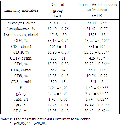
Immunological parameters were studied in 119 patients with cutaneous Leishmaniasis. Of these, 38 patients were diagnosed with a tubercular form of cutaneous Leishmaniasis, 52 – ulcerated leishmaniomas, 22 – ulcerated leishmaniomas with tubercles with lymphangitis and 7 – metalleishmaniasis. The control group consisted of data from 20 practically healthy individuals aged 25 to 40 years.The state of the immune system was assessed by the expression of CD-differentiated and activation antigens. Markers of immunocompetent cells were determined by indirect rosette formation using an immunoreagent – human erythrocytes of group 0 (I) Rh – loaded with monoclonal antibodies through a 3% solution of chromium chloride (manufactured by Sorbent LLC, Moscow, Russia) specificity CD3 – for T-lymphocyte receptors, CD4 – for T-helper/inducers, CD8 – for T-suppressors/cytotoxic lymphocytes, CD19 – for B-lymphocytes.The results of the study showed (Table 1) that in patients with cutaneous Leishmaniasis of the general group, a significant increase in the absolute number of leukocytes (5800 ± 75 cl / mcl) was observed before the start of treatment compared with the data of the control group (5380 ± 82 cl / mcl). The relative and absolute number of lymphocytes remained at the level of control values (p>0.05). The study of the state of the cellular link of the immune system showed that in patients of the general group there was a statistically significant decrease in both the relative number of CD3 cells (p<0,001) and the absolute number of CD3 cells (p<0,05) and on average they were 48,27 ± 0,40% and 881 ± 19 cells/µl, respectively, against 58,15 ± 0,74% and 1013 ± 31 cl/ml, respectively, in the control.Table 1. Indicators of the immune system in patients with cutaneous Leishmaniasis (М±m)
 |
| |
|
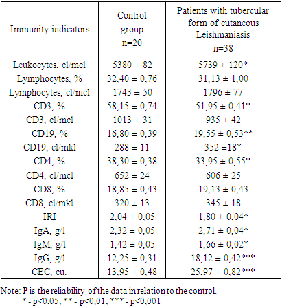
The study of the content of the subpopulation composition of T-lymphocytes showed that in patients with cutaneous Leishmaniasis before treatment, compared with the data of the control group, the relative and absolute number of CD4 cells was significantly reduced and averaged 30.23 ± 0.36% and 550 ± 12 cells/µl, respectively, versus 38.30 ± 0.38% and 652 ± 24 cells/mcl, respectively, in control. The content of another population of T-lymphocytes, CD8 cells, was inclined to increase (p>0.05) compared with the data of the control group.In patients of this group, the Immuno Regulatory Index (IRI), that is, the ratio of CD4/CD8 cells, was significantly reduced (p<0.001) and on average was equal to 1.56 ± 0.03 versus 2.04 ± 0.05 in the control.When studying the state of the humoral link of the immune system, it was revealed that in patients with cutaneous Leishmaniasis of the general group, there was a statistically significant increase in both relative (23.52 ± 0.53% at 16.80 ± 0.39% normal, p <0.001) and absolute (429 ±13 cl/µl versus 288 ± 11 cl/µl in the control, p<0.05) the number of CD19 cells. The study of the concentration of serum immunoglobulin showed that in patients of this group there was a significant increase in the level of three classes of immunoglobulin IgA, IgM and IgG relative to the control (p<0.001).The most pronounced changes were found when determining the amount of CEC in the blood serum. In the examined group of patients, there was a more than 2-fold increase in the CEC level compared to the control group and on average it was 30.43 ± 0.62 cu versus 13.95 ± 0.48 cu in the control.The data obtained by us this research showed that, in general, patients with cutaneous Leishmaniasis have a violation in the immune system, which is expressed by a decrease in cellular activity and an increase in the humoral link of the body's immunity.In further studies, we studied the state of the immune status of patients with cutaneous Leishmaniasis, depending on the clinical form of the disease.The revealed patterns in the dynamics of immunological indicators, when determining the immune status in patients with cutaneous Leishmaniasis without differentiating them by clinical forms, are preserved even when they are separated into separate groups.The results of the study showed (Table 2) that patients with the tubercular form of cutaneous Leishmaniasis had a significant decrease in the relative number of CD3 cells (p<0.05). At the same time, the absolute content of CD3 cells in patients of this group did not change much in comparison with the control (p>0.05).Table 2. Indicators of the immune system in patients with tubercular form of cutaneous Leishmaniasis (М±m)
 |
| |
|
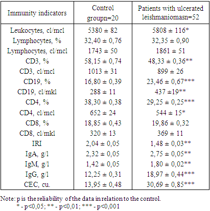
Analysis of the content of the subpopulation composition of T-lymphocytes showed that in patients of this group before the start of treatment, the relative number of CD4 cells (p<0.05) was statistically significantly reduced compared to the control group and averaged 33.95 ± 0.55% versus 38.30 ± 0.38% in the control group. The absolute number of CD4 cells remained at the level of the control group data (p>0.05). The content of another population of T-lymphocytes – CD8 cells had no statistically significant differences with the control group (p>0.05). Along with this, there was a decrease in the IRI index by 1.1 times in relation to the indicator of the control group.A study of the state of the humoral link of immunity showed that in patients of this group there was a significant increase in both relative and absolute (p<0.01 and p<0.05, respectively) CD19-cell content. The study of the level of immunoglobulin revealed that in patients with the tubercular form of cutaneous Leishmaniasis, a statistically significant increase in the concentration of IgA (p<0.05), IgM (p<0.05) and IgG (p<0.001) was observed in the blood serum.In patients of this group, compared with the control, there is a 1.9-fold increase in the content of CEC in the blood serum.A study of the state of the immune system in patients with ulcerated Leishmaniomas showed (Table 3) that in patients of this group, a significant decrease in the relative number of CD3 cells (p<0.01) was detected before the start of treatment, and on average it was 48.33 ± 0.36% versus 58.15 ± 0.74% in the control, and the absolute content of CD3 cells tended to decrease compared to the control group (p>0.05).Table 3. Indicators of the immune system in patients with ulcerated leishmaniomas (М±m)
 |
| |
|
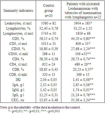
In patients of this group, the relative (p<0.001) and absolute (p<0.05) number of CD4 cells was significantly reduced compared to the data of the control group, and the content of CD8 cells was at the level of control values (p>0.05). In patients of this group, a decrease in the IRI index was revealed by 1.4 times compared with the indicator of the control group.In patients with ulcerated Leishmaniomas, a significant increase in both the relative and absolute number of CD19 cells is detected in the blood (p<0.001 and p<0.01, respectively). Against this background, there is a significant increase in the concentration of IgA (p<0.01), IgM (p<0.01) and IgG (p<0.001) compared to the control.In patients of this group, there is a 2.2-fold increase in the concentration of CEC in the blood serum. The results of the study showed (Table 4) that in patients with ulcerated Leishmaniomas with tubercles of seeding with lymphangitis, before the start of treatment, there was a significant decrease in both the relative (p<0.001) and absolute number of CD3 cells (p<0.05) and on average they were 44.50 ± 0.80% and 809 ± 31 cells /ml, respectively, at 58.15 ± 0.74% and 1013 ± 31 cl/µl, respectively, in the control.Table 4. Indicators of the immune system in patients with ulcerated leishmaniomas with tubercles of infection with lymphangitis (М±m)
 |
| |
|
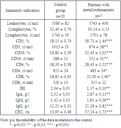
Analysis of the content of the subpopulation composition of T-lymphocytes showed that in patients of this group, both the relative (p<0.001) and absolute (p<0.01) number of CD4 cells was statistically significantly reduced compared to the control group and averaged 26.73 ± 0.74% and 489 ± 26 cells/ml, respectively, versus 38.30 ± 0.38% and 652 ± 24 cl /µl, respectively, in the control. The relative content of another population of T-lymphocytes, CD8 cells, was significantly higher than in the control (p<0.05), and their absolute number remained at the control level. Along with this, there was a decrease in the IRI index by 1.4 times in relation to the indicator of the control group.The study of the state of the humoral link of immunity revealed that in patients with ulcerated leishmaniomas with tubercles of seeding with lymphangitis, there was a significant increase in both the relative (27.68 ± 1.24% at 16.80 ± 0.39% normal) and absolute (506 ±32 cl/µl versus 288 ± 11 cl/µl in the control) number of CD19 cells (p<0.001 and p<0.01, respectively).The study of the concentration of serum immunoglobulin in patients of this group before the start of treatment revealed a significant increase in the level of IgA (p<0.01), IgM (p<0.01) and IgG (p<0.001) compared with the control values.The most pronounced changes were found when determining the amount of CEC in the blood serum. In the examined group of patients, there was a more than 2.5-fold increase in the CEC level compared to the control group and on average it was 35.36 ± 1.34 cu versus 13.95 ± 0.48 cu in the control.Studies have shown (Table 5) that in patients with metalleishmaniasis, a significant decrease in both the percentage (p<0.001) and the absolute number of CD3 cells (p<0.01) was detected compared with the data of the control group.Table 5. Indicators of the immune system in patients with metalleishmaniasis (М±m)
 |
| |
|
The study of the T-lymphocyte subpopulation showed that in patients of this group, both the relative number and the absolute content of CD4 cells were also significantly reduced compared to the control group (p<0.001 and p<0.05, respectively), and the relative content of CD8 cells significantly increased, while the absolute number remained at the control level (p>0.05). Along with this, there was a decrease in the IRI indicator by 1.5 times in relation to the indicator of the control group.A study of the state of the humoral link of immunity revealed that in the examined group there was a significant increase in both the relative and absolute number of CD19 cells (p<0.001 and p<0.01, respectively).Along with this, there is a statistically significant increase in the level of IgA (p<0.01), IgM (p<0.01) and IgG (p<0.001).In patients with metalleishmaniasis, compared with the control, there is a 2.7-fold increase in the content of CEC in the blood serum.It should be noted that the revealed changes in the immune system in patients with Leishmaniasis were more pronounced than in patients with other clinical forms of cutaneous Leishmaniasis.As you known, immune changes depend not only on the clinical form, but also on the duration of the course of the disease. In this regard, it was of interest to us to study the dependence of the intensity of the immune system on the duration of the existence of the pathological process. To do this, patients depending on the duration of dermatosis were divided into 3 groups. Group 1 was represented by 82 patients with a disease duration of up to 6 months, group 2: - 28 patients with a disease duration of 7 months to 1 one year, and group 3: - 9 patients with a pathological process duration of more than 1 one year.The results of the study showed that in all groups there was a significant decrease in the relative number of CD3 cells (p<0.001). At the same time, the absolute content of CD3 cells in patients of the second group did not change much compared to the data of healthy individuals; in patients of the first and third groups, this indicator was significantly lower than in the control (p<0.01 and p<0.05, respectively). The study of the level of CD4-cells and CD8-cells shows that in all examined groups there is a significant decrease in the relative number of CD4-cells (p<0.001), and the level of CD8-cells remained at the level of control values (p>0.05). It should be noted that the identified changes in the cellular immunity were more pronounced in patients with a disease duration of more than 1 one year.In the study of the absolute number of CD4 cells and CD8 cells, it was found that in patients of the second and third groups, the number of CD4 cells tended to decrease, and in patients of the first group this indicator was significantly lower than in the control group (p<0.05). At the same time, the absolute content of CD8 cells in all examined groups did not change much compared to the data of healthy individuals (p>0.05). IRI in all examined groups was significantly reduced (p<0.01 and p<0.001, respectively).An analysis of the state of the humoral link of immunity showed that in all the examined groups there was a significant increase in both the relative and absolute content of CD19 cells (p<0.001), as well as an increase in the concentration of IgA, IgM and IgG (p<0.05 and p<0.001 respectively).The most pronounced changes were revealed in the study of the concentration of CEC in the blood serum. In all examined groups of patients, there is a 2.1 to 2.6-fold increase in the content of the CEC compared with the control group (p<0.001). The most pronounced violation of the indicators of the humoral link and the CEC was found in patients with a disease duration of more than 1 one year.
4. Conclusions
Analysis of the data obtained indicates that the identified disorders in the immune system of patients with cutaneous Leishmaniasis are were directly dependent on the duration of the existence of the pathological process. With an increase in the prescription of the disease, the imbalance in the immune system was aggravated.Thus, the foregoing indicates that in cutaneous Leishmaniasis, disturbances in the functioning of the immune system were detected, manifested by a deficiency of the cellular and activation of the humoral parts of the immune system, which were directly dependent on the clinical form and duration of the disease.In this regard, in order to restore the identified disorders in the immune system of patients with cutaneous Leishmaniasis, it is advisable to carry out an appropriate immunocorrection.
References
| [1] | Bogadelnikov I.V., Vyaltseva Yu.V., Karimov I.Z., Degtyareva A.A., Mazinova E.R., Dyadyura E.N., Los-Yatsenko N.G. Leishmaniasis — a surprise from the East. Child's health. 2012; 3: 38. |
| [2] | Kurdina M.I. Case of tubercular leishmaniasis. Russian Journal of Skin and Venereal Diseases. 2001; 3: 27-29. |
| [3] | Pal'cev M.A., Potekaev N.N., Kazanceva I.A.,Lysenko A.I., Lysenko L.V., Chervonnaja L.V. Klinikomorfologicheskaja diagnostika zabolevanij kozhi: atlas [Clinical and morphological diagnosis of skin diseases: atlas]. 2-e izd., stereotipnoe. Moscow: Medicina, 2005 432 p. |
| [4] | Potekaev N.N., Akimov V.G. Differencial'naja diagnostika i lechenie kozhnyh boleznej [Differential diagnosis and treatment of skin diseases]. Moscow: GJeOTAR-Media, 2016 456 p. |
| [5] | Cutaneous Manifestations of Human and Murine Leishmaniasis / Breanna M. Scorza, Edgar M. Carvalho, Mary E. Wilson / International Journal Molecular Sciences – 2017 - №18. |
| [6] | Khamdamova M. T. Еchographic features of the range of variability in the size of the uterus and ovaries in women of menopausal age using oral and injectable forms of contraception // American Journal of Medicine and Medical Sciences. - 2020. - N10 (8). - P.580-583. |
| [7] | Khamdamova M. T.Еchographic features variability in the size and shape of the uterus and ovaries in women of the second period of adulthood using various contraceptives // Asian Journal of Multidimensional Research - 2020. – N9 (5). - P.259-263. |
| [8] | Vul'f K., Dzhonson R., Sjumond D. Dermatologija po T. Ficpatriku. Atlas-spravochnik [Dermatology by T. Fitzpatrick. Atlas-directory]. Moscow, Praktika, 2007 1228 p. |
| [9] | Khamdamova M. T.Somatometric characteristics of women of the first and second period of adulthood using different contraceptives with different body types // The american journal of medical sciences and pharmaceutical research - 2020. – N8 (2). - P.69-76. |
| [10] | Sokolovskij E.V., Miheev G.N., Krasnosel'skih T.V. i dr. Dermatovenerologija: uchebnik dlja studentov uchrezhdenij vyssh. prof. med. obrazovanija [Dermato-venerology: a textbook for students of institutions of higher professional medical education]. In E.V. Sokolovskiy (ed.). Saint Petersburg: SpecLIT, 2017 687 p. |
| [11] | Khamdamova M. T. Age and individual variability of the shape and size of the uterus according to morphological and ultrasound studies // Problems of biology and medicine. 2020, №1 (116). -Р.283-286. |
| [12] | Khamdamova M. T. Аge echographic characteristics of the uterus and ovaries in women of the first and second period of middle age // Biology and integrative medicine. ISSN 2181-8827 2020. №2 - March-April (42). -Р.75-86. |
| [13] | Khamdamova M. T. Аnthropometric characteristics of the physicalstatus of women in the first andsecond period of middle age // New day in medicine. 2020. - № 1 (29). - Р.98-100. |
| [14] | Khamdamova M. T.Аge echographic characteristics of the uterus and ovaries in womenof the first and second period of middle age // Biology and integrative medicine. – Bukhara. 2020. №2 (42) - Р.75-86. |
| [15] | Evropejskoe rukovodstvo po lecheniju dermatologicheskih zabolevanij [European guidelines the treatment of dermatological diseases]. In A.D. Kacambas, T.M. Lotti (ed.); per. s angl. 2-e izd. Moscow: MEDpress-inform, 2009 736 p. |
| [16] | Khamdamov I.B., Khamdamov A.B. Сlassification and properties of mesh explants for hernioplasty of hernial defects of the anterior abdominal wall (review) // Biology and integrative medicine. ISSN 2181-8827 2021. №5 – март-апрель (52). C.12-22. |
| [17] | Khamdamov I.B., Khamdamov A.B. Еndovideosurgical hernioplasty in women of fertile age // New day in medicine. 2021. №6 (38/1). P.25-27. |
| [18] | Diagnosis, Treatment and Clinical Features of Cutaneous Leishmaniasis in Saudi Arabia / Yousry A. Hawash, Khadiga A. Ismail, Maha M. Abdel-Wahab, Mahmoud Khalifa // The Korean Journal of Parasitology. |

 Abstract
 Reference
 Full-Text PDF
 Full-text HTML