-
Paper Information
- Previous Paper
- Paper Submission
-
Journal Information
- About This Journal
- Editorial Board
- Current Issue
- Archive
- Author Guidelines
- Contact Us
American Journal of Medicine and Medical Sciences
p-ISSN: 2165-901X e-ISSN: 2165-9036
2022; 12(5): 562-564
doi:10.5923/j.ajmms.20221205.22
Received: April 21, 2022; Accepted: May 12, 2022; Published: May 14, 2022

Morphological Changes of the Heart in Experimental Pneumosclerosis and the Effectiveness of Its Treatment with Pomegranate Seed Oil
 Abstract
 Reference
 Full-Text PDF
 Full-text HTML
Rajabov N. G.
Bukhara State Medical Institute, Uzbekistan
Correspondence to: Rajabov N. G., Bukhara State Medical Institute, Uzbekistan.
Copyright © 2022 The Author(s). Published by Scientific & Academic Publishing.
This work is licensed under the Creative Commons Attribution International License (CC BY).
http://creativecommons.org/licenses/by/4.0/

For the effective treatment of cardiac dysfunctions against the background of pneumosclerosis, the correction of these changes with pomegranate seed oil is currently being considered. The study of the available literature data revealed a lack of information about the effect of pomegranate seed oil on the structural and functional parameters of the heart, which is formed against the background of pneumosclerosis. This article provides an in-depth analysis of the structural and functional changes in the heart that occur against the background of experimental pneumosclerosis, and the correction of this pathological process with pomegranate seed oil.
Keywords: Experimental pneumosclerosis, Fibrotic changes in the heart, Morphological study, NO2 effects, Pomegranate seed oil
Cite this paper: Rajabov N. G., Morphological Changes of the Heart in Experimental Pneumosclerosis and the Effectiveness of Its Treatment with Pomegranate Seed Oil, American Journal of Medicine and Medical Sciences, Vol. 12 No. 5, 2022, pp. 562-564. doi: 10.5923/j.ajmms.20221205.22.
1. Introduction
- A number of pathological changes that occur during pneumosclerosis, that is, chronic and degenerative changes that develop throughout the bronchi, alveoli and interstitial tissue, have been described by many of our scientists. At the same time, pulmonary vessels, nerve fibers and lymphatic vessels are involved in this process, as well as the development of various pathological changes in a number of additional organs, especially in the heart, against the background of pneumosclerosis. At the heart of circulatory disorders are many clinical syndromes that are closely interconnected by pathogenetically different relationships. Often severe heart failure occurs with pneumosclerosis. The development of this heart failure occurs as a result of the work of the myocardium with a prolonged stress load. [1,2,3,4,5]. Pomegranate seed oil has a number of powerful regenerative, anti-inflammatory, antioxidant, antiviral, antimicrobial properties. The healing properties of pomegranate seed oil are due to the uniqueness of its fatty acid content, which means that a mixture of large amounts of tocopherols, beta-cystosterol, stigmasterol and campesterols is very useful.The purpose of research: Detection of morphological changes in the heart as a result of experimental pneumosclerosis, as well as correction of this pathological condition with pomegranate seed oil.
2. Materials and Methods
- For experimental laboratory tests, we chose white non-parody rats aged from four to nine months as the object of our research, in which an experimental model of pneumosclerosis was induced, after which it was planned to study the resulting pathological processes in the heart. To implement this plan, rats were kept in a room where this type of laboratory animals met the requirements for keeping conditions (t 20-24 CO, humidity 60%, light (12 hours) / darkness (12 hours)) and fed without restrictions. Before the start of the experiment, the animals were placed in a two-week quarantine and accustomed to stay in the experimental chamber. Nitrogen dioxide was then used to induce an experimental model of pneumosclerosis. The chemical reaction of sodium nitrite with sulfuric acid resulted in the formation of a mixture of nitrogen oxides.Staining of micropreparations was carried out by the method of hematoxylin-eosin, as well as the study of connective tissue fibers in micropreparations by the methods of Van Gieson and Weigert.In the flask, air with colorless nitric oxide was converted by oxygen into the most stable dioxide (NO2), which was introduced into the animal chamber through the outlet pipe using a rubber tube to ensure uniform distribution of the gas. The NO2 concentration was determined colorimetrically and was 30-40 mg/m3. The animals were inhaled with NO2 for 90 days three times a day for 30 minutes with an interval of 30 minutes between them. The ventilation mode was associated with the need to ventilate the chamber from carbon dioxide accumulated during the respiration of animals. To perform the experimental part of the study, 90 rats were selected, in which no signs of impaired pulmonary circulation were detected during examination by perfusion scintigraphy. Animals (n = 90) were exposed to NO2. Exposure to NO2 for 30 days revealed signs of inflammation in the lungs, after 60 days - signs of pulmonary fibrosis, but without visible complications from the cardiovascular system, and after 90 days under the influence of NO2 - pathomorphological changes were observed in the heart, i.e. pneumosclerosis of the lungs and fibrotic changes in the heart. The study was carried out on days 30, 60, and 90 after the stage of formation of pneumosclerotic changes, as well as in the control group (5 rats for each month). Morphological examination of lung tissue and heart was performed 30, 60, and 90 days after exposure to NO2.
3. Results and Its Discussion
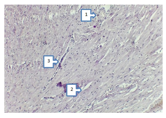
- Analysis of the results of the study showed that in the biomaterial obtained from the lungs of white mice as a result of exposure to NO2 for 30 days, signs of interstitial pneumonia with fibrosis proliferation, thickening of the alveolar septa, proliferation of myofibroblastic cells, enlarged lumen of the alveoli with rupture of their walls, bronchus wall were revealed. was with moderate infiltration, but pathological and anatomical changes in the cardiac biomaterial were not observed. On the 60th day, in the biomaterial obtained from the lungs of mice as a result of exposure to NO2, pronounced interstitial pneumonia was detected with the growth of fibrosis. Alveolar septa are thickened (1) due to proliferation, proliferation of myofibroblasts with infiltration (2, 3), lymphohistiocytic infiltration of lung tissue (4). Staining was carried out with hematoxylin-eosin entrained 20x40 times. In the biomaterial obtained from the heart, it was observed that very slight pathological changes began in this organ, but these changes had almost no effect on the clinical state of white rats.At the next stage, i.e. from 90 day 4; 5; 6; 7; eight; caused an experimental model of pneumosclerosis on 90 white non-white rats per month, after which the changes that developed as a result of pathological processes occurring in the heart were analyzed (Figures 1; 2; 3; 4; 5;).Pathological changes in the heart on the 90th day in 4-month-old white rats showed focal and diffuse infiltrates in the interstitial tissue of the myocardium, consisting mainly of neutrophils and eosinophilic granulocytes (1), dystrophic changes in muscle fibers (2). Staining was done with hematoxylin-eosin. With a magnification of 10x20 times (Fig. 1).
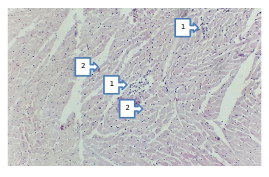 | Figure 1. Pathological changes in the heart of 4-month-old white rats after 90 days |
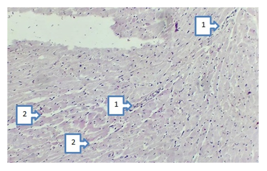 | Figure 2. Pathological changes in the heart of 5-month-old white rats after 90 days |
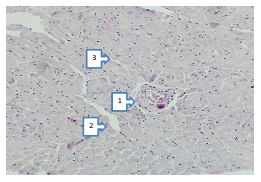 | Figure 3. Pathological changes in the heart of 6-month-old white rats after 90 days |
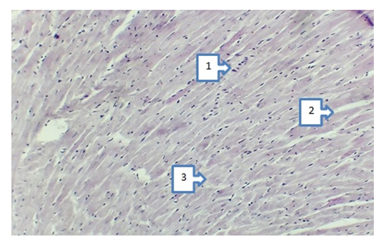 | Figure 4. Pathological changes in the heart of 7-month-old white rats after 90 days |
 | Figure 5. Pathological changes in the heart of 8-month-old white rats after 90 days |
4. Conclusions
- 1. Changes in the heart that develop as a result of experimental pneumosclerosis become more pronounced with the age of the studied animals.2. Therapy with pomegranate seed oil of pathological changes in the heart caused by experimental pneumosclerosis, depending on the dose in animals that received the drug on the 105th day of the experiment, clinical signs improved in dynamics, and in the control group that did not receive the drug in dynamics were unchanged.