-
Paper Information
- Next Paper
- Previous Paper
- Paper Submission
-
Journal Information
- About This Journal
- Editorial Board
- Current Issue
- Archive
- Author Guidelines
- Contact Us
American Journal of Medicine and Medical Sciences
p-ISSN: 2165-901X e-ISSN: 2165-9036
2022; 12(5): 495-498
doi:10.5923/j.ajmms.20221205.09
Received: April 22, 2022; Accepted: May 3, 2022; Published: May 10, 2022

Pathomorphological Changes in Spondilosis
Israilov R.1, Mamajonov B. S.2
1Andijan Medical Institute, Uzbekistan
2Republican Pathological Anatomy Center, Uzbekistan
Copyright © 2022 The Author(s). Published by Scientific & Academic Publishing.
This work is licensed under the Creative Commons Attribution International License (CC BY).
http://creativecommons.org/licenses/by/4.0/

The article discusses pathomorphological changes that develop in osteophytes of the spinal cord, annulus fibrosus and spinal cord in spondylosis. The results showed that the osteophytes arising at the anterior edge of the spine have a morphologically concentric structure, fibrous structures and disordered ground substance, consisting of tissue rich in foci of calcification and pigmentation. As a result of the penetration of osteophytes into the fibrous-annular tissue of the disc, splitting occurs, the destruction of its fibrous structures, the formation of a coarse substance, the development of calcification and chondromatous metaplasia. At first, in the vibrating nucleus, the chondroid material becomes rough, colored, the fibrous structures and the intermediate material are dispersed and thickened. was found, that the number of chondrocytes increased and they underwent processes such as dystrophy and destruction to varying degrees. In chronic spondylosis, complete destruction and necrobiosis of chondrocytes in the nucleus accumbens, their transformation into structureless substances, and the formation of foci of calcification were confirmed.
Keywords: Spine, Spinal disc, Spondylosis, Osteophyte, Fibrosis ring, Rebirth nucleus
Cite this paper: Israilov R., Mamajonov B. S., Pathomorphological Changes in Spondilosis, American Journal of Medicine and Medical Sciences, Vol. 12 No. 5, 2022, pp. 495-498. doi: 10.5923/j.ajmms.20221205.09.
1. Introduction
- Spondylosis is a chronic disease of the spine, the progression of which leads to thinning of the intervertebral disc, resulting in hernias and osteophytes. If not treated in time, the vertebrae become tangled and lose their mobility [1,2]. Its causes are the natural aging of the spine, overload and injury. Injuries play an important role for these reasons. In case of injury, the surface of the vertebrae is damaged and first defects appear, then a calyx forms in their place and osteophytes grow. Excessive stress on the spine leads to stretching and flattening of the disc. These changes ultimately lead to improper regeneration, often with growth and proliferation of connective tissue, leading to pathological changes in the tissue structures of the disc [3,4]. If an inflammatory process joins the foci, then the disease naturally passes into an inflammatory form and pathological infiltrates appear, combining both exudative and proliferative processes characteristic of inflammation in all tissue structures. Changes of this type are observed in metabolic disorders, i.e. in diseases such as diabetes mellitus, obesity, actomegaly [5,6]. With the development of spondylosis, metabolism is often disturbed and the intervertebral disc causes a lack of substances in the disc tissue. Changes of this type are observed in metabolic disorders, i.e. in diseases such as diabetes mellitus, obesity, actomegaly [5,6]. With the development of spondylosis, metabolism is often disturbed and the intervertebral disc causes a lack of substances in the disc tissue. Changes of this type are observed in metabolic disorders, i.e. in diseases such as diabetes mellitus.The pathogenesis of the development of spondylosis is associated with degenerative changes in the intervertebral disc and osteochondrosis. The risk of its development increases with scoliosis, kyphosis and lordosis [4,5,6,7]. So, spondylosis is the final stage of osteochondrosis. Under the influence of various pathological factors, biochemical changes occur in the intervertebral disc, as a result of which the amount of fluid and proteoglycans decreases in it. As a result, the collagen fibers in the disc break down and break down, reducing cushioning properties. In parallel with these changes, the tone of the spinal cord decreases, it becomes brittle. At this point, bundles of spinal nerves begin to contract.The purpose of this study was a microscopic study of osteophytes, in which the edges of the vertebrae damaged by spinal cord spondylosis, the fibrous ring wraps around the disc and the elastic core.
2. Materials and Methods
- The material of the study is surgical interventions performed in the neurosurgical department of the ASMI clinic in 2019-2022, i.e. with discectomy, laminectomy, fibrous membrane of the intervertebral disc, elastic membrane covering the spine, osteophytes on the anterior edge of the common bone, fibrous ring of the disc. These tissue fragments were solidified for 72 hours in formalin dissolved in 10% phosphate buffer. The bone fragments were decalcified in 10% nitric acid. Then all the pieces were washed with running water for 3-4 hours, dehydrated in alcohols of increasing concentration, poured with paraffin wax and prepared bricks. Histological sections 5-7 µm thick were made from paraffin bricks and stained with hematoxylin-eosin and Van Gieson stains. The preparations were studied under a light microscope.
3. Results and Discussion
- The results of the morphological study showed that the anterior edge of the vertebrae was thickened, osteophytes of various sizes in the form of tubercles were histologically concentrically arranged randomly, consisting of fibrous structures with numerous cracks and holes of various sizes. Fibrous structures are relatively sparse and scanty in color along the periphery of osteophytes, with dark calcification foci on the outer surface with hematoxylin (Fig. 1). The fibers located in the central part of the osteophytes have a relatively dense structure and are stained dark with eosin. In the center of the osteophyte is a cavity of light soft tissue with granular eosin.
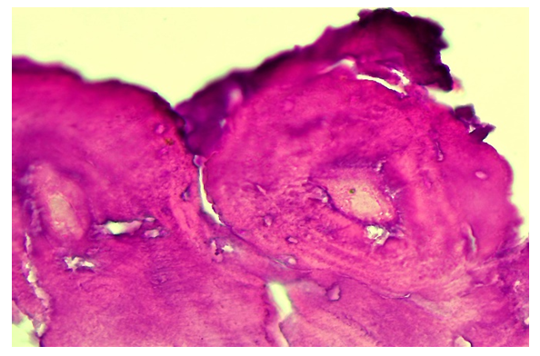 | Figure 1. Histological structure of an osteophyte formed along the edge of the spine. Stain: H.E. Size:10x40 |
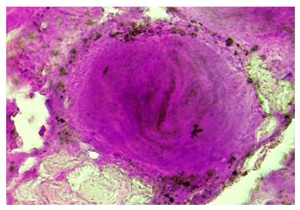 | Figure 2. Osteophyte with a relatively dense structure. Stain: H.E. Size:10x40 |
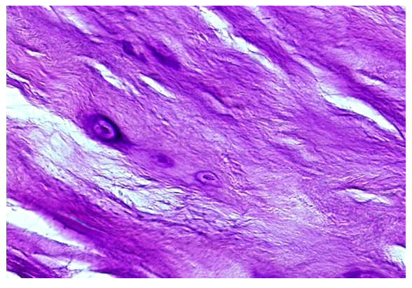 | Figure 3. Histological structure of the outer annulus of the disc in the area of osteophytes. Stain: H.E. Size: 10x40 |
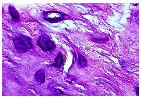 | Figure 4. Pathological changes in the fibrous ring of the disc. Stain: H.E. Size: 10x40 |
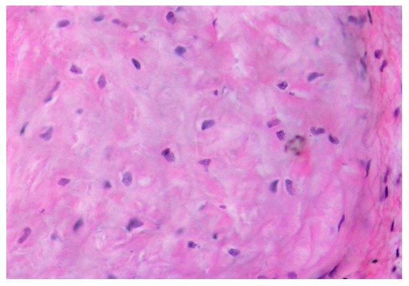 | Figure 5. The structure of the nucleus accumbens in spondylosis. Stain: H.E. Size: 10x40 |
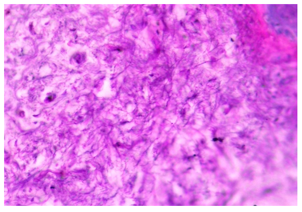 | Figure 6. Pathological changes in the nucleus accumbens in severe forms of spondylosis. Stain: H.E. Size:10x40 |
4. Conclusions
- It has been shown that osteophytes that appear on the anterior edge of the spine in spondylosis have a morphologically concentric structure, fibrous structures and a disordered structure rich in foci of calcification and pigmentation.As a result of the penetration of osteophytes into the fibrous-annular tissue of the disc, splitting occurs, the destruction of its fibrous structures, the formation of a coarse substance, the development of calcification and chondromatous metaplasia.In the living nucleus, it was initially found that the chondroid material is rough, colored, fibrous structures and intermediates are scattered and thickened, the number of chondrocytes is increased, they are subject to processes such as dystrophy and destruction to varying degrees.In chronic spondylosis, it was confirmed that chondrocytes in the nucleus accumbens are completely destroyed and necrobiose, turn into structureless substances, and foci of calcification appear.
 Abstract
 Reference
 Full-Text PDF
 Full-text HTML