-
Paper Information
- Previous Paper
- Paper Submission
-
Journal Information
- About This Journal
- Editorial Board
- Current Issue
- Archive
- Author Guidelines
- Contact Us
American Journal of Medicine and Medical Sciences
p-ISSN: 2165-901X e-ISSN: 2165-9036
2022; 12(3): 348-352
doi:10.5923/j.ajmms.20221203.25
Received: Mar. 3, 2022; Accepted: Mar. 16, 2022; Published: Mar. 24, 2022

Features of Clinical and Neurological Disorders of the Formation of Venous Encephalopathy on the Background of Phlebopathy
 Abstract
 Reference
 Full-Text PDF
 Full-text HTML
Shmyrina Ksenia Vladimirovna1, Djurabekova Aziza Takhirovn2, Umirzokov Oybek Nishonovich3, Vyazikova Natalia Fedorovna1
1Candidate Medical Scienes, Assistant of the Department of Neurology Samarkand Medical Institute, Uzbekistan
2Head of the Department of Neurology Samarkand Medical Institute, Uzbekistan
3Clinical Resident of the Department of Neurology, Samarkand Medical Institute, Uzbekistan
Copyright © 2022 The Author(s). Published by Scientific & Academic Publishing.
This work is licensed under the Creative Commons Attribution International License (CC BY).
http://creativecommons.org/licenses/by/4.0/

Chronic progressive disorder of cerebral circulation is a slow form of deterioration of microcirculation in the brain, where oxygen is not received due to a decrease in blood flow, especially during the load of the body. In recent years, more and more emphasis has been placed on venous insufficiency as one of the factors leading to an early stage of cerebrovascular pathology [1,5,8].
Keywords: Chronic, Cerebral professional, Varicose veins, Varicose veins in pregnancy, Thrombophlebitis, Phlebosclerosis venous encephalopathy, Blood flow
Cite this paper: Shmyrina Ksenia Vladimirovna, Djurabekova Aziza Takhirovn, Umirzokov Oybek Nishonovich, Vyazikova Natalia Fedorovna, Features of Clinical and Neurological Disorders of the Formation of Venous Encephalopathy on the Background of Phlebopathy, American Journal of Medicine and Medical Sciences, Vol. 12 No. 3, 2022, pp. 348-352. doi: 10.5923/j.ajmms.20221203.25.
1. Introduction
- The complexity of the anatomical structure of the venous circulation, insufficient information about the venous outflow, led many scientists to the conclusion that there is no venous encephalopathy, and even if there is a place for venous stasis, it is perfectly compensated, and cannot be considered both clinically and pathogenetically [2,6]. Therefore, for a long time, violation of venous cerebral circulation was studied as a cause of migraine, functional disorder, vegetative-vascular dystonia; and here, venous insufficiency of the lower extremities is described by vascular surgeons on a large scale, in all variations: professional varicose veins, varicose veins in pregnancy, thrombophlebitis, phlebosclerosis, etc. [3,4,7]. What is the relationship between these seemingly different forms of diseases, does the factor of venous insufficiency in the lower extremities affect the process of chronic cerebral circulation, and to what extent is it possible to examine the venous bed of the brain and peripheral circulation of the veins in the aggregate. The presented questions show the relevance and urgent need for the analysis of venous insufficiency of the brain in combination with venous insufficiency of the lower extremities.To study venous insufficiency of the lower extremities as a factor in early chronic cerebrovascular accident and optimize diagnostic tactics.
2. Materials and Research Methods
- The survey involved patients aged 45-62 years (mean age 51±7.8 years) in the amount of 36 people, 23 women, 13 men. -2022 year. The criteria for enrolling patients in the study were the initial signs of chronic cerebral circulation, venous encephalopathy, and all patients had venous insufficiency of the lower extremities to some extent (in the form of varicose veins, thrombophlebitis); The control group - 20 people, consisted of patients with early signs of chronic cerebral circulation, without changes in venous circulation in the lower extremities. The exclusion criteria were severe organic diseases of the brain, such as a brain tumor, consequences of stroke, a history of surgery on the brain (consequences of trauma), myocardial infarction. Research methods are based on the goal, of course, this is a targeted study of angio -neurological features, for this angiography and MRI of the brain were carried out, the vessels of the internal jugular veins and veins of the lower extremities, bronchocephalic veins were studied by ultrasound. The analysis was given importance, where the fact of venous circulation disorders in the legs, the timing and cause, hereditary predisposition, methods of treatment were clarified. Clinical, neurological and somatic status was examined according to standard rules. Laboratory methods included blood biochemistry, with an emphasis on hematopoietic factors. Statistical data processing was carried out according to the standard Student's indicators on an individual computer using the Microsoft software package. Excel (version 14.0).
3. Research Results
- The capabilities of the brain are multifaceted, and one of the important points is the compensatory nature of cerebral circulation, especially venous circulation, where clinical symptoms do not make themselves felt for a long time. And only when the compensatory fact reaches the limit, does congestive hypoxia begin, manifested by a headache; venous dystonia with functional complaints, usually paying little attention to themselves. In this survey, patients of the main group (MG) complained of a headache characteristic in the morning, experienced heaviness in the head, and a very specific complaint for patients with venous insufficiency, "sand in the eyes" again in the morning, in the anamnesis of these patients use a low headboard for sleep, such observations were 93% of the OG. The second most common symptom is swelling of the face, swelling of the eyelids, slight sometimes slightly noticeable cyanosis 96% OG. Less often, but there is a symptom of dizziness when standing up, only in 7 cases, the rest of the dizziness that was observed in patients was according to the patients in the anamnesis, early not at the time of the examination (perhaps these are signs of vertebrobasilar disorders, respectively, this symptom was not evidence). If we take complaints in a comparative aspect with the control group (GC), then the difference is visible, headache in this group is often closer to the second half of the day, practically does not occur in the morning. Puffiness is less common, and without a cyanotic component, in our case, only in 2 patients. But dizziness is much more common, in 12 out of 20 patients. Another feature of the detection, the presence of low blood pressure, especially diastomic in the range from 50 to 70, in cases of OH, 15 patients had such pressure, and only women. In the CG, features of high blood pressure were revealed, from 130 to 170 systonic (in several cases, patients had a hypertensive crisis). Cephalgic pain in OH patients was noted by one fact, patients tried to get out of bed faster in the morning, since the vertical position reduced tension and heaviness in the head, in the second CG patients tried to be in a horizontal position longer, since verticalization aggravated the headache.A distinctive feature of the main group was a change in the form of venous blood flow disorders in the legs, which had independent complaints. This is a venous pattern on the legs, varying in frequency and severity. Mostly in women, 16 of whom describe this sign slowly progressing after childbirth; 2 women work as hairdressers with great experience; 5 women associate the cause with hereditary maternal predisposition. It turned out to be characteristic that in women with a more significant body weight (obesity of 1-2 degrees), these signs are significantly pronounced; with the presence of venous tubercles, "snakes". For the men included in the survey, the violation of the venous outflow in the legs was accompanied by severe swelling in the legs, cyanosis of the skin, totally in the region of the lower leg, sometimes asymmetrically. 3 men had a history of varicocele, 8 patients had hemorrhoids, all these patients were observed in the surgical department (preparing for surgery). One patient had a history of acute phlebothrombosis, against the background of severe pain in the inguinal region, along the inside of the thigh, examination by a vascular surgeon and ultrasound showed the need for urgent hospitalization, surgery ended in lymphoria, the duration of which lasted more than 2.5 weeks; as a result, the process was completely restored, the signs of chronic venous insufficiency persisted. The second case is associated with a long flight in an airplane (more than 12 hours), in which the patient with excess body weight practically did not get up from her seat, her legs were swollen like “elephantiasis”, pain when touched, difficulty walking, after a few days the process recovered, but again left a complication in the form of chronic venous insufficiency.The electroencephalographic picture of the examined persons revealed a certain specificity of the results obtained. In some persons of the OG, as well as in the persons of the CG, the α -rhythm was represented by a regular component with a maximum amplitude of up to 55.0 ± 23.0 μV, interhemispheric asymmetry 12.0 ± 5.9%, a dominant frequency of 11.0 ± 0, 9 Hz, severity index 86.9±6.8%; dominated in the occipital regions of the brain; had a spindle shape without flashes of hypersynchronization and distortion.In a number of persons OG α -rhythm was unmodulated, represented by an irregular frequency component with a maximum amplitude of up to 36.0 ± 9.0 μV, interhemispheric asymmetry 20.0 ± 6.8%, a dominant frequency of 10.0 ± 1.0 Hz, a severity index of 84, 0 ± 9.0%. Zonal differences in α -rhythm were smoothed out. There was disorganization of a -activity of a mild degree due to the β-rhythm or due to single 0-waves in amplitude not higher than the background activity with a dominant frequency of 4.09±0.5 Hz and a severity index of 11.0 ± 5.9%. This type corresponded to the fourth type of EEG and prevailed in persons with an early stage of CNMC on the background of venous disorders of the lower extremities and atherosclerosis of the extremities (p <0.05), where the decrease in the amplitude-frequency parameters of the EEG spectrum from the side of the dominant hemisphere. And a decrease in amplitude indicators, on the EEG, was noted without a decrease in frequency indicators.
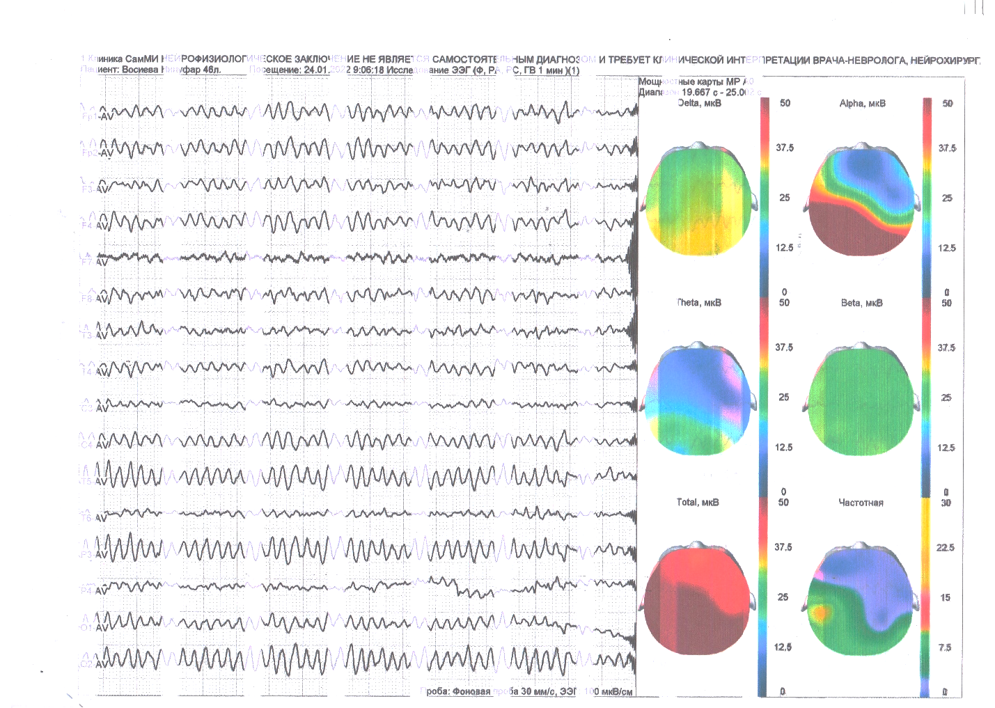 | Rice 1. EEG b-th 46 years of the main group |
|
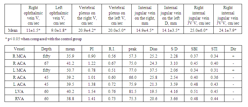 | Table 2. Ultrasound examination of venous circulation in the head in the group of examined |
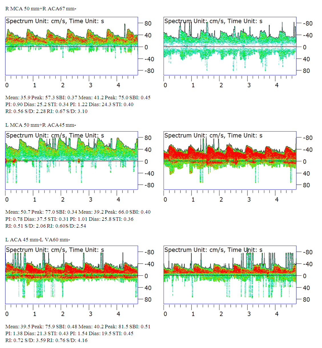 | Rice 2. TKDG b-oh 54 years of the main group |
4. Conclusions
- The analysis of the conducted study confirmed the relationship of venous disorders in the lower extremities (phlebotapia) with impaired venous cerebral circulation, which must be taken into account when examining patients for the presence of neurological symptoms.1. Knowledge of the clinical features of venous dysgemia makes it possible to diagnose chronic cerebral disorders in the early stages, where the linear blood flow velocity (according to TCDR) in the veins of the cerebral circulation is much higher than the normative data, indicating the development of venous encephalopathy and its slow progression.2. Venous dysgemia plays an important role in the formation of chronic cerebral ischemia of the brain, which requires a targeted study of the stages of the disease; differential diagnosis with other factors such as arterial hypertension, diabetes mellitus, cardiovascular insufficiency, blood disease in the formation of age-related discirculatory encephalopathy.