-
Paper Information
- Next Paper
- Previous Paper
- Paper Submission
-
Journal Information
- About This Journal
- Editorial Board
- Current Issue
- Archive
- Author Guidelines
- Contact Us
American Journal of Medicine and Medical Sciences
p-ISSN: 2165-901X e-ISSN: 2165-9036
2022; 12(3): 327-329
doi:10.5923/j.ajmms.20221203.19
Received: Feb. 19, 2022; Accepted: Mar. 11, 2022; Published: Mar. 17, 2022

Advantages of Ultrasonography in Inflammatory and Degenerative Diseases of the Hip Joint
 Abstract
 Reference
 Full-Text PDF
 Full-text HTML
Khodjibekov Marat Khudoykulovich1, Nazarova Gulchekhra Usmonovna2, Madumarova Zarnigor Shukhrat Kizi3
1Department of Medical Radiology and Oncology of the Tashkent Medical Academy
2Andijan State Medical Institute, Faculty of Advanced Training and Retraining of Physicians, Head of the Department of Family Medicine
3Andijan State Medical Institute, Faculty of Advanced Training and Retraining of Physicians, Senior Teacher at the Department of Medical Radiology and Clinical Laboratory Diagnostics
Copyright © 2022 The Author(s). Published by Scientific & Academic Publishing.
This work is licensed under the Creative Commons Attribution International License (CC BY).
http://creativecommons.org/licenses/by/4.0/

This article is devoted to the application of ultrasonography in the diagnosis of inflammatory and degenerative diseases of the hip joint. The article discusses the relevance of the use of ultrasound in the disease of ostearthrosis (OA), rheumatoid arthritis and aseptic necrosis of the femoral head, as well as the differential assessment of inflammatory and degenerative processes based on ultrasound Doppler sonography. For a long time, a simple X-ray was considered the reference technique of osteoarthritis (OA). Recently, ultrasonography is an innovative method for visualizing this disease. The use of ultrasound highlights the various anatomical structures in great detail and detects intra- and extra-articular changes.
Keywords: Coxoarthrosis, Coxitis, Rheumatoid arthritis, Osyeoarthritis aseptic necrosis, Ultrasonography, Hip joints, Diagnostics
Cite this paper: Khodjibekov Marat Khudoykulovich, Nazarova Gulchekhra Usmonovna, Madumarova Zarnigor Shukhrat Kizi, Advantages of Ultrasonography in Inflammatory and Degenerative Diseases of the Hip Joint, American Journal of Medicine and Medical Sciences, Vol. 12 No. 3, 2022, pp. 327-329. doi: 10.5923/j.ajmms.20221203.19.
1. Introduction
- Hip joint disease is one of the urgent problems of modern orthopedics, as it contributes to the rapid development of severe anatomical and functional inferiority of the musculoskeletal system, in particular in the elderly. According to WHO, more than ten percent of the world's population suffers from joint diseases. In terms of the frequency of lesions, the hip joint ranks first (42.7%), the second is the knee (33.3%), the third is the shoulder (10.8%), and the remaining joints account for 13.2% [1].The differential diagnosis of lesions of the hip joint is difficult due to its deep occurrence and the presence of large muscle masses around the joint. In this regard, it is not possible to assess the external changes in the joint, it is very difficult to perform its puncture and arthroscopy. The differential diagnosis of coxarthrosis and coxitis at an early stage is especially difficult [2]. In this regard, the issue of differential diagnosis of diseases of the hip joint of dystrophic and inflammatory origin is relevant.The ultrasound method, being non-invasive, reproducible and relatively affordable, can be widely used to assess both the accumulation of fluid inside the joint, in the joint bags, and to assess the thickness of the synovial membrane and erosive changes [3,4]. Modern ultrasound equipment makes it possible to evaluate both superficial periarticular and intra-articular tissues that make up the joint.Purpose of the study: Improving the efficiency of radiodiagnosis of inflammatory and degenerative diseases of the hip joint based on the ultrasound method.
2. Materials and Research Methods
- The total number of patients examined was 138, of which 15 (13.9%) were men and 93 were women (86.1%). The patients were divided into groups and subgroups. Group I included patients with dystrophic diseases (coxarthrosis stage I-III), a total of 82 people. Group II included 10 patients with coxarthrosis formed as a result of aseptic necrosis of the femoral head. Of these, men 5 (50%) people, women 5 (50%) people. Group III with inflammatory diseases included patients with rheumatoid arthritis, in the amount of 12 people. Of these, 4 men (33.3%) people, women 8 (66.7%) people.All patients in the period from 2018 - 2020. were treated by an orthopedist in the ASMI clinic, and an ultrasound examination was carried out in the radiology department of the ASMI clinic in Andijan.Ultrasound examination was carried out on Sonoscape S-22 (China) and Mindray DC3 (China) digital multifunctional scanners. For the most clear visualization, a study was used with convex and linear sensors, in the frequency range of 7-12 MHz.Comprehensive ultrasound examination included survey scanning of the hip joint in gray scale mode, examination of the shape and contour of the femoral head, determination of the presence of effusion, measurement of parameters of the joint capsule, thickness of the hypoechoic layer of hyaline cartilage, examination of the state of muscles, ligaments and bones adjacent to the joint.When evaluating the structures of the hip joint according to the standard method from the anterior approach, the bone landmarks were the upper edge of the acetabulum and the semicircle of the femoral head, where the hypoechoic hyaline cartilage, the articular capsule of the hip joint was visualized (Fig. 1).
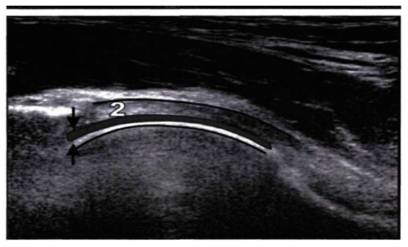 | Figure 1. Sonogram of the structural elements of the hip joint is normal. The arrows in the diagram show the measurement of the thickness of the hypoechoic layer of hyaline cartilage. 2 - fibrous capsule |
3. Research Results
- When analyzing the revealed sonographic and Dopplerographic diagnostic criteria for the stages of the pathological process, in patients of group I, it was revealed that the thickness of the hypoechoic layer of hyaline cartilage decreases from normal values of 2.4 mm to 1.3 mm as the stages of the disease increase in patients of subgroup IA, to 0.9 mm in subgroup IB and thins to 0.8 mm in subgroup I C. At stage III of coxarthrosis, the thickness of the hypoechoic layer of hyaline cartilage is 0.85 mm, and with coxarthrosis formed as a result of aseptic necrosis of the femoral head, cartilage in the necrosis zone is not visualized. In rheumatoid arthritis, there was an increase in the thickness of the fibrous capsule, up to 3.4 mm, in contrast to 2.23 mm with unchanged joints, and an important sign was the identification of normal sizes of hyaline cartilage. At the same time, the sizes of osteophytes increased as the stage of coxarthrosis increased from 4.2 mm to 10.1 mm. Thus, in stage III coxarthrosis, the size of osteophytes was 8.3 mm, and in coxarthrosis formed as a result of aseptic necrosis of the femoral head, it increased to 15.0 mm.All patients with stages I-III of coxarthrosis showed significant differences in the shape of the head of the hip joint, so if at stage I of the disease almost all (96%) of the examined patients had a spherical shape of the head, then at stage II of the disease only in 58% of cases a spherical shape was detected, and in 42% of cases - a moderately flattened shape of the head. At stage III, this sign increased and in 96% of cases it was found to be significantly flattened (Fig. 2).
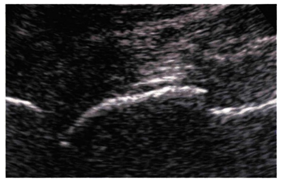 | Figure 2. Sonogram of the hip joint with coxarthrosis (significantly flattened shape of the head) |
4. Discussion
- The thickness of the hyaline cartilage and fibrous capsule, the presence or absence of intra-articular effusion, the shape of the femoral head, the size of osteophytes (if any) are the main structural parameters on which the differential diagnosis of dystrophic and inflammatory diseases of the hip joint is based. In case of an inflammatory lesion of the hip joint, the leading sonographic criteria are: an increase in PVR to numbers - 32.0 cm/s, a CDP to - 10.3 cm/s, and a decrease in IR to numbers - 0.65, combined with an increase in the thickness of the fibrous capsules while maintaining the normal thickness of the hyaline cartilage. And the signs of the presence of coxarthrosis are: a progressive decrease in the rate of blood flow in the lateral circumflex arteries of the thigh and progressive thinning of the hyaline cartilage, the presence of marginal osteophytes, a violation of the shape and contour of the femoral head. Coxarthrosis, formed as a result of aseptic necrosis of the femoral head, is characterized by a significant decrease in the rate of blood flow in the lateral circumflex arteries of the thigh (PRS up to digits - 17.8 cm/s, CDS up to digits - 3.7 cm/s) in combination with early and more complete thinning of the hyaline cartilage, more significant sizes of osteophytes, significant deformity of the femoral head, effusion in the joint cavity.A number of undoubted advantages - non-invasiveness (unlike arthroscopy), accessibility, simplicity, cost-effectiveness (compared to CT and MRI) - provided the method of hip joint ultrasound with a priority among other instrumental methods for examining joints and soft tissues [5]. Ultrasound is highly informative in reflecting small details of the surface of bones, the ligamentous-tendon apparatus, and also allows you to identify and control inflammatory changes in tissues. The advantage of ultrasound over the X-ray method is safety, polypositionality, as well as the possibility of dynamic observation.The most important features of a comprehensive ultrasound examination of the hip joints are: determination of the state of the joint capsule, the presence of effusion in the cavity of the hip joint, assessment of the sphericity and contour of the head, determination of the state of the hyaline cartilage and periarticular tissues, and using the Doppler method, the blood flow in the lateral circumflex arteries of the thigh is assessed, with the calculation of blood flow PSS, blood flow CDS and IR, which is very important in the differentiation of inflammatory and degenerative processes.