American Journal of Medicine and Medical Sciences

p-ISSN: 2165-901X    e-ISSN: 2165-9036

2022;  12(3): 243-250

doi:10.5923/j.ajmms.20221203.02

Received: Feb. 11, 2022; Accepted: Mar. 4, 2022; Published: Mar. 9, 2022


Visual Anatomical Deviations of the External Genitals in Girls. Current State of the Issue (Literature Review)

F. I. Zokirov, B. B. Negmadzhanov

Ministry of Health of the Republic of Uzbekistan, Samarkand State Medical Institute, Department of Obstetrics and Gynecology No. 2, Samarkand, Uzbekistan

Copyright © 2022 The Author(s). Published by Scientific & Academic Publishing.

This work is licensed under the Creative Commons Attribution International License (CC BY).
http://creativecommons.org/licenses/by/4.0/

Abstract

Visual anatomical (phenotypic) deviations of the external genitalia present significant difficulties for both patients and their parents, as well as medical workers who, in connection with their professional duties, advise and provide medical care, since there are many contradictions in management tactics. Visual anatomical (phenotypic) deviations of the external genitalia are any visible to the eye - ad oculus deviations from the norm of the structure of the external genitalia, such as the degree of development of the clitoris, the structure of the large and small labia, the type of introitus, the location of the external opening of the urethra.

Keywords: He location of the external opening of the urethra

Cite this paper: F. I. Zokirov, B. B. Negmadzhanov, Visual Anatomical Deviations of the External Genitals in Girls. Current State of the Issue (Literature Review), American Journal of Medicine and Medical Sciences, Vol. 12 No. 3, 2022, pp. 243-250. doi: 10.5923/j.ajmms.20221203.02.

Article Outline

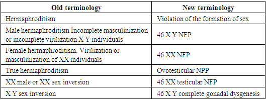
Classification of anomalies of the female genital organs: in surveys, the classification of diseases of the reproductive system is still discussed at numerous forums, congresses. But unfortunately, there is still no consensus on this matter, and various sources or authors’ own systematizations are still used in scientific research and practice, which do not always cover the entire spectrum of causes [2].
Given the above, professors A.B. Okulov and B.B. Negmadzhanov (2000) [2] systematized lesions of the reproductive system requiring surgical treatment in accordance with critical remarks and modern requirements for pediatric surgical care. A distinctive feature of this classification is the coverage of diseases of the reproductive organs of both girls and boys.
According to this systematization, lesions of the reproductive system are divided into 2 large groups of congenital diseases and acquired diseases. Below is an abbreviated form of this systematization.
Classification of surgical diseases of the reproductive system [2].
1. Congenital Diseases
1.1. Complex (“classic”) disorders of sexual differentiation
1.1.1. Gonad forms
1.1.1.1. Female type of external genitalia
1.1.1.2. Mixed (gender indefinite) type of external genitalia
1.1.1.3. Male type of external genital organs
1.1.2. Extragonadal forms (due to a genetic defect in androgen reception or high levels of extragonadal androgens)
1.1.2.1. Female type of external genitalia
1.1.2.2. Mixed (gender indefinite) type of external genitalia
1.1.3. Rare forms of disorders of sexual differentiation
1.1.3.1. Female type of external genitalia
1.1.3.2. Mixed (gender indefinite) type of external genitalia
1.1.3.3. Male type of external genitalia
1.2. Small (“non-classical”) forms of violation of sexual differentiation
1.2.1. Female type of external genitalia
1.2.2. Mixed (gender indefinite) type of external genitalia
1.2.3. Male type of external genital organs
1.2.4. Breast lesions
1.2.4.1. Female phenotype
1.2.4.2. Male phenotype
2. Acquired Diseases (postnatal impairment of sexual differentiation)
2.1. Gonadal forms
2.1.1. Female type of external genitalia
2.1.2. Male type of external genital organs
2.2. Extragonadal forms
2.2.1. Female type of external genitalia
2.2.2. Male type of external genital organs
2.3. Rare forms
2.3.1. Female and male phenotypes
2.4. Changes in the external genitalia due to previous diseases and injuries
2.4.1. Female type of external genitalia
2.4.2. Male type of external genital organs
2.5. Iatrogenic lesions of the organs of the reproductive system
2.5.1. Female phenotype
2.5.2. Male phenotype
2.6. Acute diseases of the reproductive organs
2.6.1. Female phenotype
2.6.2. Male phenotype
As can be seen, the above classification deals with disorders associated with the urogenital sinus. Separately from the above, anomalies in the development of internal genital organs in women, the so-called. anomalies in the development of derivatives of the Mullerian ducts. The Müllerian ducts, being the primordial rudiments of the female reproductive tract, differentiate to form the fallopian tubes, uterus, cervix, and upper aspects of the vagina.
To systematize the anomalies of this group, the classification of the American Society for Reproductive Medicine (formerly the American Fertility Society) is most widely used, based on the classification of V. Buttram and W. Gibbons ( Buttram V. 1983) [9], which combined the degree of developmental disorders with clinical manifestations. This ASRM classification includes 7 classes, and is given below [46]:
Class I. _ Partial (hypoplasia) or complete agenesis of the Müllerian ducts:
I.A. _ – vaginal
I.B. _ - cervical
I.V. _ - uterine
I.G. _ - pipe
I.D. _ - combined
Class II. Unicorn uterus:
II.A. - with a rudimentary horn with an endometrial cavity:
II.A.1. - communicating with the second horn
II.A.1. - not communicating with the second horn
II.B. - with a rudimentary horn without an endometrial cavity
II.B. - without rudimentary horn
Class III. Doubling uterus:
Class IV. Bicornuate uterus:
IV.A. _ – with full separation (with two cervixes)
IV.B. – with partial separation (with one cervix)
Class V. _ Uterus with septum:
V.A. _ - full baffle
V.B. _ - incomplete partition
Class VI. Arcuate (saddle) uterus
Class VII. Uterus with intracavitary changes (T-shaped, strands in the uterine cavity, expansion of the lower three-quarters of the uterus - the consequences of intrauterine exposure to diethylstilbestrol, turinal).
Along with the above, there are also so-called small forms of lesions of the organs of the reproductive system, which are not included in the above classifications. These include atresia of the hymen, synechia of small genitals.
Atresia of the hymen
Hymen serves as the boundary between the internal and external genitalia in the female. The hymen itself develops as a result of the proliferation of synovaginal bulbs and the vaginal plate (urogenital tubercle), the perforation of which occurs before or immediately after birth. Atresia of the hymen is observed when the complete sewage of this "plate" is disrupted and is the extreme degree of various disorders of the sewage process, such as annular narrowing (hymen anularis), cloisonne hymen (hymen septus), trellised (hymen cribriformis), high hymen (hymen altus) as well as differences in the rigidity and/or elasticity of the hymen tissue [2,22].
The etiological factors leading to atresia of the hymen are not known today, and according to some authors, it occurs sporadically. But in the literature there are data on family cases, and often it occurs together with other anomalies of the genitourinary system [17,43].
Epidemiology: Being the most common and distal form of obstructive pathology of the female genital organs, ADP, according to different authors, occurs with a frequency of 1 per 10,000 to 1 per 1,000 people [22].
Diagnosis: It is not uncommon for neonates with hymenal atresia to develop mucocolpos shortly after birth under the influence of maternal estrogen.
Diagnosis of mucocolpos is not difficult, and during a routine gynecological examination in the genital area, a bulging of the stretched hymen can be detected. A whitish turbid mass shines through it [2]. Despite the fact that mucocolpos is a benign congenital pathology, late diagnosis can lead to severe morbidity and additional medical interventions [39]. Thus, based on the foregoing, we can say that today, along with gynecological and / or recto -abdominal examination, ultrasound of the pelvic organs is the only method for diagnosing mucocolpos, in which a formation in the small pelvis can be detected.
Clinic: due to bursting, a formation that causes severe pain, children are often restless, poorly fed, they have frequent painful urination. In severe cases, the mucocolpos becomes so large that it may give the impression of a small pelvis. In 37-60% of these patients, urinary retention and constipation are observed. In some, urinary retention is so severe that bilateral hydronephrosis can occur, leading to acute kidney injury and fatal sepsis [29,36].
Treatment: To date, the only treatment for atresia of the hymen is surgical treatment - dissection of the hymen (hymenotomy or hymenectomy). Currently, the classical method is widely used throughout the world using a cruciform incision of the hymen, in which, after evacuation of the contents, 4 edges of the excised hymen are sutured to the vaginal mucosa using absorbable suture material to achieve hemostasis and prevent secondary closure of the hymenal opening [22,38,44].
This method, despite a good surgical outcome, has a huge drawback, since when sewing the edges of the hymen to the vaginal mucosa, the integrity of the hymen is often violated. In addition, when making an incision and suturing, there is a high risk of injury to both the hymenal ring and the urethra, and the risk of re-closure of the hymenal opening is also high. Thus, according to Lee et al. (2019), who conducted a comprehensive systematic review of the literature with the number of patients of 236 people, complications such as re-closure of the hymenal opening, cicatricial stenosis of the vaginal inlet, the need for repeated surgical intervention, etc. were observed in 15 patients (6.6%), while a satisfactory outcome was observed only in almost 60% of patients. In 75 patients (31.8%), the treatment outcome was unknown [29].
Taking into account the fact that an intact hymen is a sign of virginity, this disadvantage is unacceptable in many social cultures and religions, including in Uzbekistan. Therefore, to date, there are several descriptions of the technique of surgical treatment of hymenal atresia with preservation of the integrity of the hymen in the literature. For example, Acar Ali et al (2007) in their work described the method of surgical treatment of 65 patients with preservation of the integrity of the hymen. After the central puncture of the hymen and expansion, they evacuated the contents and, to prevent re-fusion, inserted a Foley catheter into the incision and inflated the cuff to 10 cm3. The catheter was left in place for 2 weeks and an estrogen-containing ointment was applied to the hymen for 2 weeks. A single dose of antibiotics was given prophylactically. According to the authors, the result was satisfactory in 100% of cases, the hymen remained open and intact [3]. Unlike Acar Ali, Temizkan O. _ et al (2012), use the method of excision of the central part of the hymen without the use of a Foley catheter and estrogen -containing ointments. Although they used this method in only 2 cases, they report satisfactory postoperative results [45].
C deserves interest. Cetin et al., 2016, in which 15 patients with hymen atresia were operated on by circular excision of the central part of the hymen using an electrocoagulator. The results according to the authors were satisfactory in 100% of cases. No complications or relapses were observed [11]. Also found in the literature is the work of M. Basaran et al. (2009), where they describe 2 cases of treatment of hymen atresia complicated by hematocolpos with a vertical incision in the hymen and multiple sutures on the edges of the hymen to prevent reoccurrence. The authors assure the integrity of the hymen after such treatment and recommend surgical treatment as a hymen-preserving method [6].
To date, in addition to the studies described above, there are also many modifications of surgical techniques in the literature using laser or electrosurgery or traditional methods that preserve the integrity of the hymen.
Fusion of the labia minora
Unions of the labia minora or the so-called synechia are fibrous fusion of the labia minora, which completely or partially closes the entrance to the vagina, and in severe cases to the urethra. Typically, fusion begins at the posterior commissure and continues anteriorly to the clitoris.
Etiology: The cause of the appearance of synechia is considered to be a hypoestrogenic state, therefore this pathology does not occur after the onset of menarche, when the synthesis of sex hormones (estrogens) begins. Also in newborns, due to the intake of maternal estrogen with breast milk, this pathology does not occur. However, a study by Ca ğ lar M. _ K. _ et al. in 2007, showed that there are no statistically significant differences in the level of estradiol in children with synechia and children from the control group [10]. Also, according to a number of authors, inflammatory processes (vulvovaginitis), mechanical irritation and tissue trauma (use of diapers, synthetic underwear, hematomas and injuries of the external genitalia) play an important role in the development of synechia [31]. All this is apparently due to the fact that, in a state of hypoestrogenism, inflammatory processes proceed faster and without any protective mechanisms of the body. There is hyperactivation of macrophages, which are responsible for the destruction of cells at the site of inflammation. In response, the body tries to restore damaged epithelial tissues - collagen production is underway.
Epidemiology: According to the literature, synechia of the labia minora occurs in 1.8% of girls, with a peak incidence at the age of 13-23 months [31]. And according to Dowlut - McElroy T et al (2019), can occur with a frequency of up to 22% in prepubertal girls, as many cases go unnoticed due to the absence of symptoms [13].
Diagnosis: According to the literature, in more than 35% of cases of synechia remain asymptomatic [33,42] and the discovery of this pathology by chance by parents during hygienic procedures entails great psychological discomfort in the family. Typically, such patients are subjected to many unnecessary examinations and wandering around doctors, which in turn add to the psychological burden. Synechia can be easily diagnosed during routine examination. In children under 18 months of age, examination is best done in the frog-leg position, while in children over 18 months in the dorsal lithotomy position. Usually the fusion starts from the posterior commissure and goes anteriorly towards the clitoris for some distance. In severe cases, synechia completely block the entrance to the vagina and urethra. There is no need for special research methods for the diagnosis of synechia [28].
Clinic: As it was said in 35% of cases of synechia are asymptomatic [33,42]. When synechia completely block the entrance to the vagina and sometimes the urethra begins to flow into the vagina, symptoms of urinary obstruction appear, leakage of urine after the act of urination, the so-called post- urethral leakage of urine or vaginal urination, in severe cases up to acute urinary retention and dysuric symptoms [42]. In 20-40% of patients, there are symptoms of urinary tract infections, hematuria, symptoms of vulvovaginitis due to urinary retention [28,33].
Treatment: Some authors recommend expectant management until symptom onset [5], while others recommend immediate treatment to prevent symptom onset [33,42]. To date, the method of choice for the treatment of synechia in asymptomatic course is the use of estrogen -containing ointments. Many authors recommend using the surgical manual method of treatment if conservative therapy is ineffective and/or if there are dense adhesions, if synechia is symptomatic, and if parents refuse conservative therapy [4,15,33,42].
According to various authors, the effectiveness of local estrogen -containing is 15-100% [4,27,33,42], and the recurrence rate varies within 11-41% [15,33,42,47]. And with manual treatment, the recurrence rate is 15% [33,42], i.e. and with conservative and surgical tactics, relapses are observed. With each relapse, the adhesions become even more dense and extended.
The main disadvantage of manual separation of adhesions is the painfulness of the procedure, so it is recommended to perform under general anesthesia [33]. The disadvantages and side effects of local estrogen -containing ointments are local irritant effect, vulvar hyperpigmentation, engorgement and enlargement of the mammary glands, and often spotting from the genital tract [13,28].
Recently, there have been reports in the literature about the use of betamethasone -containing ointments as an alternative method of treatment. The authors report no side effects, satisfactory results up to 90% and, of course, the availability of the latter in terms of the financial capabilities of patients [16,32,35].
Studies using betamethasone -containing ointments in the treatment of synechia are encouraging, but studies published in the literature are insufficient and require further study.
Sex formation disorders
Sex formation disorders, formerly known as hermaphroditism, intersex conditions, bisexuality, are a group of congenital diseases characterized by a clinical and biochemical discrepancy between genetic, gonadal, and phenotypic sex [30].
In 2006, at an international conference on disorders of sex formation, organized by the Lawson Society for Pediatric Endocrinology Wilkins, together with the European Society of Pediatric Endocrinologists, adopted a consensus to revise the terminology and nomenclature of hermaphroditism. It was proposed to replace both the term hermaphroditism itself, which sounds offensive to patients, and the indication of gender in the diagnosis, i.e. male or female hermaphroditism. The use of the term " Disorders of Sex Development " (D S D), in the Russian version "disorders of sex development" (NFP) is recommended. Thus. NFP is a congenital condition caused by a chromosomal, gonadal, or somatic disorder of sex formation [14,30].
Proposed new nomenclature
     
Androgen resistance syndrome
Androgen resistance syndrome (ARS), formerly known as testicular feminization syndrome, is an X-linked recessive genetic disease in which the normal masculinization of the external genitalia in a genetically male sex is impaired due to a defect in androgen receptors. Depending on the number of defective receptors, CRA can be complete or incomplete [34]. In the complete form of SRA, individuals have a female external genitalia, normal labia, clitoris, and introitus [21,37].
And with an incomplete form of androgen resistance syndrome (AFSRA), the phenotype of the external genitalia can vary from slightly virilized female genitalia (clitoromegaly without any other anomalies) - Lubs syndrome and insufficiently virilized male genitalia - Hilbert-Dreyfus syndrome to mild forms of undervirilized male genitalia (hypospadias, limited size of the penis) - Reifenstein 's syndrome.
In both cases, patients have normal testicles with normal testosterone production and conversion to dihydrotestosterone. Since they have a normal synthesis of AMH in patients, there are no tubes, uterus, proximal part of the vagina.
Etiology: CPA develops as a result of a mutation in the AR gene (androgen receptor gene), which is localized on the long arm of the X chromosome (Xq11-13). More than 1000 such mutations have been described, including complete or partial deletion of the gene, point mutations, etc. These mutations lead to various functional defects, such as the complete loss of receptors on the cell surface, due to incomplete protein synthesis, and impaired substrate binding properties. Violation of the binding properties of substrates is manifested by the loss of signal transmission, despite the normal number of receptors on the cell surface [19,23]. Those. when cells lose the function of androgen reception, despite normal levels of androgen production, post-receptor phenomena, manifested by the effects of androgens on cells and / or tissues, do not occur. It promotes prenatal undervirilization of the external genitalia, absence of pubic and axillary hair growth, absence of acne and voice changes in puberty.
Epidemiology: according to the most According to available data in the literature, the incidence of SRA is 1 in 20,400 male live births. This statistic is based on an analysis of Danish patient registration data, which included only hospitalized cases. Therefore, the real frequency can be much higher [7].
Clinical: Almost all cases of SRA are diagnosed at birth. In the complete form, unilateral or bilateral masses are more often found in the thickness of the labia majora or in the inguinal canal, with or without hernial contents, which are identified as testicles after surgical treatment [41].
In the case of NFSRA, various abnormalities in the structure of the external genitalia are found at birth, from ambivalent to almost normal male. It depends on the number of modified receptors. With the onset of puberty, sparse hair growth and progressive clitoral enlargement are found, along with other signs of masculinization.
Insufficiency of virilization of children with a genetically determined male sex is classified in accordance with the data given in Table 1 [40].
Table 1
     
Diagnosis: Diagnosis of NFSRA is not difficult. At birth or later, abnormalities in the structure of the external genitalia are found. On examination, be sure to palpate the labia majora to detect the formation (testicles) in them. An obligatory research method is ultrasound of the pelvic organs, where the absence of internal female genital organs is detected. Sometimes you can find the testicles of the fetus in the inguinal canal. The final diagnosis is made after a genetic study - determining the karyotype. Identification of the 46 XY karyotype confirms the diagnosis. [2].
Treatment: The treatment of patients with 46 XY NPP in which ambivalence of the external genitalia is observed presents significant difficulties to both parents of patients and doctors, who, by virtue of their duties, must advise and provide medical assistance. On both an individual and social basis, there are many disagreements and uncertainties, as well as a sense of insecurity and sometimes guilt in the treatment of such patients. Because after growing up, many patients may not agree with their assigned gender. Given the fact that over time, the vision of certain medical problems in the future may change, i.e. what was considered correct many years ago, today can be assessed as incorrect, and sometimes harmful [25]. Those. what is considered medically, ethically and legally correct in one society and culture may be considered wrong in another society and culture.
The difficulties of gender identification in NFSRA are especially evident. In this case, as mentioned above, with a genetic, gonadal and hormonal male field at birth, the female somatic sex is determined by the external genitalia, thereby predetermining the development of socio-psychological determinants. The gap between the physical and socio-psychological parameters of sex can also manifest itself in a longer period of time. An example is the formation of a complex of transsexualism, when the structure and functions of the gonads and genital organs correspond to the same sex, and the developing psychosexual self -identification is the opposite [2].
Therefore, today there are many disagreements about the method of surgical treatment (assigning the gender of upbringing), the timing of operations in such patients. And the available data are based on only a few published cases in practice and are mainly the opinion of individual experts in the field.
Feminizing plastic surgery of the external genitalia: the goal of feminizing operations in patients with an ambivalent structure of the external genitalia is to give the external genitalia a characteristic female appearance and functionality. Feminizing surgeries include resection or reduction of the hypertrophied clitoris/ micropenis, vulvoplasty (creation of the labia minora and labia majora), and creation of a neovagina [2].
Surgical correction of the appearance of the external genitalia in patients with disorders of sex formation is an important stage in their complex treatment.
In patients with 46 XY disorders of sex formation, feminizing operations are performed in 2 stages:
Stage 1 - feminizing plastic of the external genitalia, resection of the cavernous bodies and vulvoplasty, the latter includes the formation of the clitoris and the creation of the labia minora from the skin case of the resected cavernous bodies [2]. When the testicles are found in the thickness of the labia majora, their removal.
The 2nd stage is the creation of a neovagina and plastic surgery of the mammary glands for the implementation of a full-fledged sexual function [2].
The most important point of the 1st stage of feminizing plastic surgery is, if possible, early correction of the appearance of the external genitalia to ensure normal psychosexual adaptation of the patient. Then, before the onset of puberty, if possible, any gynecological interventions should be avoided so as not to create uncertainty in the structure of the genital organs in patients and psychological discomfort in relatives.
Assignment of the gender of upbringing: the issue of assigning the gender of upbringing should be decided by a multidisciplinary team (surgeon, endocrinologist, psychologist, urologist, gynecologist) with experience in this field and with the obligatory participation of the patient's parents. At the same time, it is necessary to take into account such points as sociocultural and religious factors, possible psychosexual outcomes after growing up, the possibilities of medicine in a given society (the presence of an experienced surgeon in the field of feminizing / masculinizing interventions), as well as the patient's fertility potential in the future [2].
Of course, many (especially in the West) understand and approve of those who stand up for tolerance in society, for ensuring that a person is treated with respect, with tolerance, regardless of origin, skin color, race and gender. Unfortunately, we do not see such tolerance either in our society or in other sociocultural environments. It is enough for a child to have a slight deviation from the norm (short stature, obesity, poor academic performance, high giftedness, bodily defect) to be singled out in the school community and in leisure groups. Children with indeterminate gender in puberty are especially traumatized. Therefore, by doing nothing, the doctor does not relieve himself of responsibility for the fate of such children.
Data on the study of long-term psychosexual outcomes of sex assignment in patients with 46 XY DFP are scarce. To date, there is no consensus on the assignment of one or another gender with 46 XY NFP. Many authors recommend assigning both female and male sex [20,24]. So according to Wisniewski A. _ b. et al. (2000) only 23% (9 out of 54) of patients were dissatisfied with the assigned sex, moreover, gender dysphoria was observed in equal proportions in both women and men [48]. It is important to emphasize the fact that patients with NFSRA often have oligospermia and, due to the risk of malignancy, it is necessary to remove both testicles.
Since the long-term results of such operations have not yet been studied, the attitude towards them should be restrained. Based on the foregoing, the volume of surgical correction of the external genital organs in children should be minimal [1,2].
Age of surgical interventions: to date, there is no consensus regarding the timing of surgical interventions in patients with 46 XY NFP. Many surgeons recommend surgery within the first 6 months after birth [2,26]. During this period, the tissues of the newborn under the influence of maternal estrogens still remain plastic and the healing processes proceed better. All this will contribute to fewer possible repeated surgical interventions. And most importantly, the normal structure of the external genitalia in the future will contribute to normal psychosexual adaptation.
In addition, there is another view that is especially popular in the West and in the USA, which states that any surgical intervention without the consent of the patient himself (not parents or guardians) infringes on ethical standards [12]. Supporters of this approach, in particular members of the "working group against violence in pediatrics and gynecology," advocate the rejection of gender as a feature that characterizes the personality, and any surgical intervention in children, since children are not yet able to express their attitude to this issue. In their opinion, this infringes on their rights and ethical standards. Therefore, they recommend that any surgical intervention be carried out with the consent of the patient himself after he reaches the age of majority. But modern medicine today cannot provide enough evidence in favor of such an attitude.
Operation technique: today there are many surgical techniques of feminizing plastics, the main stage of which is micropenis reduction. At an international conference on disorders of sex formation, organized by the Lawson Society for Pediatric Endocrinology Wilkins, together with the European Society of Pediatric Endocrinologists in 2006, adopted a consensus according to which, in surgical interventions on the genitals of patients with NFP, give preference not only to cosmetic, but also to functional results, while maintaining sensitive areas so that patients can live a full sexual life. To date, one of the popular techniques with the preservation of sensitive zones is the technique proposed by Goodwin and later modified by others, including Okulov A.B. and Negmadzhanov B.B. (2000) [2,18]. The essence of this method lies in the resection of the corpora cavernosa with the preservation of a part of the head on the ventral surface, namely its most sensitive part - the navicular fossa. The method is characterized by relatively simple technique, good cosmetic and functional results, and fewer complications.

References

[1]  Wolf A.S., Mittag Yu.E. Atlas of Pediatric and Adolescent Gynecology: Per. with him. /According to ed. V.I Kulakova.- M.: GEOTAR-MED, 2004. -304 p.: Ill.
[2]  Okulov A.B., Negmadzhanov B.B. Surgical diseases of the reproductive system and sex transformation operations - M.: Medicine. - 2000. - p. 72-95.
[3]  Acar A, Balci O, Karatayli R, Capar M, Colakoglu M. The treatment of 65 women with imperforate hymen by a central incision and application of Foley catheter. BJOG 2007; 114: 1376–1379.
[4]  Acer T, Otgun I, Ozturk O, Kocabas T, Tezcan AY, Cirak A, et al. Do hygienic factors affect labial fusion recurrence? A search for possible related etiologic factors. J Pediatric Surg. 2012; 47: 1913-8.
[5]  Bacon JL. Prepubertal labial adhesions: evaluation of a referral population. Am J Obstet Gynecol. 2002 Aug; 187(2): 327-31; discussion 332. doi: 10.1067/mob.2002.126201. PMID: 12193920.
[6]  Basaran M, Usal D, Aydemir C. Hymen sparing surgery for imperforate hymen: case reports and review of literature. J Pediatrician Adolesc Gynecol. 2009 Aug; 22(4): e 61-4. doi: 10.1016/j.jpag.2008.03.009. PMID: 19646660.
[7]  Berglund A, Johannsen TH, Stochholm K, Viuff MH, Fedder J, Main KM, et al. Incidence, Prevalence, Diagnostic Delay, and Clinical Presentation of Female 46, XY Disorders of Sex Development. J Clin Endocrinol metab. Dec 2016 101(12): 4532-4540.)
[8]  Braverman, Paula & Breech, Lesley. (2010). Clinical Report-Gynecologic Examination for Adolescents in the Pediatric Office Setting. Pediatrics. 126. 583-90. 10.1542/peds.2010-1564.
[9]  Buttram VC Jr, Gibbons WE. Müllerian anomalies: a proposed classification. (An analysis of 144 cases). fertile Steril. 1979 Jul. 32(1): 40-6.
[10]  Caglar MK. Serum estradiol levels in infants with and without labial adhesions: the role of estrogen in the etiology and treatment. pediatrician Dermatol. 2007 Jul-Aug. 24(4): 373-5.
[11]  Cetin, C., Soysal, C. and Khatib, G. (2016) Annular Hymenotomy for Imperforate Hymen. Journal of Obstetrics and Gynecology Research, 42, 1013-1015. https://doi.org/10.1111/jog.13010.
[12]  Diamond M. Pediatric management of ambiguous and traumatized genitalia. J Urol 1999; 162: 1021-8.
[13]  Dowlut- McElroy T, Higgins J, Williams KB, Strickland JL. Treatment of Prepubertal Labial Adhesions: A Randomized Controlled Trial. J Pediatrician Adolesc Gynecol. 2019 Jun; 32(3): 259-263. doi: 10.1016/j.jpag.2018.10.006. Epub 2018 Oct 29. PMID: 30385397.
[14]  Dreger AD, Chase C, Sousa A, Grupposo PA, Frader J. Changing the nomenclature/taxonomy for intersex: a scientific and clinical rationale. J Pediatrician Endocrinol metab. 2005; 18: 729–733.
[15]  Eroglu E, Yip M, Oktar T, Kayiran SM, Mocan H. How should we treat prepubertal labial adhesions? Retrospective comparison of topical treatments: estrogen only, betamethasone only, and combination of estrogen and betamethasone. J Pediatrician Adolesc Gynecol. 2011; 24: 389–91.
[16]  Ertürk N. Comparison of estrogen and betamethasone in the topical treatment of labial adhesions in prepubertal girls. Turk J Med Sci. 2014; 44(6): 1103-7. doi: 10.3906/sag-1312-3. PMID: 25552168.
[17]  Eskandar, O. and Wheble, A. (2004) Imperforate Hymen in Two Sisters Associated with Renal Angiomyolipoma and Duplex Kidney. Journal of Obstetrics and Gynecology, 24, 922-993. https://doi.org/10.1080/01443610400019054).
[18]  Goodwin W.E. Surgical revision of the enlarged clitoris. In: de la Camp HB, Linder F, Trede M, editors. American College of Surgeons and Deutsche Gesellschaft fur Chirurgie. joint meeting. New York: Srpinger-Verlag (1969).
[19]  Gottlieb B, Beitel LK, Nadarajah A, Paliouras M, Trifiro M. The androgen receptor gene mutations database: 2012 update. Hum Mutat. May 2012 33(5): 887-94. [Medline].
[20]  Hooper HT, Figueiredo BC, Pavan-Senn CC, de Lacerda L, Sandrini R, Mengarelli JK, et al. Concordance of phenotypic expression and gender identity in a large kindred with a mutation in the androgen receptor. Clin Genet. 2004; 65: 183 ---90.
[21]  Hughes IA, Davies JD, Bunch TI, Pasterski V, Mastroyannopoulou K, MacDougall J. Androgen insensitivity syndrome. Lancet. 2012 Oct 20. 380(9851): 1419-28.
[22]  Imperforate Hymen. Paula J Adams Hillard, 2016. https://emedicine.medscape.com/article/269050-overview#a8.
[23]  Jääskeläinen J. Molecular biology of androgen insensitivity. Mol Cell Endocrinol. 2012 Apr 16. 352(1-2):4-12. [Medline].
[24]  Jurgensen M, Kleinemeier E, Lux A, Steensma TD, Cohen- Kettenis PT, Hiort O, et al., DSD Network Working Group. Psychosexual development in children with disorder of sex development (DSD) --- results from the German Clinical Evaluation Study. J Pediatrician Endocrinol metab. 2010; 23: 565 ---78.
[25]  Kiess W, Penke M, Kratzsch J, Pfaeffle R. Disorders of sex development. J Pediatrician Endocrinol metab. 2017 Jan 1; 30(1): 1-2. doi: 10.1515/jpem-2016-0452. PMID: 28072568.
[26]  Krstić Z, Perovic S, Radmanović S, Nestić S, Smoljanćc Ź, Jevtić P. Surgical treatment of intersex disorders. J Pediatrician Surg 1995; (30): 1273-81.
[27]  Kumetz LM, Quint EH, Fisseha S, Smith YR. Estrogen treatment success in recur-rent and persistent labial agglutination. J Pediatrician Adolesc Gynecol. 2006; 19: 381-4.
[28]  Labial Adhesions Kenneth G Nepple, 2020. https://emedicine.medscape.com/article/953412-overview.
[29]  Lee KH, Hong JS, Jung HJ, Jeong HK, Moon SJ, Park WH, Jeong YM, Song SW, Suk Y, Son MJ, Lim JJ, Shin JI. Imperforate Hymen: A Comprehensive Systematic Review. J Clinic Med. 2019 Jan 7; 8(1): 56. doi: 10.3390/jcm8010056. PMID: 30621064; PMCID: PMC6352236.
[30]  Lee PA, Nordenström A, Houk CP, Ahmed SF, Auchus R, Baratz A, Baratz Dalke K, Liao LM, Lin-Su K, Looijenga LH 3rd, Mazur T, Meyer- Bahlburg HF, Mouriquand P, Quigley CA, Sandberg DE, Vilain E, Witchel S; Global DSD Update Consortium. Global Disorders of Sex Development Update since 2006: Perceptions, Approach and Care. Horm Res Paediatr. 2016; 85(3): 158-80. doi: 10.1159/000442975.
[31]  Leung AK, Robson WL, Tay-Uyboco J. The incidence of labial fusion in children. J Pediatric Child Health. Jun 1993; 29(3): 235-6.
[32]  Mahato, G., Palit, P. and Md Hasanuzzaman, - 2020. To Compare the Outcome of Estrogen and Betamethasone Cream in the Treatment of Labial Adhesion in Pre-pubertal Girls. Bangladesh Journal of Child Health. 43, 3 (Oct. 2020), 161-164. DOI: https://doi.org/10.3329/bjch.v 43i3.49573.
[33]  Mayoglou L, Dulabon L, Martin- Alguacil N, Pfaff D, Schober J. Success of treatment modalities for labial fusion: a retrospective evaluation of topical and surgical treatments. J Pediatrician Adolesc Gynecol. 2009 Aug; 22(4): 247-50. doi: 10.1016/j.jpag.2008.09.003. PMID: 19646671.
[34]  Mongan NP, Tadokoro- Cuccaro R, Bunch T, Hughes IA. Androgen insensitivity syndrome. Best Pract Res Clin Endocrinol metab. 2015 Aug. 29(4): 569-80.
[35]  Myers JB, Sorensen CM, Wisner BP, Furness PD 3rd, Passamaneck M, Koyle MA. Betamethasone cream for the treatment of pre-pubertal labial adhesions. J Pediatrician Adolesc Gynecol. 2006 Dec; 19(6): 407-11. doi: 10.1016/j.jpag.2006.09.005. PMID: 17174831.
[36]  Nazir Z, Rizvi RM, Qureshi RN, Khan ZS, Khan Z. Congenital vaginal obstructions: varied presentation and outcome. pediatrician Surg Int. 2006 Oct. 22(9): 749-53.
[37]  Oakes MB, Eyvazzadeh AD, Quint E, Smith YR. Complete androgen insensitivity syndrome--a review. J Pediatrician Adolesc Gynecol. Dec. 2008 _ 21(6): 305-10.
[38]  Pediatric Imperforate Hymen, Amulya K Saxena and co. 2018. https://emedicine.medscape.com/article/954252-overview/.
[39]  Posner, JC; Spandorfer, PR Early detection of imperforate hymen prevents morbidity from delays in diagnosis. Pediatrics 2005, 115, 1008–1012.
[40]  Sinnecker GHG, Hiort O., Nitsche EM et al. Functional assessment and clinical classification of androgen sensitivity in patients with mutations of the androgen receptor gene. German Collaborative Intersex Study Group // Eur. J. Pediatrician. - 1997. - Vol. 156. - P. 7-14.
[41]  Solari A, Groisman B, Bidondo MP, Cinca C, Alba L. [Complete androgen insensitivity syndrome: diagnosis and clinical characteristics]. Arch Argent Pediatrician. Jun. 2008 106(3): 265-8. [Medline].
[42]  Soyer T. Topical estrogen therapy in labial adhesions in children: therapeutic or prophylactic? J Pediatrician Adolesc Gynecol. 2007; 20: 241-4.
[43]  Stelling JR, Gray MR, Davis AJ, Cowan JM, Reindollar RH. Dominant transmission of imperforate hymen. fertile Steril. Dec. 2000 74(6): 1241-4.
[44]  Te Linde's Operative Gynecology, 10th Edition. John A. Rock, Howard W. Jones. Lippincott Williams & Wilkins. 2008.
[45]  Temizkan O, Kabil Kucur S, Agar S, Gözükara I, Akyol A, Davas I. Virginity sparing surgery for imperforate hymen: report of two cases and review of literature. J Turk Ger Gynecol Assoc 2012; 13: 278-280.
[46]  The American Fertility Society classifications of adnexal adhesions, distal tubal occlusion, tubal occlusion secondary to tubal ligation, tubal pregnancies, müller-ian anomalies and intrauterine adhesions. fertile Steril. 1988 Jun. 49(6): 944-55.
[47]  Wejde, E., Ekmark, AN & Stenström, P. Treatment with oestrogen or manual separation for labial adhesions – initial outcome and long-term follow-up. BMC Pediatr 18, 104 (2018). https://doi.org/10.1186/s12887-018-1018-x.
[48]  Wisniewski AB, Migeon CJ, Meyer- Bahlburg HF, Gearhart JP, Berkovitz GD, Brown TR, et al. Complete androgen insensitivity syndrome: long-term medical, surgical, and psychosexual outcome. J Clin Endocrinol metab. 2000; 85: 2664 ---70.