Achilova Matlyuba Mirkhamzaevna, Bayjanov Allabergan Kadirovich, Yarmukhamedova Nargiza Anvarovna
Samarkand State Medical Institute, Samarkand, Research Institute of Virology, Tashkent, Uzbekistan
Correspondence to: Yarmukhamedova Nargiza Anvarovna, Samarkand State Medical Institute, Samarkand, Research Institute of Virology, Tashkent, Uzbekistan.
| Email: |  |
Copyright © 2022 The Author(s). Published by Scientific & Academic Publishing.
This work is licensed under the Creative Commons Attribution International License (CC BY).
http://creativecommons.org/licenses/by/4.0/

Abstract
This article discusses the clinical and laboratory signs of blastocystosis in HIV-infected patients. The research was carried out in 30 patients who were treated at the Samarkand Regional Infectious Diseases Hospital in January-October 2021. The study used general clinical, parasitological, biochemical, serological, molecular- genetic, instrumental methods. In patients with the gastrointestinal tract: nausea, diarrhea or constipation, pain in the epigastrium, umbilical region and left lateral region were more often determined in the main group than in the control group. One of the clinical manifestations of blastocystosis is allergic symptoms. In the examined patients, urticaria was observed in 1 (7.14%) patient, dermatoses in 2 (14.2%), skin itching in 4 (28.5%) and allergic rhinitis in 1 (7.14%). In laboratory tests, in 12 (85.7%) cases, the number of eosinophil counts ranged from 5 to 8%. In addition, when determining the amount of IgE by the ELISA analysis method to assess the immunological status of patients, it was found that this indicator increases with association of HIV infection with blastocystosis.
Keywords:
HIV infection, Blastocystosis, Immunological status of patients
Cite this paper: Achilova Matlyuba Mirkhamzaevna, Bayjanov Allabergan Kadirovich, Yarmukhamedova Nargiza Anvarovna, Clinical and Laboratory Characteristics of Accompanied with Blastocystis and HIV Infection, American Journal of Medicine and Medical Sciences, Vol. 12 No. 2, 2022, pp. 104-107. doi: 10.5923/j.ajmms.20221202.07.
1. Introduction
HIV infection is a chronic infectious disease of a viral nature, predominantly affecting the immune system, causing the development of opportunistic diseases and tumors and ending in death without antiretroviral therapy [Raimondo M. et al., 2017; Guaraldi G., 2019]. HIV infection remains an actual health problem worldwide today. The disease mainly affects people aged 14-50 years. At the same time, HIV infection is causing great damage to the world economy. With HIV infection, along with a decrease in the activity of the immune system, the central nervous system, respiratory and digestive tract are strongly affected [Pokrovsky V.V., 2013]. From intestinal parasites, Blastocystis is found mainly in cats. The parasite has also been revealed in insects and leeches. Blastocysts enter the human body if hygiene rules are not observed [Prodeus T.V., 2014]. Currently, due to the increase in the number of people with immunodeficiency, the incidence of pathogenic intestinal simple animals Lamblia intestinalis and Entamaeba hystolitica, as well as Blastocystis spp, Cryptosporidium spp and Cyclospora spp has also increased [Bartlett D., 2012; Taylor T.H., 2016].In recent years, there has been growing interest in studying the characteristics of parasitism of Blastocystis hominis in the human body among intestinal parasites. To date, by applying molecular genetic methods in the detection of parasites, it has been found that 9 of 17 subtypes of blastocysts (1-9) are found in humans, 1-3 CTs in humans and animals, the rest mainly in animals [Bugero N.V. et al., 2011, 2012; Rule K.N., 2017]. According to the literature, Blastocystis spp is distributed among 1 billion people around the world (10% of developed countries, 80% of developing countries) [Abdiev T.A., 2013, 2018].It has been reported that blastocystis in people with immunodeficiency is often manifested by impaired intestinal function and allergic manifestations, rarely occurring without clinical symptoms [Balint A., 2014; Tamalee R., 2014; Gavrilyuk T.V., 2015].In the available literature, there is not enough information about the features of the course of HIV infection in intestinal parasitosis. At present, the high incidence of intestinal parasitosis in humans is due to the prevalence of this disease in nature, and a particular problem remains the lack of opportunities for their laboratory detection. The study of clinical and laboratory characteristics of HIV infection in intestinal parasitosis plays an important role in optimizing the treatment of this pathology. The above data indicate the actuality of this research.
2. The Aim of the Research
To study the clinical and laboratory characteristics of blastocyst invasion and HIV infection.
3. Object and Methods of Research
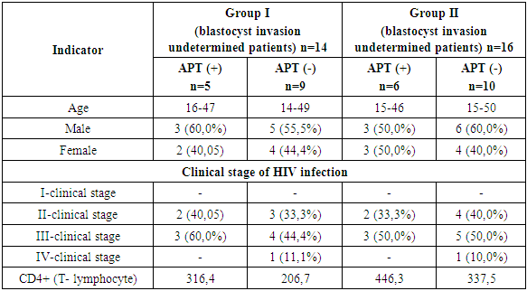
The research work was carried out at the Samarkand Regional Clinical Hospital of Infectious Diseases in 30 patients with HIV who were treated in January-October 2021. The patients were divided into 2 groups: group I (basic) included 14 patients with HIV and blastocyst invasion, and group II (control) consisted of 16 patients with undetermined blastocyst invasion (Table 1).Table 1. General characteristics of the examined patients
 |
| |
|
As can be seen from the table, the groups are suitable for mutual comparison.Group I: (patients diagnosed with blastocyst invasion) 5 (35.7%) of 14 (100%) patients receive antiretroviral therapy (ART) and 9 (64.3%) patients do not receive ART and group II: (patients with blastocyst invasion) 6 (37.5%) of 16 (100%) patients receive ART and 10 (62.5%) patients do not receive ART. According to these indicators, the groups are mutually compatible. The diagnosis of HIV infection in patients was established in accordance with the order of the Ministry of Health of the Republic of Uzbekistan dated April 30, 2018 No. 277 based on the results of enzyme immunoassay and immunoblotting at the Samarkand Regional AIDS Center. The clinical stages of HIV infection were established based on the 2012 revised classification of the World Health Organization. According to this classification, a total of I, II, III and IV clinical stages of HIV infection are distinguished. There were no patients with I-clinical stage in the study groups.Blood serum (plasma) of patients was obtained as the material for the research. The number of СD4 cells (T-lymphocytes) in the blood was determined by cytofluorimetry using Becton Dickinson FACS Calibur cytometry technology. Blastocysts were determined by methods of coproovoscopy (native/thick drop method of Kato and Miur) and formalin ether sedimentation in feces. The formalin ether sedimentation method is based on differences in the specific gravity of helminth eggs. To diagnose blastocyst invasion, the smear microscopy method was used. The smear was colored with Lugol solution by the smear native method. The result was considered positive when more than 5 parasites were determined when the fecal smear was magnified X400 times. Molecular-genetic methods used the method of polymerase chain reaction (PСR) with the use of two pairs of primers for the SSurDNA area. The detection of the blastocysts by both methods was the basis for confirming the presence of blastocyst invasion, and these patients were included in the study group. The results of the research work were collected in Microsoft Excel for statistical processing. Probability of chance (OR) and other biostatic indicators for factors were calculated using computer program EpiInfo 7 and application StatCalc (wwwn.cdc.gov/epiinfo).
4. The Results of Research
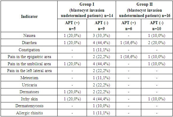
In the group where blastocyst invasion was defined nausea, diarrhea or constipation, pain in the epigastric, umbilical and left lateral area area were more common than in the group where blastocyst invasion was not defined (Table 2).Table 2. Occurrence of clinical indications in examined patients
 |
| |
|
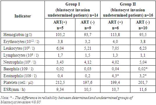
The data in the table above show that nausea, diarrhea, and pain around the umbilicus in gastrointestinal complaints were 2 to 4 times more common in the group where blastocyst invasion was determined than in the group where blastocyst invasion was not determined. It should be noted that in the study groups, subfebrile temperature and eosinophilia rates were mostly observed in people with СD4 + cells (T-lymphocytes) in the blood less than 250 cells/μl. In the group of HIV-infected patients with blastocyst invasion, subfebrile temperature (fever) was determined in 7 (50.0%) cases of 14 (100%) patients and in 3 (18.7%) cases of 16 patients in the group without blastocyst invasion.Eosinophilia indicators were determined in 6 (66.6%) of 9 (100%) patients who were not receiving antiretroviral therapy in the group of HIV-infected patients with blastocyst invasion, and in 3 (60.0%) of 9 (100%) patients who were receiving antiretroviral therapy and blastocyst invasion ranged from 0.5–1% in 2 (20.0%) patients out of 10 (100%) patients who did not receive antiretroviral therapy without blastocyst invasion.The condition of eosinophilia was not determined in patients receiving antiretroviral therapy in the group of HIV-infected patients who did not have blastocyst invasion. The laboratory indicators described above are summarized in the table below (Table 3).Table 3. Indicators of laboratory analysis
 |
| |
|
There were no significant differences between the study groups in terms of general laboratory indicators (except for the number of eosinophils).The difference in eosinophilia rates between groups of HIV-infected patients is clearly seen in the following figure (Fig. 1).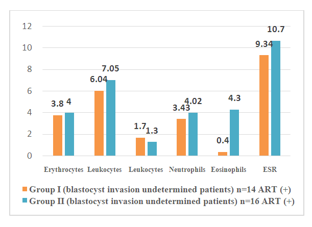 | Figure 1. Laboratory findings in groups with and without blastocyst invasion |
5. Conclusions
When HIV infection is mixed with blastocyst invasion, patients develop more clinical symptoms (nausea, diarrhea, pain around the umbilicus) due to gastrointestinal dysfunction. Allergic symptoms characteristic of blastocyst invasion (urticaria, dermatoses, itchy skin) are more common in the mixed form of the disease. Eosinophilia in patients' blood is one of the indicators of intestinal parasitosis - blastocyst invasion. Information about the source of support in the form of grants, equipment, and drugs. The authors did not receive financial support from manufacturers of medicines and medical equipment.Conflicts of interest: The authors have no conflicts of interest.
References
| [1] | Abdiev T.A., Egamberdiev O.A., Abdiev F.T., Saidakhmedova D.B., Abdusattorov M.M., Vakhobov T.A., Kovalenko D.A., Mahmudova L.B. Clinical guide to helminthiasis. - Tashkent, - 2013. |
| [2] | Abdiev T.A., Saidakhmedova D.B., Suvonkulov U.T., Akhmedova M.D., Saipov F.S., Mahmudova L.B., Abdiev F.T., Vakhobov T.A., Kovalenko D.A., Kachugina L.V., Anvarov J.A. Parasitic human disease in Uzbekistan. - Tashkent, 2018. |
| [3] | Abdurakhmanov D. S., Rakhmanov Q. E., Davlatov S. S. Clinical questions extreme currents syndrome Mirizzi // Electronic innovation bulletin. – 2021. – №. 6. – Р. 37-40. |
| [4] | Bugero N.V., Nemova I.S., Potaturkina-Nesterova N.I. Persistence factors of the simplest fecal flora in intestinal dysbiosis // Bulletin of new medical technologies. - 2011. - T. 18. - No. 3. |
| [5] | Bugero N.V., Potaturkina-Nesterova N.I. The results of determining the virulence of Blastocystis spp. By the method of restriction analysis of protozoan DNA // Fundamental research. - 2012. - No. 11-5. |
| [6] | Bushman F.D. HIV: From biology to prevention and treatment // Cold Spring Harbor, New York, USA: Cold Spring Harbor Laboratory Press. 2012. – P. 572. |
| [7] | Bаlint A. Do not forget the stool examination! - cutaneous and gastrointestinal manifestations of Blastocystis sp. infection // Parasitology research. - 2014. - Т. 113. - №4. - P. 1585-1590. |
| [8] | Coyle C.M. Blastocystis: to treat or not to treat… // Clinical infectious diseases. - 2011. - P. 810. |
| [9] | Davlatov S. S. Application hemoperfusion as the method of homeostasis protection in multiple organ failure syndrome// Collection of scientific works of students and young scientists of the All-Russian scientific-practical conference with international participation. Yaroslavl. - April 24-26. – 2013. - Р. 147. |
| [10] | Erkin M. et al. The challenge of emerging and re-emerging infectious diseases in Uzbekistan: study of rickettsiosis using pcr diagnostic method //European science review. – 2018. – №. 5-6. |
| [11] | Hameed D.M., Hassanin O.M., Zuel-Fakkar N.M. Association of Blastocystis hominis genetic subtypes with urticaria // Parasitology research. - 2011. - Т. 108. - №3. - P. 553-560. |
| [12] | Inoyatova F. I. et al. Features of doppler indices in chronic hepatitis with the transition to liver cirrhosis in children // International Journal of Pharmaceutical Research. – 2020. – Т. 12. – №. 3. – С. 4026-4029. |
| [13] | Inoyatova F. I. et al. Possibilities of modern echography technologies in the diagnostics of chronic viral hepatitises in children //International Journal of Pharmaceutical Research. – 2020. – Т. 12. – №. 3. – С. 4040-4043. |
| [14] | Inoyatova F. I., Yusupalieva G. A. Doppler researches informativeness in diagnosis of chronic viral hepatitises in children // European science review. – 2014. – №. 11-12. – С. 23-25. |
| [15] | Inoyatova F. I., Yusupalieva G. A., Inogamova G. Z. Doppler Examination Informativity in Children with Chronic Viral Hepatitis // Detskie Infekcii (Moskva). – 2015. – Т. 14. – №. 3. – С. 60-64. |
| [16] | Raimondo M., Camoni L., Suligoi B. et al. HIV-positive individuals on ART and with viral load suppressed in 12 Infectious Diseases Clinics in Italy: successes and disparities in the HIV Continuum of Care / AIDS Res Hum Retroviruses. 2017, -Р. 1056-61. |
| [17] | Saag M.S., Chambers H.F., Eliopoulos G.M., Gilbert D.N. The Sanford guide to HIV/AIDS therapy 2012. - Sperryville, VA, USA: Antimicrobial Therapy Inc., 2012. –Р. 214. |
| [18] | Tukhtaeva Kh. Kh., Khamdamov B.Z. Features of the influence of chronic irradiation on bone marrow cells in the experiment // Problems of biology and medicine. Samarkand. - 2021. - No. 6 (132). – P. 200-203. |
| [19] | Yusupalieva G. A. et al. Role of Echography in Diagnosis of Chronic Kidney Disease in Children // Annals of the Romanian Society for Cell Biology. – 2021. – С. 1895-1899. |
| [20] | Yusupalieva G., Vaxidova N., Abzalov A. Informative value of sonography in children with pneumonia // International Journal of Applied and Fundamental Research. – 2016. – №. 6. – С. 2-5. |

 Abstract
 Reference
 Full-Text PDF
 Full-text HTML