American Journal of Medicine and Medical Sciences

p-ISSN: 2165-901X    e-ISSN: 2165-9036

2022;  12(1): 40-45

doi:10.5923/j.ajmms.20221201.08

Received: Jan. 2, 2022; Accepted: Jan. 22, 2022; Published: Jan. 24, 2022


May-Thurner Syndrome Complicated by Deep Vein Thrombosis of Left Lower Extremity: Clinical Study on the Optimal Timing to Perform Stenting

Sardor Kuchkorov1, Tang Li Ming2, Liu Chun Jiang3, Nasritdinov Umid4

1Student of Master's Degree, Shaoxing University, Zhejiang Province, China

2Head of the Vascular and Hernia Department, Shaoxing People’s Hospital, Zhejiang Province, China

3Secretary of the Vascular and Hernia Department, Shaoxing People’s Hospital, Zhejiang Province, China

4Head of the Emergency Surgical Department, 2nd Clinic of the Tashkent Medical Academy, Tashkent, Uzbekistan

Correspondence to: Tang Li Ming, Head of the Vascular and Hernia Department, Shaoxing People’s Hospital, Zhejiang Province, China.

Email:

Copyright © 2022 The Author(s). Published by Scientific & Academic Publishing.

This work is licensed under the Creative Commons Attribution International License (CC BY).
http://creativecommons.org/licenses/by/4.0/

Abstract

May–Thurner syndrome (MTS) is a rare clinical entity featuring venous obstruction of the left lower extremity. The aim of the present study was to report our experience with MTS complicated by deep vein thrombosis (DVT) and to evaluate the utility of tactically new approach, in which balloon angioplasty and stenting precede catheter- directed thrombolysis (CDT) therapy.

Keywords: May–Thurner syndrome, Deep vein thrombosis, Iliac vein stenting, Catheter directed thrombolysis

Cite this paper: Sardor Kuchkorov, Tang Li Ming, Liu Chun Jiang, Nasritdinov Umid, May-Thurner Syndrome Complicated by Deep Vein Thrombosis of Left Lower Extremity: Clinical Study on the Optimal Timing to Perform Stenting, American Journal of Medicine and Medical Sciences, Vol. 12 No. 1, 2022, pp. 40-45. doi: 10.5923/j.ajmms.20221201.08.

1. Introduction

May-Thurner syndrome (MTS) or iliac vein compression syndrome is associated with deep vein thrombosis (DVT) resulting from chronic compression of the left iliac vein against lumbar vertebrae by the overlying right common iliac artery. MTS refers to chronic compression of the left iliac vein against the lumbar spine by the overlying right common iliac artery. The compression may be asymptomatic. The syndrome is a clinical spectrum of physical findings and history plus the lesion. It is characterized by the varying degrees of venous hypertension. This can be non-thrombotic, combined with acute DVT or post-thrombotic. Traditionally, acute DVT was treated with standard anticoagulation and sometimes, thrombectomy. However, these measures do not address the underlying culprit lesion of mechanical compression. Furthermore, if managed only with anticoagulation, patients with residual thrombus are at risk for developing recurrent DVT or post-thrombotic syndrome (PTS). Both retrospective and prospective studies have shown that endovascular management should be the preferred approach to dissolve proximal thrombus and also to treat the underlying compression with endovascular stent placement. We propose a tactically new approach, in which balloon angioplasty and stenting precede catheter-directed thrombolysis (CDT) therapy. The results of this approach are studied and presented in this article.

2. The Main Results and Findings

To investigate the effect of the timing of stent implantation on the curative effect of thrombolytic therapy for acute deep venous thrombosis (DVT) of left lower extremity secondary to May-Thurner syndrome.
We retrospectively analyzed data collected between January 2017 and January 2019 on 26 MTS complicated by DVT patients treated using endovascular techniques. We reviewed patient demographics, symptoms, procedural features, radiological and clinical outcomes. Written informed consent was obtained from each patient after the purpose and the risks of treatment were fully explained.
All patients exhibited radiographic evidence of iliac vein stenosis caused by MTS upon computed tomography (CT) venography or a combination of duplex ultrasound and venography.
Patient selection criteria:
- Ultrasound or angiography-confirmed DVT of the left lower extremity;
- Onset time ≤14 d;
- Luminal stenosis of the left iliac vein >70%;
- No history of cerebral haemorrhage, no history of gastrointestinal and other internal organ bleeding within 1 month.
Technical success was defined as successful restoration of antegrade flow, with <30% residual stenosis or thrombus. Iliac vein patency was defined as uninterrupted flow apparent on Color Doppler Ultrasound (CDU) or venography. Clinical success was defined as improvement in and resolution of symptoms on follow-up visits.
Of our patients, 18 (69.2%) were female and 8 male. Mean patient age was 67.23 ± 7.37 years (range, 18–73 years). All patients were symptomatic. 24 patients presented with acute DVT (16 iliofemoral; eightiliofemoropopliteal); two with subacute DVT (one iliofemoral, one iliofemoropopliteal) chracterized by swelling of and pain in the left lower extremity. Six patients exhibited risk factors for DVT development. These were a previous history of DVT (n = 1); postoperative status (n = 3); postpartum status (n = 1); oral contraceptive (n = 1) and the Factor V Leiden mutation (n = 1). No patient had a symptomatically indicated pulmonary embolism. Onset time was 4 hours~10 days, average 3.65±2.71 days.
An antegrade venography of the lower limbs was performed through the left (affected) dorsal vein of the lower extremity to determine the location, extent, degree of obstruction, and collateral circulation of the thrombus. The right (healthy) femoral vein approach was taken 0.5 to 1.0 cm below the renal vein to place the Aegisy disposable filter (Lifetech Shenzhen Technology Co., Ltd.); the patient was in the prone position, and vascular access was established via ultrasonography-guided puncture.
After establishment of deep venous guidewire access and introduction of a balloon catheter (diameter 8-10 mm) to expand the deep vein thrombosis in sequence andto crush the thrombus. Subsequently, the left iliac vein was found to be severely narrowed after balloon dilation, and the Wallstent (Boston Technology Company, USA) /SMART (Cordis, US) stent with a diameter of 12 to 14 mm and a length of 80 to 100 mm was implanted into iliac vein, expanded after balloon dilatation. After that we inserted thrombolysis catheter (effective perfusion length 20-50 cm, subject to complete coverage of thrombus), connected to a micro pump, and performed continuous infusion of urokinase (dissolved in 100 mL of 0.9% sodium chloride solution) at 60×104 U/24 h, supplemented with low molecular weight heparin sodium (5 000 U/12 h). After the venous lumen is unobstructed and the thrombolysis was stopped, cava filter was removed through the right femoral vein. Subsequent 24-hour angiography was done to review thrombus clearance: no residual filling defects in the popliteal, femoral, iliac, and inferior vena cava, and stop thrombolysis after the lumen is unobstructed. All patients received 3000 units of heparin at the start of each procedure. Heparin was stopped when the international normalized ratio (INR) reached 2. We maintained the activated partial thrombin time (APTT) at a normal value 2~2.5 times, while adjusting the warfarin medication. Regular warfarin anticoagulant therapy was prescribed for at least 6 months. All patients were instructed to wear graduated compression stockings.
The circumference difference (the circumference of crus 10 cm below the knee and the circumference of thigh 15 cm above the knee) was calculated to determine the detumescence rate of affected limb. The detumescence rate of the affected limb was calculated according to the circumference difference of the crus of the affected side and the healthy side and the circumference difference of the thigh of the affected side and the healthy side respectively. The detumescence rate of affected limb = (the circumference difference before treatment-the circumference difference after treatment) / the circumference difference before treatment × 100%. Thrombus clearance rating: Thrombus clearance rate >90% is grade III, 50%~90% is grade II, <50% is grade I, thrombus clearance rate = (pre-treatment score-post-treatment score)/pre-treatment score×100%. We evaluated the veins of the lower extremities (popliteal vein, lower superficial femoral vein, upper superficial femoral vein, common femoral vein, external iliac vein, common iliac vein, inferior vena cava, etc.) according to angiographic images. Complete obstruction is 3 points, subtotal obstruction (50%~99%) is 2 points, partial obstruction (<50%) is 1 point, 0 points for complete patency.
During treatment we focused on observation of symptomatic pulmonary embolism and bleeding complications (exudation, congestion, hematoma at the puncture point, bleeding from the gums or bleeding from other organs).
Follow-up clinical and imaging evaluations were initially performed at 1, 3, 6 and 12 months, and yearly thereafter. All patients had greater than two-year follow-up. Follow-up imaging featured primarily CDU. A few patients were re-imaged using CT or repeat venography because it was difficult to obtain adequate images of the stents using CDU alone.
SPSS 22.0 software was used for data analysis. The counting data were expressed as percentage and compared by chi square test. The measurement data were expressed as mean ± standard deviation (x ± s) and compared by t test. Rank sum test was used for rank data. A P value < 0.05 indicated a significant difference.
Venography performed during endovascular treatment of MTS revealed significant stenosis of the left common iliac vein in 18 of 26 (69.2%) patients. In 8 of the 26 (30.7%) patients, total occlusion of the left common iliac vein (LCIV) was evident. All patients presenting with venous varices exhibited common iliac vein stenosis.
26 cases were implanted with 26 left iliac vein stents (14 mm×100 mm – 7 cases, 14 mm×80 mm - 13 pieces, 14 mm×60 mm - 1 piece, 12 mm×80 mm - 4 pieces, 12 mm×60 mm - 1 piece), the patency rate of the deep vein and iliac vein stent was 100%. No symptomatic pulmonary embolism occurred. The average dosage of thrombolytic drugs (urokinase) was 3.27±1.41million U. Thrombolysis was performed for 43-169 h (mean 78.69 ± 34.33 h) in 26 patients with acute and subacute DVT. After the treatment, the temporary filters of the inferior vena cava were taken out.
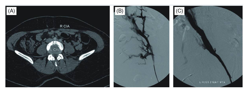
Figure 1. A 44-year-old female who presented with the left lower extremity and inguinal varicosities. (A) A transverse computed tomography image shows the right common iliac artery (single arrow) compressing the left common iliac vein (double arrow). (B) Initial venography revealed significant stenosis of the left common iliac vein with extensive cross-pelvic and hypogastric collateralization. (C) Venography performed after thrombolysis with following stent implantation and angioplasty revealed complete resolution of collateral filling with good flow through the femoral and iliac system into the IVC
Thigh circumference difference improved from preoperative 5.43±1.16 cm to postoperative 1.59±0.84 cm, there is a significant statistical difference (t=18.11, P<0.001). And the calf circumference difference improved from preoperative 3.81±0.81 cm to postoperative 1.26±0.81 cm, there is a significant statistical difference (t=13.50, P<0.001).
The clinical and radiological mean follow-up time was 43.8 ± 7.8 months (range, 32–56 months). Upon follow-up, complete symptomatic regression was observed in 22 of the 26 patients (84.6%). Four patients who could not be completely recanalized to relieve femoral vein thrombosis exhibited moderate levels of symptomatic improvement. Follow-up performed in the first postoperative month showed that leg swelling decreased in DVT patients, and pain was relieved. Leg edema in those with chronic venous hypertension decreased and varicose veins collapsed. 2 patients had minor puncture site bleeding, which was stopped. Follow-up CDU and CT venography revealed patent lumen of the stents (92.3%) in 24 of the 26 patients who received stents. Restenosis (7.6%) of stents occurred in two patients at 6 and 12 months postoperatively. In one of the patients in which restenosis was developed Factor V Leiden mutation which is known to be a risk factor for DVT was present. In the other patient postoperative irregular anticoagulant medicament usage story was present. One patient was treated via repeat balloon dilatation; the symptoms improved. The other patient received no intervention as no significant clinical finding was apparent. There were no complications such as stent fracture or displacement. No patient experienced PE during follow-up. No procedure-related mortality or major complication was noted.
May–Thurner syndrome, also termed the “syndrome of compression of the iliac vein”, is caused by an anatomical anomaly in which the left common iliac vein is compressed by the overlying right common iliac artery. In 1957, May and Thurner described three varieties of intra-luminal spurs occurring in the left common iliac vein close to its junction with the inferior vena cava (IVC). [1] The incidence of May-Thurner syndrome is unknown and ranges from 18%–49% among patients with left-sided lower extremity DVT. [2]
The cited authors concluded that the obstructive lesion, which they termed a “spur”, was analogous to a callus in that formation thereof probably required chronic repetitive irritation, identified to be intermittent compression of the LCIV region lying between the pulsating right common iliac artery (RCIA) and the vertebral body. Chronic vibratory pulsation of the vein may damage the intimal wall, in turn causing intimal proliferation and venous thrombosis. [1,2] Further work showed that patients with venous spurs could remain asymptomatic for extended periods of time because collateral forms of venous drainage developed. [9] Thus, spur formation was considered to be irreversible. Extensive pelvic venous collateral flow circumventing the abnormal LCIV segment is a hallmark of MTS. [9,10] Several anatomical variants of MTS have been described; these include LCIV compression by the left common iliac artery (LCIA); right common iliac vein (RCIV) compression by the right internal iliac artery; and right-sided MTS developing where the LCIA compresses the RCIV. [11,12] Cadaveric studies showed that the prevalence of LCIV compression ranged from 14–32%, [1,13] but was present in 2–5% of patients evaluated after complaining of lower limb venous disorders. [14] This difference between postmortem and symptomatic compression levels clearly shows that LCIV compression is necessary, but not sufficient, to trigger MTS. [15]
In the past, MTS was thought to be rare, but the reported frequency of the condition has risen upon development of catheter-directed endovascular treatment of lower-extremity DVT. The likely reason is that the iliac veins are better visualized when venography is performed using vascular sheaths placed in increasingly central veins. [6]
MTS has been reported to be more frequent in females, as, indeed, we found. No explanation of the female predominance of IVCS has yet been advanced. [11,16]
MTS most frequently presents as chronic venous insufficiency, and DVT, in young-to- middle-aged females, and is diagnosed in 18–49% of cases presenting with left lower limb DVT. [4,5,6,11] The presence of MTS is suggested by a history of recurrent left lower-extremity DVT and symptoms associated with chronic venous hypertension including varicosities; venous ulcers; lower limb swelling; and pain in the inguinal, calf, or upper leg regions. Also, acute thrombosis may develop in patients at high risk of DVT. DVT attributable to IVCS was reported to occur predominantly in young-to-middle-aged females (aged 20–40 years). Pregnancy and postpartum status are known risk factors for DVT. [15] In our present study, 14 were of older age. One patient was of postpartum status and had a history of eclampsia, and two were of early postoperative status, having undergone surgery for removal of lower-extremity venous varices. Also, patients with IVCS are at increased risk of DVT. [15] Significant iliac vein compression may thus be but one risk factor of many favouring DVT development.
Several imaging modalities are useful in MTS diagnosis. CDU is the first-line method used to screen patients with CVI disorders, and can detect DVT. [17,18] However, the technique cannot accurately identify venous spurs or compression of the LCIV. If MTS is suspected in patients subjected to ultrasonography, cross-sectional imaging (CT venography, magnetic resonance angiography) or venography should be used to accurately visualize the pelvic region. [19,20]
Surgical techniques have had mixed success in management of MTS because of high rates of morbidity and variation in patency rates. Surgical options include Palma’s crossover technique; relocation of the RCIA behind the LCIV or inferior vena cava; and vein-patch angioplasty with removal of intraluminal obstructions and encasement of the LCIV in polytetrafluoroethylene (ePTFE) grafts. [21] Jost, et al. (2001) showed that surgical reconstruction techniques were associated with primary and secondary 3-year patency rates of 54% and 62%, respectively. [5,10,21,22]
The surgical techniques used in another study were both diverse and complicated, including multiple treatments of autologous veins. However, despite such diversity, similar primary and secondary 5-year patency rates (42% and 59%, respectively) were reported in a larger patient group. [22]
Today, MTS patients rarely undergo highly invasive venous surgical reconstructions because management using endovascular techniques has been relatively successful, and is associated with fewer operative risks. [4-8]
DVT treatment aims to alleviate acute or chronic symptoms, and to prevent recurrence, PE development, and development of post-thrombotic syndrome. The standard treatment for acute or subacute DVT is anticoagulation therapy, which reduces the rates of recurrence and PE but does not effectively remove clots. [23] However, in MTS cases, such management is unlikely to entirely prevent the development of chronic thrombi caused by venous spurs. [4,10,24] This is because anticoagulation therapy does not directly treat iliac vein compression or the resulting venous spur; these are the most probable underlying causes of MTS.
Treatment of MTS patients who present with thrombosis has changed dramatically over time; CDT has found increasingly wider applications. Catheter-directed endovascular treatment is more effective in terms of clot removal and symptom improvement, compared to anticoagulation therapy. [25] Local infusion of thrombolytic agents at a site of thrombotic occlusion maximizes therapeutic effects while minimizing the risk of major bleeding. Such methods also reduce post-thrombotic morbidity and improve health-related quality-of-life. [26]
PTA procedures lacking subsequent stent placement have been associated with low patency rates. A 73% recurrence rate was noted in patients with acute left-sided iliofemoral DVT when the underlying obstruction was not treated via stent placement. [4] The chronic venous compression occurring in MTS patients cannot be relieved via temporary balloon angioplasty. Stents are effective to treat venous obstructions, and are superior to balloon dilatation alone. [27] Thus, MTS treatment almost always features the placement of stents exhibiting high radial force. [28] Study series featuring iliac vein stenting in MTS patients have increased in number over the past decade and have generally advocated the use of endovascular techniques. [5,6,29] Recently, a retrospective review by Titus, et al. identified MTS patients treated via endovenous stenting and showed that the primary patency rates were 88%, 78%, and 78%, respectively, at 6, 12, and 24 months postoperatively. [30] O’Sullivan, et al. reported that the overall patency rate at 1 year was 79%. [5] Stent placement in iliac veins in combination with thrombus removal affords good short- and mid-term patency especially upon treatment of DVT developing secondarily to MTS. Stent insertion may be important if organic abnormalities are to be treated. [10] The possible problems of valve destruction by stents, with subsequent development of venous insufficiency, does not apply to femoral veins because iliac veins lack valves. It has been reported that venous claudication improved after successful stent recanalization in limbs, despite deterioration of the reflux that normalizes venous outflow. Also, calf muscle pump function was enhanced and significant improvements in clinical outcomes were apparent. [31]
Today, the guidelines of both the Society for Interventional Radiology and the Society of Vascular Surgery recommend iliac vein stenting in the context of external compression. [32]

3. Conclusions

In this study, patients with severe iliac vein stenosis (>70%) were treated according to new tactic: iliac vein stent implantation followed by CDT. Compared with the traditional stent implantation model after CDT, the dosage of urokinasewas significantly reduced, and the indwelling time of thrombolytic catheter was significantly shortened. And there are possible reasons:
1. Implanting a stent to open the iliac vein will help reduce the venous pressure of the lower extremities, which strengthen the diffusion of urokinase from the distal end to the proximal end, and reduce transcollateral reflux;
2. Increase blood flow to make more plasminogen, activated by urokinase, thereby enhancing the thrombolytic effect;
3. Balloon expansion before stent implantation has an auxiliary effect on thrombolysis.
The scientific significance shows that the new model improves the efficiency of thrombolysis while satisfying the curative effect. In addition, the use of the new model has certain advantages:
- The iliac vein stent implantation and CDT can be completed in one operation at the same time, which can reduce the number of consumables such as guide wires, catheters, and balloons;
- The efficiency of thrombolysis is improved, the number of re-examinations and the dosage of thrombolytic drugs are reduced, and the course of the disease is shortened.
The limitations of the present study were the retrospective nature of the work and the limited number of MTS patients evaluated. Follow-up periods and control numbers were not standardized, and follow-up periods were relatively short. Another potential limitation is the variability in stent type and size. No clinical scale was used for symptomatic evaluation; this was a significant limitation.
As a result, recent reports have shown that endovascular treatment of symptomatic MTS patients with thrombotic or nonthrombotic presentations is both feasible and safe, and improves symptoms. On long-term follow-up, stent patency does not seem to be affected by ongoing extrinsic compression of a vein by the adjacent iliac artery. We obtained excellent immediate results and early patency, with minimal complications, using localized CDT, angioplasty, and stent implantation.
The authors declare that they have no conflicts of interest.
MTS – May-Thurner syndrome
DVT – Deep vein thrombosis
LCIV – Left common iliac vein
RCIV – Right common iliac vein
RCIA – Right common iliac artery
IVC – Inferior vena cava
CVI – Chronic venous insufficiency
IVCS – Iliac vein compression syndrome
CDT – Catheter directed thrombolysis
PTS – Post-thrombotic syndrome
CT – Computed tomography
CDU –Color Doppler Ultrasound

References

[1]  May R, Thurner J. The cause of the predominantly sinistral occurrence of thrombosis of the pelvic veins. Angiology 1957; 8: 419-27. [PubMed] [Google Scholar]
[2]  Cockett FB, Thomas ML, Negus D. Iliac vein compression.–Its relation to iliofemoral thrombosis and the post-thrombotic syndrome. BrMed J 1967; 2: 14-9. [PMC free article] [PubMed] [Google Scholar]
[3]  Kibbe MR, Ujiki M, Goodwin AL, et al. Iliac vein compression in an asymptomatic patient population. J VascSurg 2004; 39: 937-43. [PubMed] [Google Scholar]
[4]  Mickley V, Schwagierek R, Rilinger N, et al. Left iliac venous thrombosis caused by venous spur: treatment with thrombectomy and stent implantation. J VascSurg 1998; 28: 492-7. [PubMed] [Google Scholar]
[5]  O’Sullivan GJ, Semba CP, Bittner CA, et al. Endovascular management of iliac vein compression (May-Thurner) syndrome. J VascIntervRadiol 2000; 11: 823-36. [PubMed] [Google Scholar]
[6]  Oguzkurt L, Tercan F, Ozkan U, et al. Iliac vein compression syndrome: outcome of endovascular treatment with long-term follow-up. Eur J Radiol. 2008; 68: 487-92. [PubMed] [Google Scholar]
[7]  Hager ES, Yuo T, Tahara R, et al. Outcomes of endovascular intervention for May-Thurner syndrome. Journal of Vascular Surgery: Venous and Lymphatic Disorders, Volume 1, Issue 3, Pages 270-275. [PubMed] [Google Scholar]
[8]  Heijmen RH, Bollen TL, Duyndam DAC, et al. Endovascular venous stenting in May-Thurner syndrome. J CardiovascSurg 2001; 42: 83-7. [PubMed] [Google Scholar]
[9]  Cockett FB, Thomas ML, Negus D. Iliac vein compression.– Its relation to iliofemoral thrombosis and the post-thrombotic syndrome. Br Med J 1967; 2: 14-9. [PMC free article] [PubMed] [Google Scholar]
[10]  Patel NH, Stookey KR, Ketcham DB, et al. Endovascular management of acute extensive iliofemoral deep venous thrombosis caused by May-Thurner syndrome. J VascInterv Radiol 2000; 11: 1297-302. [PubMed] [Google Scholar]
[11]  Hassell DR, Reifsteck JE, Harshfield DL, et al. Unilateral left leg edema: a variation of the May-Thurner syndrome. CardiovascInterventRadiol 1987; 10: 89-91. [PubMed] [Google Scholar]
[12]  Abboud G, Midulla M, Lions C, et al. “Right-sided” May-Thurner syndrome. CardiovascInterventRadiol. 2010; 33: 1056-9. [PubMed] [Google Scholar]
[13]  Ehrich WE, Krumbhaar EB. A frequent obstructive anomaly of the mouth of the left common iliac vein. American Heart Journal 1943; 26: 18-31. [Google Scholar]
[14]  Posner C, Owen M, Melhem N, et al. An acutely swollen leg. Clin Med 2010; 10: 484-6. [PMC free article] [PubMed] [Google Scholar]
[15]  National Institute of Health. Prevention of venous thrombosis and pulmonary embolism. NIH Consensus Development. JAMA 1986; 256: 744-9. [PubMed] [Google Scholar]
[16]  Brazeau NF, Harvey HB, Pinto EG, et al. May-Thurner syndrome: diagnosis and management. Vasa. 2013; 42: 96-105. [PubMed] [Google Scholar]
[17]  Ahmed HK, Hagspiel KD. Intravascular ultrasonographic findings in May-Thurner syndrome (iliac vein compression syndrome). J UltrasoundMed 2001; 20: 251-6. [PubMed] [Google Scholar]
[18]  Chung JW, Yoon CJ, Jung SI, et al. Acute iliofemoral deep vein thrombosis: evaluation of underlying anatomic abnormalities by spiral CT venography. J VascIntervRadiol 2004; 15: 249-56. [PubMed] [Google Scholar]
[19]  Oguzkurt L, Tercan F, Pourbagher MA, et al. Computed tomography findings in 10 cases of iliac vein compression (May-Thurner) syndrome. Eur J Radiol. 2005; 55: 421-5. [PubMed] [Google Scholar]
[20]  Gurel K, Gurel S, Karavas E, et al. Direct contrast-enhanced MR venography in the diagnosis of May-Thurner syndrome. Eur J Radiol 2011; 80: 533-6. [PubMed] [Google Scholar]
[21]  Jost CJ, Gloviczki P, Cherry KJ, Jr, et al. Surgical reconstruction of iliofemoral veins and the inferior vena cava for nonmalignant occlusive disease. J VascSurg 2001; 33: 320-7; discussion 327-8. [PubMed] [Google Scholar]
[22]  Garg N, Gloviczki P, Karimi KM, et al. Factors affecting outcome of open and hybrid reconstructions for nonmalignant obstruction of iliofemoral veins and inferior vena cava. J VascSurg 2011; 53: 383-93. [PubMed] [Google Scholar]
[23]  AbuRahma AF, Perkins SE, Wulu JT, et al. Iliofemoral deep vein thrombosis: conventional therapy versus lysis and percutaneous transluminal angioplasty and stenting. Ann Surg 2001; 233: 752-60. [PMC free article] [PubMed] [Google Scholar]
[24]  Nazir SA, Ganeshan A, Nazir S, et al. Endovascular treatment options in the management of lower limb deep venous thrombosis. Cardiovasc Intervent Radiol 2009; 32: 861-76. [PubMed] [Google Scholar]
[25]  Mewissen MW, Seabrook GR, Meissner MH, et al. Catheter-directed thrombolysis for lower extremity deep venous thrombosis: report of a national multicenter registry. Radiology 1999; 211: 39-49. [PubMed] [Google Scholar]
[26]  Comerota AJ, Throm RC, Mathias SD, et al. Catheter-directed thrombolysis for iliofemoral deep venous thrombosis improves health-related quality of life. J VascSurg 2000; 32: 130-7. [PubMed] [Google Scholar]
[27]  Kim JY, Choi D, GukKo Y, et al. Percutaneous treatment of deep vein thrombosis in May-Thurner syndrome. CardiovascInterventRadiol 2006; 29: 571-5. [PubMed] [Google Scholar]
[28]  Neglén P, Hollis KC, Olivier J, et al. Stenting of the venous outflow in chronic venous disease: long-term stent-related outcome, clinical, and hemodynamic result. J VascSurg 2007; 46: 979-90. [PubMed] [Google Scholar]
[29]  Binkert CA, Schoch E, Stuckmann G, et al. Treatment of pelvic venous spur (May-Thurner syndrome) with self-expanding metallic endoprostheses. Cardiovasc Intervent Radiol 1998; 21: 22-6. [PubMed] [Google Scholar]
[30]  Titus JM, Moise MA, Bena J, et al. Iliofemoral stenting for venous occlusive disease. J VascSurg 2011; 53: 706-12. [PubMed] [Google Scholar]
[31]  Delis KT, Bountouroglou D, Mansfield AO. Venous claudication in iliofemoral thrombosis: long-term effects on venous hemodynamics, clinical status, and quality of life. AnnSurg 2004; 239: 118-26. [PMC free article] [PubMed] [Google Scholar]
[32]  Meissner MH, Gloviczki P, Comerota AJ, et al. Early thrombus removal strategies for acute deep venous thrombosis: clinical practice guidelines of the Society for Vascular Surgery and the American Venous Forum. J VascSurg 2012; 55: 1449-62. [PubMed] [Google Scholar]