-
Paper Information
- Next Paper
- Previous Paper
- Paper Submission
-
Journal Information
- About This Journal
- Editorial Board
- Current Issue
- Archive
- Author Guidelines
- Contact Us
American Journal of Medicine and Medical Sciences
p-ISSN: 2165-901X e-ISSN: 2165-9036
2021; 11(12): 875-881
doi:10.5923/j.ajmms.20211112.07
Received: Nov. 25, 2021; Accepted: Dec. 3, 2021; Published: Dec. 15, 2021

Radial Visualization of the Foramen Arcuale Atlantis
 Abstract
 Reference
 Full-Text PDF
 Full-text HTML
Yanova Elvira Umarjonovna
Samarkand State Medical Institute, Samarkand, Uzbekistan
Correspondence to: Yanova Elvira Umarjonovna, Samarkand State Medical Institute, Samarkand, Uzbekistan.
| Email: |  |
Copyright © 2021 The Author(s). Published by Scientific & Academic Publishing.
This work is licensed under the Creative Commons Attribution International License (CC BY).
http://creativecommons.org/licenses/by/4.0/

Introduction. The posterior parts of the brain are nourished by the vertebral arteries passing in the canal of the transverse processes of the cervical vertebrae. Blood circulation in the vertebrobasilar region often can be impaired and the reason for this is mainly the influence of the bone structures of the cervical spine (spondylogenic factor). The consequence of such changes can be caused a slight decrease in cerebral circulation to ischemic complications in the vascular bed (vertebrobasilar insufficiency). Objective of the study: to assess the effect of Kimmerle's anomaly on blood circulation in the vertebrobasilar zone. Materials and methods. Of the 381 patients examined from January 2020 to March 2021, 62 were diagnosed with a Kimmerle anomaly by cervical spine X-ray. All participants in the study underwent computed tomography (CT) of the brain with the capture of the upper cervical spine, ultrasound Doppler and transcranial Doppler (TCD) of the vertebral arteries (PA). The exclusion criteria from the examination were patients with tumors, cerebral hemorrhages, with manifestations of severe arterial hypertension. Statistical data were obtained using “Statistic 6.0” software. Results. Studies of the variant of the development of the first cervical vertebra with the presence of a bony bridge have shown that Kimmerle's anomaly can occur in all age groups. Most often, bilateral Kimmerle anomaly was detected, which, according to TCD, affects the blood flow. In the unilateral variant, the anomaly was observed mainly on the left side. Conclusions. The frequency of detection of Ponticulus posticus was within 16.3% of cases, that is, it occurs in every 5-6 people and is common in all age groups, with a prevalence in young and middle age. It was found that the main structural change in the cerebral vascular bed, according to ultrasound diagnostics, is a change in the straightness of the vertebral arteries in the transverse processes of the cervical spine, including the atlas. Recommendations. For the patients with various complaints such as pain in the cervical region, headache, dizziness, flashing of flies in front of the eyes, it is recommended to conduct an X-ray of the cervical spine in 2 projections or at least in the lateral one. It is necessary to perform CT to clarify the state of the morphology of the vertebrae and their possible effect on the blood flow in the vertebral arteries. In patients with diagnosed Kimmerle's anomaly using ultrasound duplex sonography and transcranial Doppler sonography. It is necessary to assess the blood flow in the V3 and V4 segments of the vertebral arteries.
Keywords: Vertebral artery, Vertebrobasilar insufficiency, Computed tomography, Transcranial Doppler ultrasonography, Kimmerle's anomaly
Cite this paper: Yanova Elvira Umarjonovna, Radial Visualization of the Foramen Arcuale Atlantis, American Journal of Medicine and Medical Sciences, Vol. 11 No. 12, 2021, pp. 875-881. doi: 10.5923/j.ajmms.20211112.07.
Article Outline
1. Introduction
- Сonsidering that increase in the percentage of cerebral vascular lesions in the structure of the causes of morbidity and mortality in the population, the problem of reducing blood flow in the craniovertebral zone occupies a special place. The pathology of the cerebral vessels leading to a slowdown in cerebral circulation in the vessels of the vertebral and basilar zones is diagnosed in 20-30% of people and occupies one of the leading positions in the overall structure of morbidity and disability standing out in the group of people of working age [1-3,6-8,15,23]. According to scientific works of recent years, the rates of morbidity and mortality in vascular pathologies of the brain are invariably high [6,7,15,23]. According to the observations of researchers, bone abnormalities, as well as arterial abnormalities, or a combination of both, can cause a decrease in cerebral blood flow [6,15,24]. There is evidence that one of the main reasons leading to changes in the straightness of the blood flow of the cerebral arteries is a change in the course of the VA [4,9,10] in the canal of the transverse processes of the cervical vertebrae of the atlas (C1). Based on scientific data in recent years, high importance is attached to the presence of an arcuate foramen of the first cervical vertebra. Arcuate foramen of atlas is an ossification of the oblique atlanto-occipital ligament superior to the vertebral artery groove of the atlas. The vertebral artery, which passes under these bony projections, can be compressed along with the suboccipital nerve causing a variety of symptoms. According to some resources this change in C1 was diagnosed in 37-80% of the surveyed (Fig. 1). This change in atlas is found in literary sources under the names: arched foramen of the atlas, superior posterior articular foramen, bony bridge or arcuate foramen of the first cervical vertebra, but is more often referred to as Kimmerle's anomaly (KA).
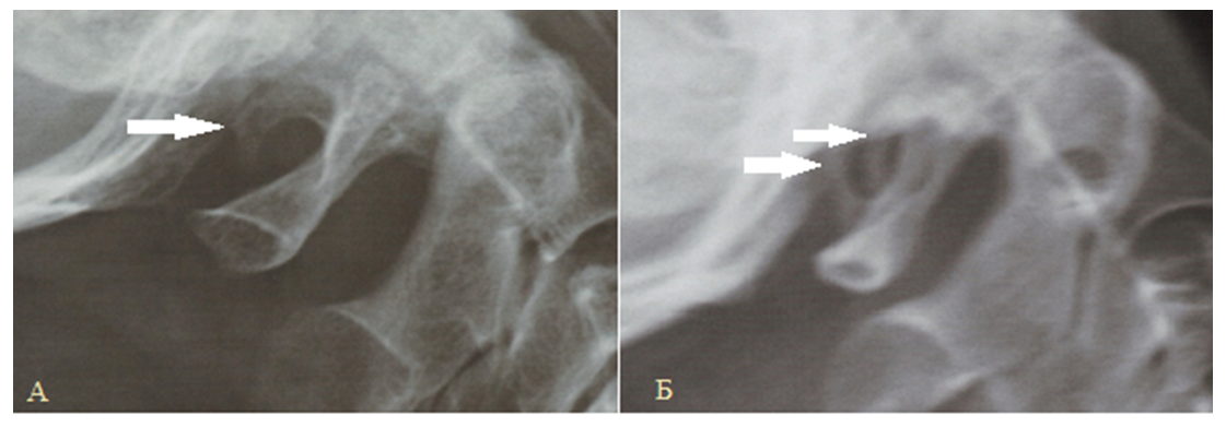 | Figure 1. Radiography of the vertebrobasilar zone, lateral projection A - unilateral posterior incomplete bone bridge of the first cervical vertebra (Kimmerle anomaly) Б - bilateral posterior complete bone bridge of the first cervical vertebra |
2. Purpose of the Study
- To assess the effect of Kimmerle's anomaly on blood circulation in the vertebrobasilar zone.
3. Materials and Methods
- Was carried out the complex radiation diagnostics of the craniovertebral region of patients with verified Kimmerle's anomaly. We analyzed the data of X-ray examination of patients who complained for headaches and cervicalgia from January 2020 to March 2021. Kimmerle's anomaly was detected In 62 out of 381 examined and computed tomography (CT) of the brain was performed with the capture of the upper cervical spine for these 62 patients. The examination was carried out on a GE-Optima 520 apparatus with 16 rows of slices (manufactured in the USA). CT scan parameters: tube current - 249 mA for a CT head, voltage - 120 kV, tube rotation speed - 1.0 s, pitch - 0.85, slice thickness 1.25 mm. The radiation exposure at CT was 2 mSv. Ultrasound duplex sonography of the V3 vertebral artery was performed using ESoate Mylab class C (linear transducer - 5-7.5 MHz, convex transducer - 2-5 MHz). Transcranial Doppler sonography was performed using the “EDAN instruments” version 1.2 system using a phased transducer with a frequency range of 3-7 MHz. The exclusion criteria from the examination were patients with tumors, cerebral hemorrhages, with manifestations of severe arterial hypertension. Statistical data were obtained using “Statistic 6.0” software.The study was carried out on the basis of the medical clinic of the Samarkand State Medical Institute and agreed with the management. All patients consented to this examination. Bioethics committee conclusion (ClinicalTrials.gov): ID NCT04562259.
4. Results
- Clinical complaints in 62 examined patients manifested themselves in the form of dizziness, various types of headache, mainly localized in the occipital region, nausea or flashing of flies infront of the eyes, in the rest - only cervicalgia. The pain was paroxysmal. The examinees associated the appearance of pain and discomfort mainly with the "uncomfortable" position of the head or neck during sleep, a sudden movement, and a change in position. Palpation reveals tension in the occipital muscles. The vertebrobasilar region is a complex department of the anatomical functions of the muscular frame and the location of the vessels.A diagnostic examination was carried out: 11 patients under the age of 30 years, 15 patients aged 31 to 40 years, 18 patients aged 41 to 50 years, 13 - from 51 to 60 years old and 5 patients over 61 years old. The age period of the patients ranged from 18 to 85 years. The average age in the male part of the patients was 47.3 years, in the female - 54.1 years, while the Kimmerle anomaly was almost equally noted in both men and women.When evaluating computed tomograms of the brain of patients with diagnosed Kimmerle's anomaly 41.9% of cases showed signs of vascular encephalopathy in 19.4% of cases signs of cerebellar atrophy and in 9.7% of cases single cysts. In the prevailing percentage of patients with Kimmerle's anomaly examined by us the posterior arch of the atlas was visualized on the screen as a high-density band on computed tomography, over which an annular hypodense zone was determined, bordered by a partially or completely closed hyperdense ring - a bone "bridge" (ponticulus posticus) (Fig. 2).The results of our studies showed that in 48 patients (77.4%) with diagnosed Kimmerle's anomaly, there is a bilateral location of the arcuate foramen of the first cervical vertebra. In 14 cases, the Kimmerle anomaly was one-sided, and almost all of them (85.7%) were located on the left.Ponticulus posticus, located on the right, was closed in 75.8% (complete Kimmerle anomaly) and open in 27.4% (incomplete Kimmerle anomaly). The arcuate foramen of the first cervical vertebra above the left arch was closed in 47 cases and open in 13 cases.
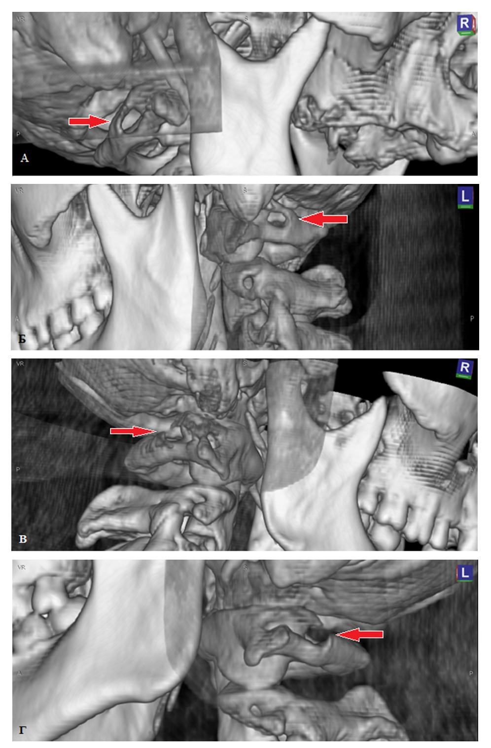 | Figure 2. Computed tomography with 3D reconstruction. Craniovertebral articulation: A) rear complete Kimmerle ring on the right, B) posterior complete ring C1 on the left, C) posterior incomplete ring C1 on the right, D) posterior incomplete ring C1 on the left (indicated by arrows) |
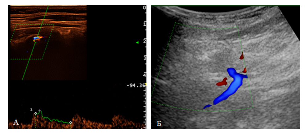 | Figure 3. Doppler ultrasonography of the vertebral arteries Craniovertebral department: A - visualization using a linear sensor, B - visualization using a cavity sensor |
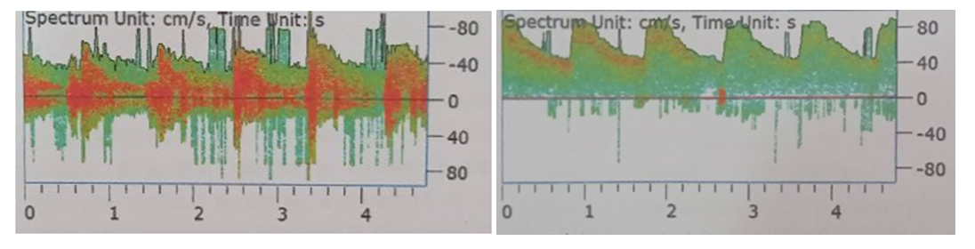 | Figure 4. Transcranial Doppler ultrasonography of the vertebral artery. Vertebrobasilar department |
5. Conclusions
- Summing up we can say that the relevance of studying the effect of craniovertebral insufficiency on the occurrence of blood flow disturbances in the vertebrobasilar vascular bed, the search for reliable and, at the same time, available methods for identifying, optimizing therapeutic methods for this pathology require appropriate studies. The main structural change in the cerebral vascular bed, according to ultrasound diagnostics, is a change in the straightness of the vertebral arteries in the transverse processes of the cervical spine.In our study, the frequency of the vaulted opening is close to the average occurrence of this feature (16.7%) [4,21], however, according to some authors, ponticulus posticus is diagnosed from 8.3% [6] to 12% of the occurrence. Interestingly, in some studies, these changes were found in one third of the examined and even from 37 to 80% of patients [7,17]. In the studies of individual authors, it is said that incomplete forms with many different variations are often encountered, as a result of which there is no consistent classification in the literature [3,5] and they may not be taken into account.Nowadays the ability of computed and magnetic resonance imaging to obtain detailed anatomical information about the structure of the spine and craniovertebral region provides a unique opportunity to renew attention to this complex area of the skeleton [24]. However, for assessing the morphology of bones and their trabecular structure including the bone bridge of the atlas, CT has better visualization than MRI.Statistics say that the incidence of the Kimmerle anomaly is 16-20% and this is certainly a frequent change in C1. This change correlates with the types of pathological abnormalities of the vascular bed of the brain and the main arteries of the head. A review of the literature once again proves the importance of vascular changes in the onset of cranialgia and requires special treatment and prevention tactics. It must be admitted that today doctors do not think, and some do not even know about the possibility of the existence of this pathology and for months and sometimes for years they carry out symptomatic therapy not suspecting that the Kimmerle anomaly can be confirmed or excluded by routine methods.In order to predict the impact of the presence of ponticulus posticus several questions need to be resolved:- to establish the ratio of the diameters of the vessels to each other and to the diameter of the C1 bone bridge,- determine full or incomplete arcuate foramen,- unilateral or bilateral pathology,- options for the structure of the great vessels in each individual case.The combination of these data can predict possible causal factors that can cause a decrease in cerebral blood flow, which in turn leads to early disability and loss of working capacity.
6. Recomindations
- Patients with complaints of pain in the cervical region, headache, mainly in the occipital region, dizziness, flashing of flies in front of the eyes and if a certain pathology is not identified that causes these symptoms we recommend performing an X-ray of the cervical spine in frontal and lateral projections or although would be in the side.If Kimmerle's anomaly is found, at the next stage, the study must be supplemented with CT to clarify the state of the morphology of the vertebrae and their possible effect on blood flow in the vertebral arteries.It is necessary to assess the blood flow in the V3 and V4 segments of the vertebral arteries by using ultrasound duplex sonography and transcranial Doppler sonography.The results of this study emphasize the importance of the role of the spondylogenic factor, which can cause or contribute to the development of circulatory disorders in the vertebrobasilar zone. Arterial and venous discirculation with prophylactic detection by radiation research methods, by means of CT and transcranial Doppler ultrasonography, has a positive prognosis for the therapy of cerebrovascular changes in patients with KA.Funding source and conflict of interest. This article does not have financial support for the study and conflicts of interest of the authors, which must be reported.