-
Paper Information
- Next Paper
- Paper Submission
-
Journal Information
- About This Journal
- Editorial Board
- Current Issue
- Archive
- Author Guidelines
- Contact Us
American Journal of Medicine and Medical Sciences
p-ISSN: 2165-901X e-ISSN: 2165-9036
2021; 11(12): 847-850
doi:10.5923/j.ajmms.20211112.01
Received: Nov. 11, 2021; Accepted: Nov. 26, 2021; Published: Dec. 8, 2021

Influence of Alloxan Diabetes During Pregnancy on the Morphological Formation of the Sterno-Costal Complex
 Abstract
 Reference
 Full-Text PDF
 Full-text HTML
Lobar Ibrohimovna Ibrohimova 1, Hamidulla Abdullaevich Rasulov 2
1Tashkent Pediatric Medical Institute, Tashkent, Uzbekistan
2Sc.D., Tashkent Pediatric Medical Institute, Tashkent, Uzbekistan
Copyright © 2021 The Author(s). Published by Scientific & Academic Publishing.
This work is licensed under the Creative Commons Attribution International License (CC BY).
http://creativecommons.org/licenses/by/4.0/

Diabetes mellitus is on the list of social problems and is accompanied by profound changes in the human body. Disturbances of peripheral microcirculation and innervation are observed with a long course of the disease. The morphofunctional active point in the formation of the chest is the sterno-costal complex. Changes in the shape and functional structure of the chest affect the functional state of the chest cavity organs. The lack of data on the morphofunctional properties of the sterno-costal complex leads to serious shortcomings and errors in the prevention and treatment of injuries and deformities in certain areas. Conducting scientific research in this area has not only scientific, but also practical importance. The study was carried out on 42 white laboratory rats weighing 150-200 g. The animals were kept in living conditions according to a standard diet (with food and water). For the study, the animals were divided into 2 groups. In the control group, there were 10 rats, male rats were injected in a ratio of 3: 1 and 0.5 ml of 0.9% sodium chloride solution was injected once. The research group consisted of 32 rats, the male had a ratio of 3: 1. The material for the study was the breast components of 7–14–21–1–45–45 days of age in young rats born to mothers with experimental diabetes. For histological analysis in the study, it was necessary to obtain a sterno-costal combination of rats of the experimental group. The results of control and morphological studies of the sterno-costal complexes in the offspring of experimental rats in the dynamics of growth of early postnatal ontogenesis showed that maternal alloxan diabetes leads to a change in the mechanism of development of the sternum during pregnancy and lactation of a mother with diabetes.
Keywords: Experimental diabetes, Alloxan, Cartilage, Chondrocyte, Sternocostal complex
Cite this paper: Lobar Ibrohimovna Ibrohimova , Hamidulla Abdullaevich Rasulov , Influence of Alloxan Diabetes During Pregnancy on the Morphological Formation of the Sterno-Costal Complex, American Journal of Medicine and Medical Sciences, Vol. 11 No. 12, 2021, pp. 847-850. doi: 10.5923/j.ajmms.20211112.01.
1. Introduction
- According to the World Health Organization, more than 100 million people worldwide have been diagnosed with diabetes. Diabetes mellitus is on the list of social problems and is accompanied by profound changes in the human body. Disturbances of peripheral microcirculation and innervation are observed with a long course of the disease [1]. The morphofunctional active point in the formation of the chest is the sterno-costal complex. Changes in the shape and functional structure of the chest affect the functional state of the chest cavity organs. Congenital and acquired diseases of the musculoskeletal system account for 94–99% of a person's life expectancy, of which the incidence of congenital defects is 32: 1000 [2] Therotologically, defects in the development of the chest make up 4–12% of all anomalies [3]. The lack of data on the morphofunctional properties of the sterno-costal complex leads to serious shortcomings and errors in the prevention and treatment of injuries and deformities in certain areas. Conducting scientific research in this area has not only scientific, but also practical importance.All of the above allows us to draw conclusions about the problem we have developed and its relevance in connection with the prevalence of deformities and injuries of the sternocleidomastoid complex and their morphological and functional substantiation.The purpose of the study the dynamics of morphological changes in the thoracic-costal complex of rats with alloxan diabetes mellitus.
2. Materials and Methods
- The study was carried out on 42 white laboratory rats weighing 150-200 g. The animals were kept in living conditions according to a standard diet (with food and water). For the study, the animals were divided into 2 groups. In the control group, there were 10 rats, male rats were injected in a ratio of 3: 1 and 0.5 ml of 0.9% sodium chloride solution was injected once. The research group consisted of 32 rats, the male had a ratio of 3: 1. On the fifth day of gestation, the rats were induced experimental diabetes mellitus using the alloxan model. In the experimental group, a mixture of alloxan 150 mg / kg was injected intraperitoneally by a single intraperitoneal injection. An increase in blood glucose up to about 350 mg / dL (Plus Satellite, Russia) confirms hyperglycemia.In the rats in our experiment, sucking was observed after 30 minutes. Within 5-7 hours, the tails began to turn blue. In the following days, thirst, polyuria, a decrease in wheezing and tachycardia were observed. Follow-up 20-24 days later followed by observation in rats with low mobility, weight loss, prolonged wound healing, and hair loss. Of the 22 rats taken for the experiment, 40% died. The material for the study was the breast components of 7–14–21–1–45–45 days of age in young rats born to mothers with experimental diabetes. For histological analysis in the study, it was necessary to obtain a sterno-costal combination of rats of the experimental group.The decapitation of animals was carried out under general ether anesthesia using a guillotine knife in accordance with ethical rules. For histological examination, an incision was made in the sternocleidomastoid joint. The samples taken for the experiment were immersed in 10% formalin. The sample was dried in increasing percent alcohol and processed histologically. Then paraffin was poured. The incisions were made on a microtome with a thickness of 4-5 µm. The prepared sections were stained by the Mason method for the study of hematoxylin-eosin and connective tissue, cells and its intermediate elements, as well as a histochemical reaction was performed using the PIC methods. The finished slides were examined under a SARL Zeiss Microscopy GmbH microscope and photographed with an Axio Lab.A1 camera (Germany).Blood was taken from rats to determine mineral metabolism and markers of bone tissue (indices of resorption and remodulation), as well as fragments of organs and tissues for morphological examination during subsequent periods of observation.Electrolytes Ca, P, Mg in blood serum were determined using a HUMAN colorimetric kit (Germany).Bone resorption marker - alkaline phosphatase and remodulation marker - b-STX were investigated using ELISA reagents.
3. Results and Discussion
- In fact, the growth zone in all bones plays an important role in the formation and growth of bones, so the state of the structures of the sternocleidomastoid ridge has been studied.In morphological examination for histological examination of the thorax regions of the sternum complex of rats in the initial period of observation, the normative formation of ribs in the control animal during this period corresponds to the observation period (Fig. 2).During this period, patients showed symmetrical basophilia of the sternum along the ossification line, relative intensity of proximal ossification, chaotic formation of the distance between the columns of bone tissue, uneven distribution of the proliferative stratum corneum, and an abundance of fatty pads in the surrounding soft tissue interval. experimental group of animals (Fig. 6).On the 14th day of observation, when examining the breast tissue, linear differentiation at the junction of the chest with the chest in the control animal began on the periphery, and bruises on the periphery were found in the cervical matrix. It was found that the epichondral film became thinner, relatively thickened in the corners of the cartilage-costal film (Fig. 3).The results of control and morphological studies of the sterno-costal complexes in the offspring of experimental rats in the dynamics of growth of early postnatal ontogenesis showed that maternal alloxan diabetes leads to a change in the mechanism of development of the sternum during pregnancy and lactation of a mother with diabetes. [4] On day 21 of the experiment, the main side effects of diabetes mellitus were observed in connective tissue in zone 4 of the growth of the sternum and in synchondrosis at the junction of the ribs (Fig. 4).By day 30, the transitional tissue in all four growth zones has the same architecture. The tubules on both sides (proximal and distal) are separated from the bone growth zones by the same mechanism of bone formation. Isogenic chondrocytes are not observed in the main group of the study, in the early stages (7-14 days) they are chaotic, single, in pairs, in later periods (14-30 days) small isogenic groups, consisting of an average of 3 people. There are 4 cells of different sizes [5]. The shape of these chondrocytes is elliptical or cylindrical, in some places round-shaped cells are revealed (Fig. 5).At a later date (45 days), chondrocytes have a spherical or round nucleus surrounded by cytoplasm. The growth zone is represented by proliferative cells, which are columnar and consist of 10-12 cells in each column. These chondrocytes differ from the adjacent columns by a clear set of fibrils oriented longitudinally along the layers of the main matrix material. In some chondrocytes of this zone, mitotic patterns were revealed. Specific cases of mitosis were revealed in rats of the main group [6]. Cartilage cracks are found between the columns of chondrocytes. In places, local dystrophic changes in chondrocytes are detected, the vascular wall thickens, fibrous structures decrease, and plasmorrhagic foci appear around the vessels (Fig. 1).
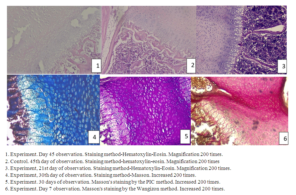 | Figure 1-6 |
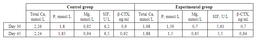 | Table 1. Indicators of mineral metabolism and markers of bone tissue in experimental DM |
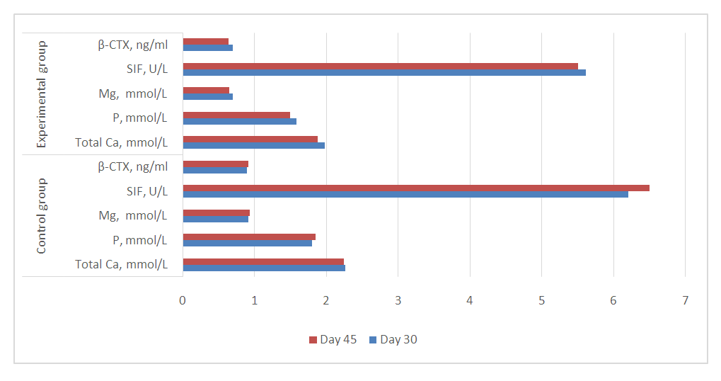 | Diagram 1. Dynamics of mineral metabolism and markers of bone tissue in the EQD on the 30-45th day of observation (R≤0.005) |
4. Conclusions
- The greatest changes in rats under conditions of diabetes mellitus of a pregnant mother occur in the connective tissue of the growth zones, where it becomes significantly thinner and the number of cells in it decreases.It was found that significant changes in markers of mineral metabolism and ossification in the advanced generation as a result of the adverse effects of experimental diabetes during pregnancy were more pronounced in the late (30–45 days) follow-up period.During the observation periods, the process of differentiation in the bone trabeculae slows down sharply, and therefore the range of the growth zone of the sternum expands in the distal and proximal directions.The data obtained indicate that during pregnancy in rats with maternal alloxan diabetes, the development of all structural structures of the growth zones of the sternocostal complex is delayed.