-
Paper Information
- Next Paper
- Previous Paper
- Paper Submission
-
Journal Information
- About This Journal
- Editorial Board
- Current Issue
- Archive
- Author Guidelines
- Contact Us
American Journal of Medicine and Medical Sciences
p-ISSN: 2165-901X e-ISSN: 2165-9036
2021; 11(11): 774-779
doi:10.5923/j.ajmms.20211111.06
Received: Oct. 22, 2021; Accepted: Nov. 5, 2021; Published: Nov. 15, 2021

Clinical and Epidemiological Features of Dishormonal Diseases and Breast Cancer in Men in the Aral Sea Region
 Abstract
 Reference
 Full-Text PDF
 Full-text HTML
Alimkhodjaeva L. T.1, Norbekova M. Kh.2
1Republican Specialized Scientific and Practical Medical Center of Oncology and Radiology (RSSPMCO&R), Tashkent, Uzbekistan
2Department of Oncology of Tashkent Medical Academy, Tashkent, Uzbekistan
Copyright © 2021 The Author(s). Published by Scientific & Academic Publishing.
This work is licensed under the Creative Commons Attribution International License (CC BY).
http://creativecommons.org/licenses/by/4.0/

A significant number of scientific papers have been devoted to breast cancer in the female population, while insufficient attention has been paid to cancer and dyshormonal diseases of the mammary glands in men. Nevertheless, this issue is becoming more urgent every year. Although breast cancer in men is a relatively rare disease (1-2% of the number of these tumors in women), it is not inferior to “female” cancer in terms of malignancy, and sometimes even surpasses it. Changes in the mammary gland in men are primarily associated with hormonal imbalance, the manifestation of which is gynecomastia. Until now, there is no consensus on the relationship between gynecomastia and breast cancer, although it has been established that the incidence of malignant tumors against the background of gynecomastia makes up 3.9 - 40.0% of cases.
Keywords: Breast cancer, Gynecomastia, Oncoepidemiology, Hormone therapy
Cite this paper: Alimkhodjaeva L. T., Norbekova M. Kh., Clinical and Epidemiological Features of Dishormonal Diseases and Breast Cancer in Men in the Aral Sea Region, American Journal of Medicine and Medical Sciences, Vol. 11 No. 11, 2021, pp. 774-779. doi: 10.5923/j.ajmms.20211111.06.
1. Introduction
- The issue of precancerous diseases and breast cancer is very relevant today not only in Uzbekistan but throughout the world. The increased interest to breast cancer, first of all, is associated with the increasing number of diseases observed in recent years in the world [1-4].On average, male breast cancer occupies a leading position in the structure of the incidence of malignant neoplasm of this organ in both sexes [5-8]. Breast cancer averages 0.2% in the structure of the incidence of men with MN. On average, about 26 males die from breast cancer in Uzbekistan, accounting for 1.3% of all patients who died from breast cancer and 0.18% of all men who died from MN.Until now, there is no consensus on the relationship between gynecomastia and breast cancer, although it has been established that the incidence of malignant tumors against the background of gynecomastia makes up 3.9 - 40.0% of cases.Men, as well as women, have a genetically determined predisposition to the development of breast cancer. More than 40% of breast cancer cases in men are hereditary [9-11]. Breast cancer genes BRCA-1 and BRCA -2 were studied in more than 21 families. Mutations in BRCA – 2 have been identified in men with breast cancer [12]. The complexity of diagnosis is aggravated by the fact that male breast cancer has to be differentiated from a large group of this organdiseases, united by the common name "gynecomastia". Statistical data on the frequency of gynecomastia are few and contradictory and occur between the ages of 17 and 80 years [14-18]. Breast cancer in men can be developed both against the background of gynecomastia and without it. Proof of gynecomastia transformation into cancer can only be the detection of pre-invasive cancer in the duct epithelium.Currently, the uneven spread of malignant neoplasms in various territories is largely due to the influence of carcinogenic and modifying environmental factors, such as anthropogenic pollution, occupational hazards, as well as a number of climatic and geographical features. Geochemical factors (both natural and techno genic) are among the important components of the external environment, which cannot but influence the development of cancer-epidemiological processes. According to a number of authors, the leading role in oncogenesis is played by seven bioelements, which include copper, manganese, zinc, cobalt, iron, 7 molybdenum, iodine [19-22]. The influence of these elements on the growth of malignant neoplasms in the gastrointestinal tract, maxillofacial region, hormone-dependent organs, including the mammary gland, has been determined.Analysis of the incidence rates of dyshormonal diseases and breast cancer in men in the Aral Sea region revealed their growth. Studies carried out in this region have shown that the spread of malignant tumors of the female mammary gland in different landscape zones is heterogeneous and this is due to the uneven distribution of individual microelements in the soil and in water sources. Taking into account the heterogeneity of the biogeochemical structure of the Aral Sea region and also the increase in the incidence of gynecomastia and breast cancer among the male population, it should be noted that the study of this pathology spread features and the identification of factors contributing to its development is relevant not only in terms of expanding knowledge on the occurrence of this pathology, but also provides new materials for scientifically based measures of anticancer control.Aim of the study was to study the peculiarities of the spread of the mammary glands diseases in men living in various landscape zones of the Aral Sea region and to determine the relationship between the indicators of morbidity, the trace element composition of the blood, the level of sex hormones.
2. Material and Methods
- In order to determine the reaction of the male body to the geochemical composition of the environment, we examined 342 men with dyshormonal hyperplasia and breast cancer at the age from 12 to 80 years and older: 130 cases— with diffuse gynecomastia, 114 — with nodulargynecomastia, 76 — with mixedgynecomastia, 15 — with breast cancer and 7 people with other pathologies. The diagnosis was determined on the basis of objective data, additional examination methods, which included X-ray and ultrasound mammography and mandatory cytomorphological verification.The groups were formed with maximum homogeneity. The studied clinical groups consisted of the indigenous newcomer population living in the Aral Sea region for at least 10 years. The majority of the surveyed were represented by 135 workers (39.5+2.6%), employees and intellectuals - 75 (21.9+2.2%), schoolchildren and students - 58 (16.9+2.0%), collective farmers - 43 (12.6+1.8%), entrepreneurs - 13 (3.8+1.0%), unemployed - 18 (5.3+1.2%). We did not find the reliability of the difference in the zonal distribution of the professional factor. Consequently, the professional employment of men is evenly represented in the northern, transitional and southern zones. The control group included 50 men of the corresponding age groups, living in the Aral Sea region for at least 20-30 years, professionally employed and who, after a preventive examination, were found to be practically healthy. The food ration of the indigenous newcomer population in the Aral Sea region in all clinical groups mainly consisted of locally produced products.Taking into account the objective of the study and the accepted division of the entire territory of the Aral Sea region, depending on the soil structure into zones (northern, transitional, southern), the role and place of the environment in the diagnosis and assessment of the main parameters of homeostasis in men with dishormonal diseases and breast cancer were determined as factors of increased cancer risk. The incidence of breast cancer in the districts was studied using the registration form 030-6 / U for the period 2010-2020.Determination of trace element analysis of patients’ blood with dyshormonal diseases of the mammary glands and breast cancer was carried out by atomic absorption spectrophotometry. Determination of the hormonal profile of peripheral blood was carried out on the basis of the laboratory of radioisotope diagnostic methods. 295 samples were analyzed. Determination of folicle-stimulating hormone (FSH), luteinizing hormone (LH) was carried out by radioimmunoassay (RIA). Prolactin and testosterone were determined using CIS International kits. In parallel, the hormone determination of FSH, LH, prolactin, testosterone, estradiol was carried out by the method of enzyme-linked immunosorbent assay (ELISA).X-ray examination of the mammary glands (mammography) was performed using a mammographic X-ray apparatus mammo DIAGNOST UG manufactured by PHILIPS. Ultrasound examination was carried out on Fukuda Denshi 5000 and SonoAce 4800 HD devices using a 7.5 linear transducer; 10 MHz. We performed puncture of the mammary glands according to the generally accepted technique with a thin needle 6-10 cm long and 1 mm in diameter. The puncture site was determined on the basis of clinical, sonographic and radiological data. From the punctate and secretions from the nipples of the mammary glands, preparations were made - smears, which were subsequently stained using the Pappenheim method. The removed breast tissue was fixed in 10% neutral formalin and embedded in paraffin using a standard technique. The prepared paraffin sections with a thickness of 5 μm were stained with hematoxylin and eosin. The hormonal profile of peripheral blood was determined by RIA and ELISA. The immunological basis of the in vitro radio test (RIA) reaction is similar to the reactions occurring in the human immune system "antigen - antibody". The hormonal profile ELISA technique is based on the principle of a two-site (sandwich) test system.The elemental composition of blood samples was determined using a Hitachi atomic absorption spectrophotometer (Japan). Taking into account the possibility of daily fluctuations in the amount of mineral elements in the blood, it was taken in the morning until 10 o'clock on an empty stomach from a vein on an outpatient basis. The technique of the atomic absorption method for the determination of elements consisted in the decomposition of the analyzed sample, spraying the resulting 41 solution in an air-acetylene flame or in a flame of a mixture of nitrous oxide and acetylene, depending on the element to be determined, measuring the value of atomic absorption of resonance radiation by neutral atoms of the determined elements formed during the atomization of the sample. Calculations were performed according to the third category of accuracy, for which its margin is measured by the formula: 1 <Z.
3. Results
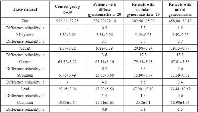
- As a result of studies by atomic absorption spectrophotometry, the maximum amount of the trace element zinc (559.8 + 39.1 μg / g) and manganese (5.54 + 0.06 μg / g) was found in the peripheral blood of patients with diffuse gynecomastia. They also had the minimum content of cobalt (9.88 + 0.39 μg / g) and copper (63.17 + 3.16 μg / g) (Tab. 1).
|
|

|

|
4. Discussion
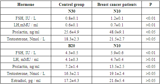
- Thus, it is possible to assume that the changes found by us in the content of trace elements zinc, manganese, cobalt, copper, strontium, lead and cadmium in the blood of the examined men can lead to dysfunction of the pituitary gland and be accompanied by changes in the secretion of FSH, LH, prolactin, testosterone and estradiol. This imbalance, in turn, determines the morphological and functional state of the mammary glands, its clinical manifestations and, possibly, acts as a factor predisposing to the development of dyshormonal hyperplasia and breast cancer in men.Comparison of the primary morbidity with the geochemical situation in three zones of the Aral Sea region revealed the following pattern: an increase in the content of copper and cobalt occurs in parallel with an increase in the incidence of gynecomastia and breast cancer in the direction from north to south. The concentration of zinc and manganese decreases in the same direction. Besides, in the transitional and southern provinces of the Aral Sea region an increased content of toxic elements (strontium, lead, cadmium) has been noted. This confirmed the relationship between the primary incidence of gynecomastia and malignant tumors of the mammary glands in men with a trace element composition of soils, water sources and food. The average values of the levels of copper, zinc, manganese and cobalt in soils, surface waters and food products were used.The coefficient of canonical analysis reflects the general relationship between the incidence of breast cancer in men and the microelement background of the biosphere. Thus, a significant dependence of the incidence rate was determined with a high level of cobalt content (K = 2.822 + 0.780, p). Against the background of the unfolding imbalance of hormones in men with dyshormonal diseases of the mammary glands, the activity of male sex hormones increases, which, apparently, indicates the work of compensatory mechanisms that require the involvement of the trace element zinc (R = 0.178 + 0.015) involving in the production of testosterone. In conditions of increased lead content in the transitional and southern zones of the Aral Sea region, a high correlation coefficient was noted between lead and the studied hormonal complex in patients with gynecomastia (R = 0.263 + 0.220).The relationship between each of the studied bioelements and indicators of the hormonal profile in the peripheral blood of men with breast cancer was traced. The accumulation of zinc and manganese in the tissue of patients with breast cancer is apparently a protective reaction, since during the development of the tumor process we noted a decrease in the content of these biometals in the peripheral blood. It creates conditions for hyperestrogenemia (21.6 + 5.4 pg / ml), and as a consequence, an increase in proliferative processes in the mammary glands.When studying the content of cobalt, copper, lead, strontium and cadmium in peripheral blood and in mammary glands tissue, their promoter activity was found, possibly leading to the development of breast cancer in men, while zinc and manganese act as inhibitors. However, it would be wrong, in our opinion, to approach the issue of the correlation between the microelement composition of the biosphere and the incidence of breast cancer purely mechanically, without taking into account the interaction of bio elements with each other and with a group of toxic microelements 98 (strontium, lead and cadmium). Excessive intake of cadmium and lead in the human body leads to a deficiency in zinc intake. The transitional and southern biogeochemical zones are already deficient in the content of trace elements zinc and manganese in mobile forms, and the layering increased content of trace elements strontium, lead and cadmium further aggravates this condition, increases the incidence of nodular gynecomastia and breast cancer among the male population.
5. Conclusions
- Thus, the revealed features of hormonal and mineral homeostasis disorders in men are closely related to the features of the microelement composition of the environment. They, in turn, have an impact on the different incidence of dyshormonal hyperplasia and breast cancer in the Aral Sea region. Therefore, for the formation of groups of increased risk for the development of dyshormonal diseases and breast cancer in men of the Aral Sea region, along with genetic and modifying factors, it is necessary to take into account the microelement composition of the environment.