American Journal of Medicine and Medical Sciences

p-ISSN: 2165-901X    e-ISSN: 2165-9036

2021;  11(10): 699-704

doi:10.5923/j.ajmms.20211110.07

Received: Sep. 29, 2021; Accepted: Oct. 19, 2021; Published: Oct. 30, 2021


The Features of Microsurgery of Cerebral Arteriovenous Malformation

Khadjibaev A. M., Makhkamov K. E., Makhkamov M. K.

Republican Research Center of Emergency Medicine (RRCEM), Tashkent, Uzbekistan

Copyright © 2021 The Author(s). Published by Scientific & Academic Publishing.

This work is licensed under the Creative Commons Attribution International License (CC BY).
http://creativecommons.org/licenses/by/4.0/

Abstract

Aim. Description of the features of the microsurgical technique of resection of cerebral arteriovenous malformation (AVM). Material and methods. A retrospective analysis of 26 patients with cerebral AVM was carried out. Of these, 15 patients were females and 11 males, aged 5 to 46 years. All patients underwent neurological examination according to generally accepted standards. After studying the history and physical examination, all patients underwent computed tomography (CT), magnetic resonance imaging (MRI), CT-angiography, and digital subtraction angiography (DSA). The microsurgical technique of access, isolation and microsurgical AVM resection is described in detail. Results. The overwhelming majority of patients with AVM (61.5%) were admitted to a rupture clinic. Of these, with intracerebral hemorrhage (ICH) accounted for 56.3% of cases and 43.7% of cases with subarachnoid hemorrhage (SAH). It was noted that microsurgical resection of complicated AVMs with low grades (grades I and II) is the method of choice. For AVMs with grade III and more methods of choice is a combined treatment method. Conclusion. Increased resectability without imploring postoperative AVM results depends on factors such as a detailed understanding of the AVM anatomical structure and presentation in three-dimensional space, as well as correct exclusion of the adductor vessels and subpial dissection with a combination of endovascular embolization followed by surgical resection.

Keywords: Microsurgical resection, 3D reconstruction, Endovascular embolization, Combined technique

Cite this paper: Khadjibaev A. M., Makhkamov K. E., Makhkamov M. K., The Features of Microsurgery of Cerebral Arteriovenous Malformation, American Journal of Medicine and Medical Sciences, Vol. 11 No. 10, 2021, pp. 699-704. doi: 10.5923/j.ajmms.20211110.07.

1. Introduction

The incidence of intracranial arteriovenous malformations (AVM) is about 1 per 100,000 per year, in people of working age it is 18 per 100,000. AVMs are characterized by significant morbidity and mortality, with a risk of rupture of about 3% per year [1]. The mortality rate due to AVM rupture is 30%, of which half of the patients have moderate or deep disabling [2]. The high medical and social significance of this disease is determined not only by its prevalence, but also by significant economic damage due to the high frequency of occurrence in people of working age from 20 to 50 years old [3]. Despite the development of endovascular methods of AVM treatment, microsurgical resection is the gold standard. However, to this day there is no consensus on the optimality of the type, stages and timing of surgical treatment, which determines the relevance and disputability of AVM surgery in modern neurosurgery.

2. The Aim of the Study

The aim of our study was to describe the features of the microsurgical technique of AVM of cerebral vessels.

3. Material and Methods

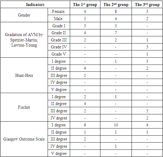
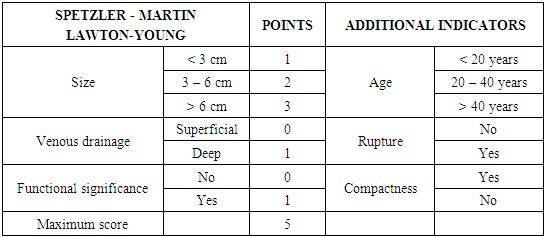
We performed a retrospective analysis of 26 patients with cerebral AVM. (Table 1). 15 patients were females and 11 males, aged from 5 to 46 years. All patients underwent neurological examination according to generally accepted standards. After studying the history and physical examination, all patients underwent CT, MRI, CT-angiography, and DSA. The application of these research methods is justified by the fact that they are all interconnected. With the help of CT, we evaluated the signs of rupture, the presence of hemorrhage and the volume of hematomas, as well as the presence of calcified areas. MRI was performed to assess the compactness of the AVM. CT angiography and DSA were used to visualize not only the nucleus itself - the "nidus" of the AVM, but also the supplying vessels and the participation of the blood supplying arterial pools, the size and course of the vessels, as well as the points of connection with the "nidus", the nature of the draining veins, their size and flow blood in the draining vein, in particular, inside the "nidus" (Fig. 1). The relationship of AVM with surrounding anatomical structures was also studied, the presence of thrombosis of the AVM was determined, which is extremely important for planning surgery. In addition, 3D modeling of intracerebral vessels was of decisive importance in preoperative planning, which made it possible to visualize the relationship of microanatomical structures, i.e. pathological vascular plexus in three-dimensional space. For the purpose of a more detailed study of the results of surgical treatment of AVMs, a modified Spetzler-Martin & Lawton-Young classification was used [4] (Table 2).
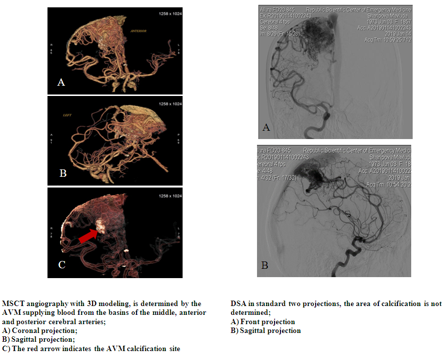
Table 1
     
Table 2
     
Surgical technique
Anatomical features of AVM from a surgical point of view.
Arteriovenous malformations come in various shapes: sphere, ellipse, cylinder, and classic cone. The conical base of the AVM is located on the surface of the cortex and tapers with a tip in the ependyma region. Although AVMs are never cube-shaped, Lawton et al. Recommends presenting them as a "box" with six sides: one upper (or superficial side), four sides further the lower (or deep side), which gives the AVM a definable side that is medial and lateral, anterior and posterior, superior and inferior [5,6]. These distinctive features can then be characterized by connections to dural venous structures, cortical landmarks, feeding arteries, draining veins, and cranial anatomy. The "box" allows the neurosurgeon to present the anatomical features of the AVM from a surgical point of view. As you collect information from angiograms, MRI scans, CT angiography and native CT scans, you need to sequence by sequence and run after run, go through each scan to get acquainted with the AVM and see the relationships between arteries and veins, analyze bifurcations or tortuosities that can be submerged in the sulcus, and identify embolic agents that can serve as landmarks during surgery. MRI images can be scrolled up and down to locate functionally significant areas of the brain, identify clear pial planes in a sulcus, or display an adjacent clot on CT scans as a pathway to an AVM. And also, the use of 3-D AVM modeling in preoperative planning and intraoperative period makes it possible to compare and evaluate in detail the structure of the angioarchitectonics of the pathological area, the leading vessels, the draining vein and to visualize in all planes the relationship of microanatomical structures in the virtual three-dimensional space of the operating field and thereby maximize the angle of attack of the operating microscope to the pathological focus (Fig. 1).
Figure 1
Craniotomy and selection of the surgical angle of attack.
One of the features of AVM surgery is the choice of the size of the craniotomy, the size of which depends on the length of the AVM area. It should be noted that many authors always recommend a sufficiently wide craniotomy to cover the entire surface of the AVM and avoid the use of minimally invasive approaches, which at one time makes it possible to more correctly understand the intraoperative angioarchitectonics of the AVM [5,6]. When it comes to osteoplastic craniotomy, the more the better. A minor craniotomy can obstruct parts of the AVM, create an obstructing bone protrusion, force additional dissection in the wrong plane and direction, limit the use of the full scope of microsurgical views and microsurgical instruments, and cause unnecessary errors. In this regard, the widest craniotomy is recommended, taking into account the shape and size of the AVM, which makes it possible to trace both the feeding vessels and the draining system of the pathological vascular plexus. Also provides sufficient area for adequate dissection and disclosure of the subarachnoid space and subpial dissection along the border of the pathological vascular plexus.
One of the important points in AVM surgery, and in all microneurosurgery, is the correct choice of the angle of attack, i.e. operating microscope viewing angle and microsurgical actions. The optimal angle of attack for convexitally located AVMs is the parallel passage of the visualization axis of the microscope optics with respect to the axis of AVM. At the same time, it becomes possible to visualize all sides of the pathological area in a layer-by-layer and stepwise sequence with a clear view of both the adducting vessels and the draining veins, which allows AVM resection with minimization of damage to the surrounding tissue, thereby reducing the risk of possible intra- and postoperative complications. For deeply located AVMs, the use of a phased dissection parallel to the axis is impractical due to the fact that the surgical corridor is lengthened. In such cases, it is recommended to use the so-called "perpendicular" access. This access provides as far as possible, phased devascularization on one side of the pathological site. The trajectory of microsurgical manipulation to turn off the blood supply for deep AVMs should be changed in stages, turning off one side and with the transition to the other sides to completely disconnect the pathological focus from the blood supply [5,6]. This angle of attack has the advantage of avoiding transcortical routes through healthy brain tissue. However, imaging restriction and reduced maneuverability around the AVM appear, and as the AVM is mobilized with this technique, the risk of intraoperative complications such as bleeding increases. Application of this method is more appropriate for small AVMs.
Devascularization and subpial AVM dissection.
From the moment the AVM was exposed, i.e. after subarachnoid perimeter dissection and after identification of the adductor vessels using bipolar coagulation, devascularization begins. It should be noted that vascular coagulation should be carried out as close as possible to the AVM coil, since with more proximal coagulation, the risk of vascular shutdown in the areas of normal brain tissue involved in nutrition and postoperative zones of infarction increases. And also, on the contrary, it should be noted that during coagulation of the feeding vessels in contact with the AVM “nidus” itself, the risk of arterial bleeding from the coil itself sharply increases. In this regard, it is recommended to maintain a distance from the AVM coil from 3 mm to 7 mm [5]. Devascularization of AVMs should be started with large, namely, feeding vessels, which are the initial goal of the very idea of devascularization due to the fact that the inflow into the AVM in these vessels is greater than in others. From the moment of excretion of the adducting vessel, a temporary clip is installed, retreating by 7 mm proximally from the AVM coil. This maneuver allows squeezing the vessel lumen and stopping the blood flow, which increases the efficiency of coagulation. After bipolar coagulation of this area, the proximally applied clip is removed and then dissection is performed using microscissors (Fig. 2).
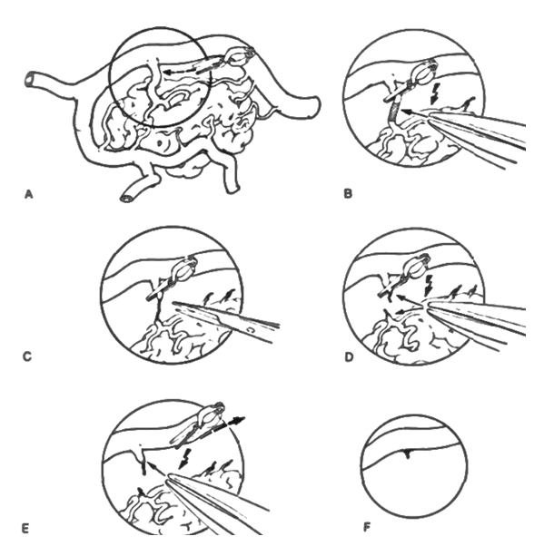
Figure 2. Schematic representation of the AVM devascularization stage
Further, devascularization should be continued along the entire perimeter, and as this maneuver proceeds, it is necessary to continue devascularization deep into the medulla along the perimeter of the AVM. It should be noted that deeply located small vessels have their own special structure, which consists in the fact that their walls are thin with poorly developed layers of elastic and smooth muscle cells and this anatomical structure makes it difficult to adequately perform bipolar coagulation. In order to perform an adequate shutdown of these small adducting vessels, we recommend using the "dirty coagulation" method. The peculiarity of this technique is that subpial AVM dissection should be carried out around these vessels with the capture of the surrounding brain tissue up to 5 mm. Since the perimeter of the AVM has a glially altered area and the method of "dirty coagulation" does not cause the risk of postoperative neurological complications and thereby avoids uncontrolled intraoperative bleeding. This technique is used to devascularize the AVM along the entire perimeter and only after that the microscope optics is focused to the AVM drainage system (Fig. 3). The main indicator, which means complete devascularization of AVM, is a change in the color of the draining vein in blue from scarlet arterial to the so-called "blue tail". This means that the AVM is completely devascularized and can be disconnected from the draining vein and the nidus may be removed from the bed.
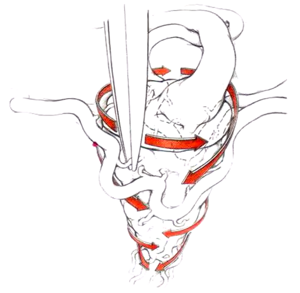
Figure 3. Schematic representation of the AVM devascularization technique

4. Results

The results of our study showed that microsurgical interventions were performed in 34.6% (n = 9) cases, 46.2% (n = 12) endovascular (embolization) interventions and in 19.2% (n = 5) cases, there was a combination of partial embolization followed by microsurgical resection. Our study was observed in 61.5% (n = 16) cases with ruptured AVM’s. From them in 56.3% of these, (n = 9) cases - intracerebral hemorrhage (ICH) and in the remaining 43.7% (n = 7) cases, SAH. Among ICH, in 33.4% (n = 3) cases, the hematoma was located in the frontal lobe, in 11.1% (n = 1) cases, in the temporal lobe, in 22.2% (n = 2) cases, in the parietal lobe with spread to the mediobasal regions and the ventricular system, in 22.2% (n = 2) cases in the occipital lobe and in 11.1% (n = 1) was subtentorial hemorrhage. Of the patients who underwent a rupture, 25% (n = 4) cases were admitted in the acute period, in 43.7% (n = 7) cases in the subacute period and in 31.3% (n = 5) cases in the cold period. In the remaining 38.5% (n = 10) cases, the clinical picture was presented by a pseudotumorous course (general cerebral symptoms, convulsions and focal deficit). From 34.6% (n = 9) cases, in the 22.2% (n = 2) showed signs of partial thrombosis of the malformation after the previous hemorrhage. Considering these signs of a partially thrombosed node, it was decided to limit ourselves to microsurgical resection. In one case, there was a postoperative complication in the form of hemorrhage in the bed of the resected malformation with the emergence of persistent neurological deficit in the form of deep hemiparesis, and in another case, in one patient, elements of motor aphasia. 19.2% (n = 5) of patients with grades III and IV, one patient in the postoperative period had a slight neurological deficit in the form of foot monoparesis, which regressed within 10 days.

5. Discussion

Surgical treatment of AVMs should be adapted depending on the AVMs’ gradation. Despite the fact that surgical resection is the gold standard, with the development of minimally invasive interventions, the use of microsurgery has become more rational for small (I and II grades), convexitally located AVMs. For AVM grade III and more, one-stage total embolization is, in most cases, hardly possible. Partially embolized AVMs of grade III and higher in the early postoperative period may be complicated by rupture and ICH from the residual part of the AVM. To date, the most optimal choice of AVM treatment method is a combination of partial endovascular embolization and microsurgical resection [7].
The incidence of vascular catastrophes due to AVM rupture is 1-2% among all strokes, in 4% of cases accompanied by ICH and in 9% of cases with SAH [2]. In our results the incidence of ICH was 61.5%.
In the studies of carried out by Hamilton M.G. and Spetzler R.F, it was noted that the risk of developing neurological deficits and deaths in the postoperative period after microsurgical resection for AVMs of I and II grades is 1%, 3% for AVMs of grades I and II, 31% and 50% - for AVMs IV and V grades, respectively [8,9]. The presence of hemorrhage or a history of early hemorrhage is a criterion for active surgical tactics. However, there are strict indications of the patient selection criterion for microsurgical resection, such as the age and general condition of the patient, AVM size, localization, angioarchitectonics of malformation, surgical accessibility, and cerebral circulation [10]. Acute hemorrhage has a number of advantages and disadvantages in AVM surgery. The advantage is that the hematoma envelops the malformation around its perimeter and thus subpial dissection is achieved. Also, as the hematoma evacuates, a residual space and a corridor for manipulation appears, minimizing trauma to the already injured medulla. Despite this, one of the most negative effects of acute hemorrhage is a significant decrease in the visualization of the AVM “nidus” due to the staining of the imbibed medulla and the adductor and outlet vessels [8,9,11,12]. Analysis of the literature also showed that the risk of hematoma in the bed in the postoperative period is about 40% [4], which was 11.1% in our research.
Despite the fact that microsurgical resection is the most radical method of treatment, the majority of patients (42.5%) require a combined method of treatment, i.e. the combination of microsurgical resection and preliminary preoperative embolization [13]. (Fig. 4).
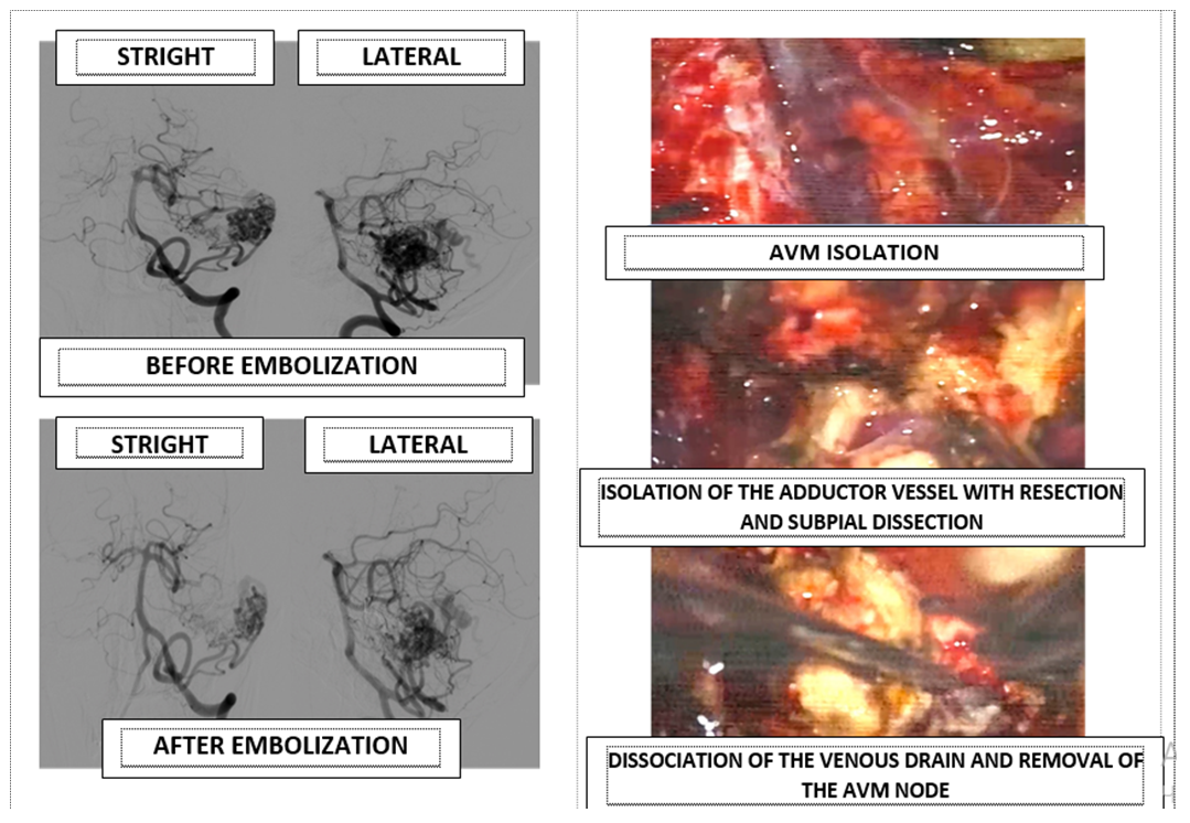
Figure 4. Combined technique of AVM treatment, endovascular embolization of the adductor vessels and partly of the AVM “nidus” followed by microsurgical resection
Preoperative embolization can increase resectability and reduce the risk of complications in patients with AVM grade III and higher, which is achieved by turning off hard-to-reach sources of blood supply and corresponding intra- and postoperative complications [14]. The advantage of the combined technique of surgical treatment of AVM is a decrease in the risk of complications that are observed with the use of each method separately [13]. It should also be noted that the risk of hemorrhage in the AVM bed in the postoperative period is sharply reduced when the two methods are combined from 40% to 18%, respectively [13,15,16].

6. Conclusions

Thus, a detailed study of the anatomical structure of the AVM, and representation in three-dimensional space using three-dimensional reconstructive anatomy, the correct choice of craniotomy and angle of attack depending on the localization of the AVM, as well as the correct shutdown of the adductor vessels and subpial dissection, as well as a combination of endovascular embolization with subsequent surgical resection, which allows not only to increase the resectability of the AVM, to minimize the trauma of the medulla and surrounding vessels in the surgical corridor, without begging for postoperative results of surgery.

References

[1]  Al-Shahi R., Warlow C. A systematic review of the frequency and prognosis of arteriovenous malformations of the brain in adults. Brain 2001; 124: 1900-26.
[2]  Laakso A., Dashti R., Seppänen J., Juvela S., Väärt K., Niemelä M., Sankila R., Hernesniemi J. Long-term excess mortality in 623 patients with brain arteriovenous malformations. J Neurosurg. 2008; 63(2): 244 – 255.
[3]  Pasqualin A., Zampieri P., Nicolato A., Meneghelli P., Cozzi F., Beltramello A. Surgery After Embolization of Cerebral Arterio-Venous Malformation: Experience of 123 Cases. Acta Neurochirurgica. 2014; 119: 105-111.
[4]  Lawton M.T., Kim H., McCulloch C.E., Mikhak B., Young W.L. A supplementary grading scale for selecting patients with brain arteriovenous malformations for surgery. Neurosurgery. 2010; 66(4): 702-713.
[5]  Lawton M. Seven AVMs Tenets and Techniques for Resection. Thieme New York • Stuttgart 2014.
[6]  Yasargil M.G. Microneurosurgery vol. 3B Stuttgart Georg Thieme Verlag.; 1988.
[7]  Redekop G., TerBrugge K., Montanera W., Willinsky R. Arterial aneurysms associated with cerebral arteriovenous malformations: classification, incidence, and risk of hemorrhage. J Neurosurg. 1998; 89(4): 539-546.
[8]  Hamilton M.G., Spetzler R.F. The prospective application of a grading system for arteriovenous malformations. J Neurosurgery. 1994; 34: 2-7.
[9]  Rennert R., Steinberg J., Cheung V., Santiago-Dieppa D., Pannell J., Khalessi A. Comprehensive endovascular and open surgical management of cerebral arteriovenous malformations. J Visualized experiments 2017; 128: 1-7.
[10]  Drake CG. Arteriovenous malformations of the brain. The options for management. N. Engl. J. Med. -1983; 309(5): 308-310.
[11]  Hartmann A., Mast H., Mohr J.P., Koennecke H.C., Osipov A., Pile-Spellman J., Duong D.H., Young W.L. Morbidity of intracranial hemorrhage in patients with cerebral arteriovenous malformation. Stroke. 1998; 29(5): 931-934.
[12]  Kretschmer T., Heros R.C. Microsurgical Management of Arteriovenous Malformations. Neurological Surgery. 2011.
[13]  Beltramello A., Zampieri P., Riccardi G.K., Piovan E., Pasqualin A., Nicolato A., Foroni R., Gerosa M. Combined Treatment of Brain AVMs: Analysis of Five Years (2000-2004) in the Verona Experience. Interventional Neuroradiology. 2005; 11(1): 63-72.
[14]  Han P.P., Ponce F.A., Spetzler R.F. Intention-to-treat analysis of Spetzler-Martin grades IV and V arteriovenous malformations: natural history and treatment paradigm. J. Neurosurg. 2003; 98: 3-7.
[15]  Thapa A., Chandra P.S., Sinha S., Gupta A., Singh M., Suri A., Sharma B.S. Surgical interventions in intracranial arteriovenous malformations: Indications and outcome analysis in a changing scenario. J Neurology India 2009; 57(6): 749-755.
[16]  van Beijnum J., van der Worp H.B., Buis D.R., Al-Shahi Salman R., Kappelle L.J., Rinkel G.J., van der Sprenkel J.W., Vandertop W.P., Algra A., Klijn C.J. Treatment of brain arteriovenous malformations: a systematic review and meta-analysis. JAMA. 2011; 306, (18): 2011-2019.