American Journal of Medicine and Medical Sciences

p-ISSN: 2165-901X    e-ISSN: 2165-9036

2021;  11(5): 419-425

doi:10.5923/j.ajmms.20211105.13

Received: May 1, 2021; Accepted: May 21, 2021; Published: May 26, 2021


Clinical-Laboratory Markers of Progression of Non-Alcoholic Fatty Liver Disease

Abror Asrorovich Khamraev1, Dilnavoz Khasanovna Yuldasheva2, Zarifa Ramazonovna Mukhamedova3, Vokhid Zokhidovich Zokirov3

1Head of the Department of Internal Medicine, Tashkent Medical Academy, Tashkent, Uzbekistan

2Department of Propaedeutics of Internal Medicine, Bukhara Medical Institute, Uzbekistan

3Department of General Hygiene and Ecology, Bukhara Medical Institute, Uzbekistan

Copyright © 2021 The Author(s). Published by Scientific & Academic Publishing.

This work is licensed under the Creative Commons Attribution International License (CC BY).
http://creativecommons.org/licenses/by/4.0/

Abstract

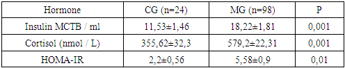
Non-alcoholic fatty liver disease (NAFLD) high prevalence, a tendency to an increase in morbidity, disease progression, coverage of the most efficient part of the population, the severity of clinical manifestations only in the late stages makes it possible to classify NAFLD as socially significant, of course, makes NAFLD one of the urgent problems of clinical medicine. The main feature of NAFLD is the absence of symptoms; the disease is often detected by chance, based on laboratory or instrumental tests. This article studied clinical and laboratory analyzes of 98 patients with non-alcoholic fatty liver disease. With steatohepatitis, higher ALT and AST values are noted than in patients with hepatic steatosis. The main differential difference between non-alcoholic steatosis and steatohepatitis, available in clinical practice, may be the severity of the biochemical syndrome of cytolysis. The article contains clinical features and prognosis in patients with fatty hepatosis and non-alcoholic steatohepatitis (NASH). Dyslipidemia (hypertriglyceridemia, decreased HDL, increased LDL) occurs in about 65-85% of patients. The basal insulin level, according to the results of our studies, in persons with NAFLD at the stage of steatosis and steatohepatitis was significantly increased.

Keywords: Non-alcoholic fatty liver disease, Liver steatosis, Non-alcoholic steatohepatitis, Insulin resistance, Obesity, Lipoprotein, Cholesterol, Alanine aminotransferase

Cite this paper: Abror Asrorovich Khamraev, Dilnavoz Khasanovna Yuldasheva, Zarifa Ramazonovna Mukhamedova, Vokhid Zokhidovich Zokirov, Clinical-Laboratory Markers of Progression of Non-Alcoholic Fatty Liver Disease, American Journal of Medicine and Medical Sciences, Vol. 11 No. 5, 2021, pp. 419-425. doi: 10.5923/j.ajmms.20211105.13.

1. The Urgency of the Problem

Currently, non-alcoholic fatty liver disease (NAFLD) is one of the most common diseases in hepatology, leading to a deterioration in the quality of life, disability and death. The overall prevalence of NAFLD in the population ranges from 10 to 40% [2,4,11,20,26,33,42]. NAFLD is in the focus of attention not only of general practitioners and gastroenterologists, but also of cardiologists, endocrinologists, and nephrologists, which is associated with the existence of a close relationship between the presence of NAFLD and a high risk of cardiovascular events, type 2 diabetes mellitus, chronic kidney disease [1,5,8,11,15,34,35,39]. The main feature of NAFLD is the absence of symptoms; the disease is often detected by chance, on the basis of laboratory or instrumental tests. NASH symptoms are nonspecific and do not correlate with its severity [38,42].
In the normal course of NAFLD, 12-40% of patients with hepatosis develop non-alcoholic steatohepatitis after 8-13 years. Of these, 15% of patients develop liver cirrhosis and liver failure. Of 7% of patients with liver cirrhosis, hepatocellular carcinoma develops within 10 years. The heterogeneity of NAFLD leads to the absence of a single generally accepted standard of treatment for such patients. Of course, all overweight patients should be advised to reduce their weight by eating a low-calorie diet and exercising regularly, and the effect of the latter seems to be more significant. Unhealthy lifestyles are the leading cause of NAFLD [2,3,11,12,18,24,27,28,30,33,36,41,43].
NAFLD occurs in all age groups, with a greater frequency in the 40-60 age group. Most often, NAFLD was detected in the following age groups: 50–59 years old (31.1%), 40–49 years old (23.6%), 60–69 years old (18.1%). The problem of an increase in the incidence of NAFLD is associated with a significant increase in the number of obese people. Moreover, the severity of the disease increases with an increase in the degree of obesity [4,11,16,17,23,33]. In obese individuals, NAFLD occurs in 30-100% of cases [16,22]. The prevalence of NAFLD among patients with type 2 diabetes mellitus (DM) reaches 70% [13,31]. NAFLD is a very diverse group of diseases, essentially differing in the combination of etiopathogenetic factors, the rate of progression and prognosis. In some cases, there is a steadily progressive course with the development of liver cirrhosis, in some cases - hepatocellular carcinoma.The most common risk factors in the NAFLD population were dyslipidemia (type 2 according to Friedrichsen) - 75.9% of patients, arterial hypertension - 69.9% and hypercholesterolemia - 68.8% [4,33,39,41,42]. NAFLD is a chronic multifactorial liver disease with a tendency to progression, which can be presented as hepatic steatosis, steatohepatitis, or liver cirrhosis. The prevalence of dyslipidemia among patients with NAFLD reaches 75%, and these figures are twice as high as in the population [5,9,21]. In 2003, at the I World Congress on Insulin Resistance in Los Angeles, it was suggested that NAFLD, along with obesity, type 2 diabetes, dyslipidemia, arterial hypertension, is a component of metabolic syndrome. The reasons for the development of NAFLD are varied, but more often there is a combination [8,10,19,33,37,40,42]. The main etiological factors of NAFLD are: eating foods high in saturated fat; low physical activity; hormonal disorders; primary and secondary insulin resistance (IR); obesity [4,7,34,38]. Thus, the high prevalence, the tendency to an increase in the incidence, the progression of the disease, the coverage of the most efficient part of the population, the severity of clinical manifestations only in the late stages allows us to classify NAFLD as socially significant, of course, makes NAFLD one of the urgent problems of clinical medicine [6,33,41,42,43].
Purpose of the study: To determine the clinical and laboratory features of non-alcoholic fatty liver disease at the stage of fatty hepatosis and steatohepatitis.

2. Materials and Research Methods

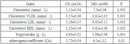
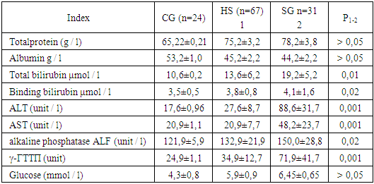
To solve the set tasks, 98 patients with NAFLD were examined, including 67 (68.3%) patients at the stage of hepatic steatosis (HS) and 31 (31.6%) patients with steatohepatitis (SH). Of these, 45 (46%) men and 53 (54%) women aged 20 to 75 years (average age 49.2 ± 4.2). The research results were recorded in the developed clinical information cards (questionnaire). The consent of the participants and members of the ethical committee for human rights in biomedicine at the Bukhara medical institute was obtained for the study. When selecting patients, we took into account the criteria for including and not including patients in the study. Criteria for the inclusion of patients in the study: - men and women aged 20 - 75 years; - the presence of fatty hepatosis and steatohepatitis; - the presence of a signed informed consent. The exclusion criteria were suspicion of alcohol or drug dependence, drug, viral, autoimmune liver damage, storage diseases, oncological diseases, severe diseases (uncorrected arterial hypertension (AH), type 2 diabetes in the stage of decompensation, chronic heart failure (CHF) III – IV functional class, heart attacks, strokes), pregnancy, lactation and low compliance. The criterion for not including from the survey was alcohol consumption in patients with fatty liver disease. We took into account the data of the anamnesis (absence of alcoholic beverages consumption regularly). We also used a special CAGE questionnaire [12,29].
We compared the results obtained in the course of the study with the indicators of the control group, formed of 24 apparently healthy individuals aged 20 to 65 years, who had no abnormalities in the hepatobiliary system. The diagnosis of non-alcoholic fatty liver disease was made on the basis of anamnesis, laboratory tests, and ultrasound examination of the liver. To detect NAFLD, ultrasound of the hepatobiliary system and liver elastography were performed. Ultrasound examination of the hepatobiliary system was performed in 500 patients with risk factors for NAFLD: obesity, dyslipidemia, impaired carbohydrate tolerance. Ultrasound of the liver revealed steatosis and steatohepatitis in 98 patients with NAFLD. The following signs of hepatic steatosis were noted: an increase in the size of the liver, an increase in its echogenicity, a relatively reduced density of the liver compared to the spleen (hepatic-splenic index less than 1), a decrease in sound conductivity, and a deterioration in visualization of the branches of the portal and hepatic veins. Ultrasound elastography was performed in 98 patients in order to exclude fibrosis in the liver parenchyma. Lipid metabolism was studied in terms of serum cholesterol (CS), high density lipoprotein (HDL), low density lipoprotein (LDL), very low density lipoprotein (VLDL) and triglycerides (TG). The LDL and VLDL values were calculated using the formula: VLDL = TG / 2, LDL = CHCR- (VLDL + HDL). Based on the results obtained, the atherogenic coefficient (CA) was calculated using the formula:
CA= CS LDL + CS VLDL/ HDL. Determination of the degree of obesity was carried out according to the Quetelet index, calculating it by the formula: BMI = weight (kg) / height (m2).
The results obtained were processed statically using the Student's t-test and the difference was considered significant in those cases when p <0.05 was expressed.

3. Results and Discussion

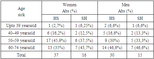
According to the results of our study, the ratio of women to men was 1.3: 1. The distribution of patients with hepatic steatosis and steatohepatitis by age is shown in Table 1.
Table 1. Distribution of steatosis and steatohepatitis among patients with NAFLD depending on age, n (%)
     
Analyzing the age criterion that SP occurs at any age, the able-bodied population is most susceptible to it (in persons from 40-59 years old - 55.2%, of them in women 34.3%, in men 20.8%; over 60 years old - in women 19%, in men over 60 years old - 20.8%), and FH occurs more often in older age (40-49 years old - 13%, in people over 60 years old - 45%). Demographic and anthropometric parameters were studied in all patients included in the study with non-alcoholic fatty liver disease (Table 2). When questioning patients, errors in nutrition were noted by 74 (76.5%) patients (irregular nutrition, abundant food, the presence of fatty and fried foods).
Table 2. Comparison of demographic and anthropometric parameters of indicators of the main and control groups
     
Patients in the observation group had increased body weight (Quetelet body mass index up to 30) DP in 29 (43%); SG 8 (26%) cases. Obesity I degree SP (body mass index (BMI) 30 - 34.9) was observed in 17 (25%), FH in 14 (45%) patients. Obesity II degree (BMI from 35 to 39.9) - in SP 15 (22.3%); SG 5 (16%). Obesity III degree (BMI 40 or more) - in SP 6 (9%); SG 4 (12.9%) patients. It is difficult to determine the duration of the onset of symptoms of liver pathology in a number of patients, since they did not present complaints from the hepatobiliary system and changes in the liver were revealed by chance during examination. Complaints that forced to see a doctor were associated with concomitant pathology (Table 3).
Table 3. The incidence of concomitant diseases of other digestive organs
     
We revealed the presence of 2-4 concomitant diseases of the digestive system in patients with fatty hepatosis and steatohepatitis. More common pathology of the gallbladder and pancreas, duodenum, gastroesophageal reflux disease (GERD), which can be explained by the anatomical and morphological features of the hepatopancreatoduodenal zone. Such an arrangement can cause damage and progression of pathological changes in these organs. The identified concomitant diseases were in remission.
To assess the characteristics of clinical manifestations, the first duty was to outline the circle of the leading symptoms of NAFLD, which constitutes the essence of the disease. Only then did they proceed to the analysis of each symptom of the disease. At the same time, two of the most important, in our opinion, qualities of clinical signs of NAFLD were subjected to a more thorough analysis: the frequency of occurrence and the degree of their perception. The results are shown in Table 4.
Table 4. Comparative assessment of the frequency of manifestations of clinical signs of NAFLD
     
The complex of the main clinical signs characteristic of NAFLD consisted of the following factors: aching pain, discomfort in the right hypochondrium, heartburn; nausea, belching, flatulence, constipation, mushy stools, fatigue, general weakness. As can be seen from Table 4, clinical signs of steatohepatitis in non-alcoholic fatty liver disease occur with a high frequency. Of these, aching pain, discomfort in the right hypochondrium 58.06%. The frequency of occurrence of another no less important clinical sign of SH, belching, turned out to be impressive. If, it was detected, in almost all (67.7%) patients with NAFLD with SH, then in patients with HS only 47.7%% of them. The detected contrast in the indicators of the spread of the eructation symptom in the presence of appropriate laboratory and instrumental supporting facts can serve as an important clinical criterion for differentiating SH and HS. As follows from the data presented in the table, the frequency of manifestations of NAFLD symptoms was different and clearly dependent on the stage of the disease. This dependence was more related to the manifestations of belching, aching pain, discomfort in the right hypochondrium, flatulence, constipation, to a lesser extent - bitterness in the mouth, mushy stools.
Among all the clinical signs of NAFLD, heartburn turned out to be more characteristic and at the same time stable. She, regardless of the stage of the disease, dominated the spectrum of clinical manifestations of NAFLD. So, if the frequency of manifestation of this symptom in patients with SH was 61.2%, then with HS it turned out to be slightly less and looked like 52.2%. The next NAFLD symptom worthy of attention was constipation. This clinical sign was observed much more often in patients with NAFLD (54.8%) with SH. At the same time, in NAFLD patients with HS, the prevalence of this symptom was an order of magnitude lower and amounted to 31.3%.
When studying the functional state of the liver, we were interested in the state of lipid metabolism of NAFLD. The level of total cholesterol (TCS) was assessed according to the classification of the European Atherosclerotic Society [17]: up to 5.2 mmol / l - normal level; 5.3-6.5 mmol / l - mild hypercholesterolemia (HCS); 6.6-7.8 mmol / l - moderate; more than 7.8 mmol / l - high. Determined the expanded lipid profile: triglycerides (TG), cholesterol (CS) low density lipoprotein (LDL) and high density lipoprotein cholesterol (HDL). The content of very low density lipoprotein cholesterol (VLDL) was calculated. According to the Russian recommendations (V revision) "Diagnostics and correction of lipid metabolism disorders for the prevention and treatment of atherosclerosis" [17], the normal TG level did not exceed 1.7 mmol / L, the target LDL cholesterol value was less than 2.6 mmol / L, cholesterol HDL is higher than 1.15 mmol / L. Lipid metabolism indicators are presented in Table 5.
Table 5. Indicators of lipid metabolism in patients of the surveyed groups
     
Disorders of lipid metabolism in NAFLD are one of the cardinal signs of the disease [17,33]. According to our data, severe HCS (more than 6 mmol / L) was recorded more often. Dyslipidemia in NAFLD was characterized by an increase in the level of triglycerides more than 1.9 mmol / L and in which the level of HDL cholesterol is <1 mmol / L. These disorders turned out to be more noticeable, which indicated more severe disorders of lipid metabolism. Lipid metabolism indicators are presented in the table. Judging by the data in Table 5, in patients with NAFLD at the stage of steatosis and hepatic steatohepatitis, significant changes in lipid metabolism were revealed towards an increase in cholesterol (p = 0.005), VLDL cholesterol (p = 0.001), LDL cholesterol (p = 0.001), TG (p = 0.001), CA (p = 0.03) and a decrease in HDL (p = 0.001). The results obtained indicate the presence of atherogenic dyslipidemia in NAFLD patients at the stage of steatosis and steatohepatitis. Atherogenicity is a concept that reflects the relationship between bad and good fats. The blood serum from the vein is examined, the indicators for calculating the coefficient are determined by the colorimetric photometric method [17,21,33]. Normally, the value ranges from 2.2 to 3.5. When calculating the coefficient of atherogenicity, experts use a simple formula:
Atherogenic coefficient (Atherogenic index) = (Total cholesterol - HDL) / HDL.
According to our data, severe coronary artery (more than 6 mmol / L) was more often recorded. The atherogenic index markedly exceeded the permissible values in all examined patients. In order to assess the functional state of the liver in NAFLD at the stage of fatty hepatosis and steatohepatitis, the parameters of pigment metabolism, cytolysis and cholestasis were studied (Table 6). Biochemical studies were carried out to determine the activity of alanine aminotransferase (ALT) and aspartate aminotransferase (AST), γ-glutamyltranspeptidase (GGTP), alkaline phosphatase (ALP), the content of total bilirubin and its fractions.
Table 6. Indicators of transaminase levels in the group of examined
     
The level of bilirubin was significantly increased relative to the indicators of the control group. The activity of HS cytolysis indices, the AST level reached 20.9, the ALT - 27.6. With steatohepatitis, there are higher ALT values 88.6 and AST 48.2 than in healthy individuals and patients with hepatic steatosis, so ALT in NASH exceeds 6-8 norms, AST exceeds 3-4 norms, with HS ALT exceeds 1- 2 norms, AST does not change significantly. The ALP activity in the SP was 132.9 U / L, which corresponded to the standard values (Table 6). The increase in alkaline phosphatase activity is 1.5-2.5 higher in patients with SH. Indicators of carbohydrate metabolism: the level of glucose in the blood serum was significantly increased (p> 0.05) in the patients we observed, since in the observation group in 25 patients (25.51%), among the comorbidities, there was a violation of tolerance to carbohydrates. In order to determine the degree of compensatoryness of the increased insulin level in NAFLD patients at the stage of fatty hepatosis and steatohepatitis, the HOMA-IR index was determined. The HOMA-IR score is a homeostasis assessment model for insulin resistance. Normally, the HOMA index does not exceed 2.7, and this indicator is the same for men and women, and after 18 years it does not depend on age either. During adolescence, the HOMA index slightly increases due to physiological insulin resistance at this age. Insulin resistance is a decrease in the susceptibility of insulin-sensitive tissues to the action of insulin when its concentration in the blood is sufficient. Insulin resistance has no specific symptoms. Insulin resistance can appear even in a person without obesity and diabetes - this happens in about 25% of cases. The indicator was calculated by the formula: [fasting insulin (IU / ml) × fasting glucose (mmol / l)] / 22.5. An indicator of less than 2 is considered normal [8,33,41]. In our study, the HOMA-IR insulin resistance index in patients was significantly increased (p = 0.01) in comparison with the control (Table 7).
Table 7. Dynamics of indicators of the level of hormones in blood serumin patients with NAFLD
     
The basal insulin level, according to the results of our studies, in individuals with NAFLD was significantly increased (p = 0.001) (Table 7).
Thus, on the basis of the studies carried out, it can be concluded that NAFLD is high prevalence, a tendency to an increase in the incidence, disease progression, coverage of the most efficient part of the population, the severity of clinical manifestations only in the late stages makes it possible to classify NAFLD as socially significant, of course, makes NAFLD one of the most relevant problems of clinical medicine. Clinical signs of aching pain, discomfort in the right hypochondrium, belching, heartburn, increased fatigue, general weakness and laboratory for the activity of cytolysis indicators in steatohepatitis, there are higher ALT, AST, ALP values than in patients with hepatic steatosis. The main differential difference between non-alcoholic steatosis and steatohepatitis, available in clinical practice, may be the severity of the biochemical syndrome of cytolysis. The activity of alkaline phosphatase (ALP) and gamma-glutamyltranspeptidase (GGTP) (its isolated increase occurs) are moderately increased in patients with SH.Dyslipidemia (hypertriglyceridemia, decreased HDL, increased LDL) occurs in about 65-85% of patients. The basal insulin level, according to the results of our studies, in persons with NAFLD at the stage of steatosis and steatohepatitis was significantly increased. Our correlation analysis revealed a positive relationship between hypercortisolemia in patients with NAFLD at the stage of fatty hepatosis, steatohepatitis and the level of total cholesterol and LDL, a negative relationship with HDL values. The presence of a connection between hypercortisolemia and hyperinsulinemia with atherogenic dyslipidemia and hyperinsulinemia with glucose parameters was shown. In order to determine the compensation of hyperinsulinemia in NAFLD patients at the stage of fatty hepatosis and steatohepatitis, the HOMA-IR index was determined. In our study, in patients with NAFLD at the stage of fatty hepatosis, the index of insulin resistance HOMA-IR was significantly increased in comparison with the control (p = 0.01).

References

[1]  Aijaz Ahmed M.D. Ryan B., Perumpail M.D., Stephen A., Harrison M.D. Hige prevalence of hepatic fibrosis in the setting of coexisting diabetes and hepatic steatosis: A case for selective screening in the general population? // Hepatology. – 2016. – № 63 (1). – P. 20 – 22. doi:10.1002/hep.28277.
[2]  Blachier M. LeleuH., PeckRadozavljevicM., ValvaD.C., RoudotThoraval F. The burden of liver disease in Europe: a review of available epidemiological data // J. Hepatol. – 2013. – № 58. – P. 593 – 608.
[3]  Byrne C.D.,Targher G. NAFLD: A multisystem disease // J. of Hepatology. – 2015. – №62. – P. 47 – 64.
[4]  Drapkina O.M., Ivashkin V.T. Epidemiological features of non-alcoholic fatty liver disease in Russia (results of an open multicenter prospective observation study DIREGL 01903) // RosZhurngastroenterolhepatolcoloproctol. - 2014. - T. 24. - No. 4. - P. 32-38.
[5]  Drapkina O.M., Deeva T.A., Volkova N.P., Ivashkin V.T. // Modern approaches to the diagnosis and treatment of non-alcoholic fatty liver disease // Therapeutic Archives 10, 2014. - P.116-123.
[6]  DoychevaI. Cui P., Nguyen E.A., Costa J., Hoocer H., Hoffich R., Bettencourt S., Brouha C.B., Sirlin R. Loomba Non-invasive screening of diabetics in primary care for NAFLD and advanced fibrosis by MRI and MRE // Aliment. Pharmacol. Ther. – 2016. – № 43(1). – P. 83 – 95.
[7]  EASL-EASD-EASO. Clinical Practice Guidelinesfor management of non-alcoholic fatty liver disease / J. of Hepatology. – 2016. – №64. – P. 1388 – 1402.
[8]  Eliseeva A. Yu. Non-alcoholic fatty liver disease in obesity and cardiovascular risk factors author. dis. PhD. Sciences: 14.01.02 - endocrinology / Minnullina Z.Sh - Moscow, 2012. -- 26 p.
[9]  Ivashkin V.T., Drapkina O.M., Maev I.V. et al. Prevalence of non-alcoholic fatty liver disease in outpatient patients in the Russian Federation: results of the Direg 2 study // Russian Journal of Gastroenterology, Hepatology and Coloproctology. - 2015. -- T.XXV. - No. 6. - S. 31 - 41.
[10]  Ivashkin V.T., Mayevskaya M.V. Lipotoxicity and other metabolic disorders in obesity // RosZhurngastroenterol, hepatol, coloproctol. - 2010. - T. 20, No. 1. - P. 4.
[11]  Ivashkin VT Diagnostics and treatment of non-alcoholic fatty liver disease // Methodical recommendations for doctors // Moscow. - 2015. -P. 38.
[12]  Ilchenko A.A., Dolgasheva G.M. Obesity as a factor in non-alcoholic fatty disease of the gallbladder (cholecystosteatosis, steatocholecystitis) // Experimental and Clinical Gastroenterology. - 2009. - No. 8. - S. 80 - 93.
[13]  Karpen S.J., Arrese М. Nuclear receptors, inflammation and liver disease: insights for cholestatic and fatty liver diseases // ClinPharmacolTher. – 2010. – Vol. 87, №4. – P. 473 – 478.
[14]  Kehiopulo K.F. // The relationship of hormonal and metabolic factors in type 2 diabetes mellitus against the background of obesity and non-alcoholic fatty liver disease // Journal of Grodno State Medical University № 3 2013. 33. - pp. 33-35.
[15]  Komshilova K.A., Troshina E.A. // Obesity and non-alcoholic fatty liver disease: metabolic risks and their correction // Obesity and metabolism. 2015; 12 (2): 35-39.
[16]  Komova A.T., Mayevskaya M.B., Ivashkin V.T. Principles of effective diagnosis of diffuse liver diseases on an outpatient basis // Ros journal gastroenterol., Hepatol., Coloproctol. - 2014. –T. 24, no. 5. - P. 36 - 41.
[17]  Krivosheev A.B., Kuimov A.D., and et al. // Disorders of lipid metabolism peculiaritiesin nonalcoholic fatty liver disease // Siberian Medical Review 04, 2016. - P.48-57.
[18]  Lazo M., Hernaez R., Bonekamp S Et al. Non-alcoholic fatty liver disease and mortality among US adults: prospective cohort study // BMJ. – 2011 – №343. – Р. 6891.
[19]  Lazebnik L.B., Zvenigorodskaya L.A. Metabolic syndrome and digestive organs // M.: Anacharsis, 2009. -184 p.
[20]  Makarov I.O., Borovkova E.I., Kazakov R.D. Prevalence of non-alcoholic fatty liver disease in obese pregnant women // Obstetrics, gynecology and reproduction. - 2012. - No. 4. - p. 18 - 21.
[21]  Minnullina Z.Sh. // Clinical - diagnostic value of bile acids in blood serum in liver stetosis // abstract of thesis. dis. PhD. Sciences: 14.00.05 / Minnullina Z.Sh - Kazan, 2018. - 24 p.
[22]  Micolasevic I., Orlic L., Franjic N., Hauser G., Stimac D. Milic Transient elastography (FibroScan) with controlled attenuation parameter in the assessment of liver steatosis and fibrosis in patients with nonalcoholic fatty liver disease – Were du we stand? // World J. Gastroenterol. – 2016. – № 22(32). – P. 7236 – 7251.
[23]  Mc Cullough A.J. The epidemiology and risk factors of NASH // Hepatology. – 2013. – Vol. 58, N 5. – P. 1644 – 1654.
[24]  Pavlov S.G., Glushenkov D.V., Bulichenko M.A. Non-alcoholic fatty liver disease in the clinic of internal diseases // BC. - 2010. - No. 28. - P. 1742 - 1749.
[25]  Palgova L. K., Baranovsky A. Yu., Ushakova T. I., Yurkina A. S., Blinov D. V. Epidemiological features of non-alcoholic fatty liver disease in the North-West region of Russia (Results of an open multicenter prospective study DIREG 2). VestnikSPbSU. Medicine, 2017, vol. 12, issue 2, pp. 118–135. DOI: 10.21638/11701/spbu11.2017.201.
[26]  Pacana T. The cardiovascular link to nonalcoholic disease // Clin. Liver Dis. – 2012. – №16. – Р. 599 – 613.
[27]  Podymova S.D. Modern view of the pathogenesis and the problem of treatment of non-alcoholic fatty liver disease // Experimental and clinical gastroenterology. - 2016. - No. 5. - P. 74 - 82.
[28]  Polunina T.E. Fatty infiltration of the liver // Effective pharmacotherapy. Gastroenterology. - 2014. - No. 3. - S. 32 - 40.
[29]  Plavinsky S.L., Boyarsky S.G., Barinova A.N. // Comparison of versions of the audit questionnaire for assessing alcohol consumption // Original scientific research. // Russia, 2012. -S. 41-46.
[30]  Ratziu V. Position statement on NAFLD/NASH based on the EASL 2009 Special Conference // – 2010. – Vol. 53. – P.372 – 384.
[31]  Rinella M.E. Nonalcoholic fatty liver disease: a systematic review // JAMA. – 2015. – №313. – P. 2263 – 2273.
[32]  Suchkova E.V. Khokhlacheva N.A., Vakhrushev Ya.M., Gorbunov A.Yu. Investigation of blood lipids in cholelithiasis // Gastroenterology of St. Petersburg. - Materials of the XIII Congress of the NOGR with international participation on March 11-12, 2013. - No. 1. - 2013. -- M28.
[33]  Suchkova E.V. Non-alcoholic fatty liver disease: clinical and laboratory features of the function of the liver and biliary tract, the effectiveness of combination therapy: author. dis. ... doc. Sciences: 14.00.05 / Suchkova E.V. - Izhevsk, 2017. - 43 p.
[34]  Suchkova E.V., Vakhrushev N.A., Khokhlacheva et al. // Assessment of the functional state of the liver in non-alcoholic fatty liver disease // Materials of the Twenty-second United Russian Gastroenterological Week, October 3-5, 2016, Moscow. - Russian Journal of Gastroenterology, Hepatology, Coloproctology - Appendix No. 48. - 2016. - T. 26. - No. 5. - P. 72.
[35]  Stepanova M.,Younossi Z.M. Independent association between nonalcoholic fatty liver disease and cardiovascular disease in the US population // ClinGastroenterolHepatol. – 2012. – №10. – Р. 646.
[36]  Tsochatzis E.A., Papatheodoridis G.V. Is there any progress in treatment of non-alcoholik fatty liver disease? // World J. Gastrointest. Pharmacol. Ther. – 2011. – Vol. 2. – №1. – P. 1 – 5.
[37]  Tarasova L.V., Tsyganova Yu.V., Opalinskaya I.V., Ivanova A.L. // Review of laboratory diagnostic methods used in non-alcoholicof fatty liver disease (NAFLD) and alcoholic liver disease (ALD) at the present stage // experimental and clinical gastroenterology | issue 164 | No. 4 2019. - P.72-76.
[38]  Cherkashina E.A. // Topical issues of diagnosis and treatmentnon-alcoholic fatty liver disease // Medical Council. 2015 | No. 4. –P.67 - 70.
[39]  Horoshinina L.P. Fatty degeneration of the liver and coronary heart disease. Geriatric aspects: monograph - Moscow: OOO Concept Design, 2014. - 346p.
[40]  Khuzhamuradov [et al.] Cholelithiasis in non-alcoholic fatty liver disease / M.N. // Russian journal of gastroenterology, hepatology, coloproctology. - No. 5. - 2015. - Materials of the Twenty-first United Russian Gastroenterological Week. - S. 90.
[41]  Хamrayev A.A., Yuldasheva D.H. Clinical, laboratory and molecular-genetic markers of the progression of non-alcoholic fatty liver disease (literature review and own data) // Society and innovations // Special Issue –2 (2021). – P. 399 – 406.
[42]  Yuldasheva D.H., Zokirov V.Z., G`ulomova Sh.Q. Non-alcoholic fatty liver disease: Modern view of the problem // A Multidisciplinary Peer Reviewed Journal. Vol.6. Issue 12. Dec.2020. – P. 286 – 292.
[43]  Yuldasheva D.H. Shadjanova N.S., Oltiboyev R.O. Non-alcoholic fatty liver disease and modern medicine // Academicia an international multidisciplinary research journal // Vol.10. Issue 11. Nov.2020. – P. 1931 – 1937.