-
Paper Information
- Previous Paper
- Paper Submission
-
Journal Information
- About This Journal
- Editorial Board
- Current Issue
- Archive
- Author Guidelines
- Contact Us
American Journal of Medicine and Medical Sciences
p-ISSN: 2165-901X e-ISSN: 2165-9036
2021; 11(4): 356-358
doi:10.5923/j.ajmms.20211104.20
Received: Apr. 2, 2021; Accepted: Apr. 27, 2021; Published: Apr. 30, 2021

Morphological Features of Thymus in Normality and with the Influence of a Gene-Modified Product in the Experiment
Dilnoza Khasanova Ahrorovna
Ph.D., Head of the Department of Anatomy, Bukhara State Medical Institute Named after Abu Ali Ibn Sino, Bukhara, Uzbekistan
Correspondence to: Dilnoza Khasanova Ahrorovna, Ph.D., Head of the Department of Anatomy, Bukhara State Medical Institute Named after Abu Ali Ibn Sino, Bukhara, Uzbekistan.
| Email: |  |
Copyright © 2021 The Author(s). Published by Scientific & Academic Publishing.
This work is licensed under the Creative Commons Attribution International License (CC BY).
http://creativecommons.org/licenses/by/4.0/

Changes in the immune system play one of the main roles in the aging of the body and the growth of particular age-related diseases. In the process of normal aging and, to a greater extent, pathological aging, the thymus-dependent link of the immune system, this includes both the thymus itself and the populations of T-cells developing in it, changes most strongly. The thymus is an important link in the immune defense, the function of which is aimed at maintaining the pool of peripheral T-lymphocytes. However, like no other organ of the immune system, it is susceptible to evolutive processes, and atrophy of the thymus can be acute or prolonged, chronic [1-4]. Regardless of the etiology of involution, there is a general pattern of pathological processes occurring in the thymus gland. Thymic atrophy is accompanied by a restructuring of the lobular architecture, a decrease in the amount of thymic parenchyma, its replacement with adipose and fibrous tissue, and a decrease in the number of peripheral thymocytes. Many of the contaminants that pose a potential risk in foods produced by new technologies using new or unconventional ingredients are contaminated, and their identification and safety assessment is essential. The study of food contamination with chemical and biological pollutants remains relevant [5-9]. Improving the technology of obtaining food products, obtaining products of a new generation and ensuring their long-term storage is one of the important tasks facing mankind today. Therefore, in subsequent years, genetically modified organisms (GMOs) appeared, with the help of genetic engineering, making changes in the genome of food products and creating products with new properties [10-13]. However, their influence on the organs and systems of the human body, the consequences of these influences are not fully understood. Taking this into account, the need to continue morphological, experimental studies on this problem has not lost its relevance.
Keywords: Thymus, Genetically Modified Organisms (GMO), Morphology, Experiment
Cite this paper: Dilnoza Khasanova Ahrorovna, Morphological Features of Thymus in Normality and with the Influence of a Gene-Modified Product in the Experiment, American Journal of Medicine and Medical Sciences, Vol. 11 No. 4, 2021, pp. 356-358. doi: 10.5923/j.ajmms.20211104.20.
1. Introduction
- The morphological changes in the thymus were studied experimentally in 65 white outbred female rats weighing 170-210 g at the Bukhara Medical Institute at the Central Scientific Research Laboratory. The material for the study was taken biopsies. For general morphology, pieces were excised from each thymus and solidified in 10% neutral formalin. After washing for 2–4 hour in running water, it was dehydrated in concentrated alcohol and chloroform, then embedded in paraffin and prepared blocks. On paraffin blocks, sections of 5-7 μm were cut, stained with hematoxylin and eosin. Semi-thin 1 µm sections were obtained from Epon bricks on a Leykaultramicrotomy. Histological preparations were examined under 10, 20, 40 lenses of a light microscope and the necessary areas were photographed.
2. Results and Discussion
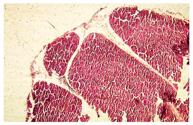
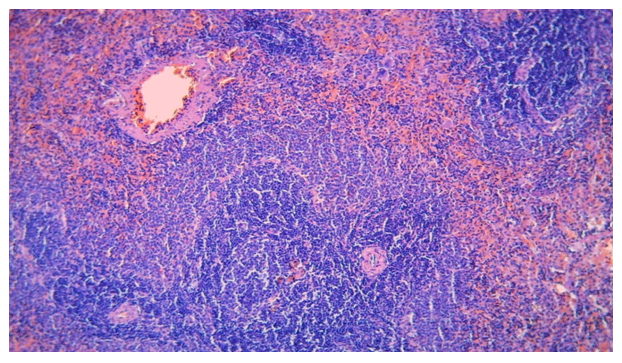
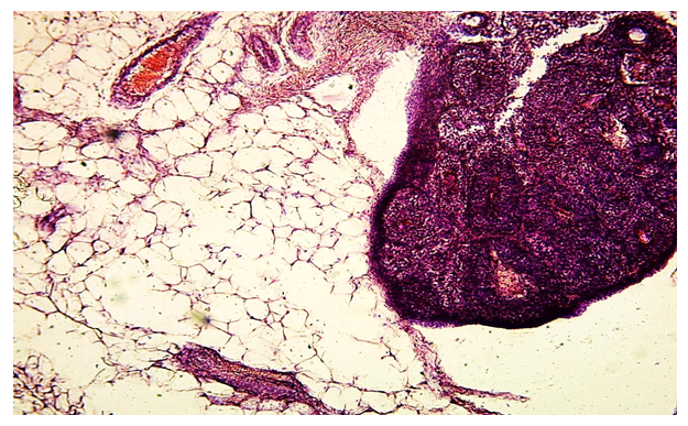
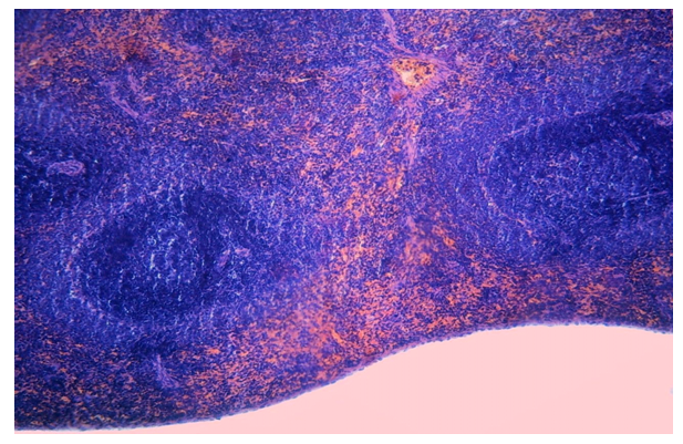
- Biopsy materials of 3-month-old female outbred rats were examined. These laboratory rats were kept in a vivarium, cared for in accordance with the "Rules for work using experimental animals." The rats were divided into two groups: intact animals (n = 25), rats with a course of GMO administration (n = 40). GMOs were prescribed at the rate of 0.1 per 1 kg of rat weight (corresponds to the average therapeutic dose for humans in terms of animal weight) 3 times a week for 3 weeks. Withdrawn from the experiment after 1.2.3 months after the end of the course of introducing GMOs by decapitation. The thymus was the object of the study.Histologically, as in the thymus, large foci consisting of thymocytes were determined, and in some places the absence of lymphocytes. The lesions had two components, both stromal and glandular structures: the ratio of these components varied depending on the types of these lymph nodes. With age, rats show involution of the thymus with the replacement of its parenchyma with adipose connective tissue, while the lobular structure of the thymus is preserved. Microscopic examination of the thymus after feeding with GMOs in animals showed that after a certain period of time after administration, a sharp reaction develops, i.e., underdevelopment, which progresses with an increase in the duration of the experiment. Thus, the results of morphological studies have shown that pathological changes are often found in rats when using GMOs. The existence of various changes in the thymus when using GMOs must be taken into account when choosing a treatment, as well as a program for the prevention of both immunodeficiency states and its complications.
 | Figure 3. A lobule of the thymus with a limited connecting septum, numerous blood vessels in the septa were also prominent. Stained with hemotoxylin and eosin. 10x10 |
3. Conclusions
- In white outbred rats, with the use of GMOs with prolonged use, there was a change in the cellular composition, T and B lymphocytes. When studying the immune status in rats under the influence of GMOs, significant violations were revealed in the form of a sharp decrease in the number of T-lymphocytes, which would allow a characteristic long-term asymptomatic or low-symptom course with the subsequent rapid development of the clinical picture and the appearance of indications for immunodeficient states. Thus, the effect of GMOs on the thymus leads to selective damage between the parenchyma and the stroma, probably the basis of these changes was the specific reaction of the thymus in response to local introduction of GMOs, which led to a weakening of the body's immunity, the emergence of allergic reactions, the formation of resistance to antibiotics, and a decrease in efficiency of the treatment of diseases, the development of pathologies associated with the process of cumulation in the human body after consumption.
 Abstract
 Reference
 Full-Text PDF
 Full-text HTML

