-
Paper Information
- Next Paper
- Paper Submission
-
Journal Information
- About This Journal
- Editorial Board
- Current Issue
- Archive
- Author Guidelines
- Contact Us
American Journal of Medicine and Medical Sciences
p-ISSN: 2165-901X e-ISSN: 2165-9036
2021; 11(4): 293-296
doi:10.5923/j.ajmms.20211104.08
Received: Feb. 6, 2021; Accepted: Mar. 29, 2021; Published: Apr. 2, 2021

Results of Morphological Features of the Structure of the Kidneys after Severe Craniocerebral Injury
 Abstract
 Reference
 Full-Text PDF
 Full-text HTML
Khuseynova Gulshan Khuseynovna
Bukhara State Medical Institute Named after Abu Ali ibn Sino, Department of Clinical Anatomy and Forensic Medical Examination
Correspondence to: Khuseynova Gulshan Khuseynovna, Bukhara State Medical Institute Named after Abu Ali ibn Sino, Department of Clinical Anatomy and Forensic Medical Examination.
| Email: |  |
Copyright © 2021 The Author(s). Published by Scientific & Academic Publishing.
This work is licensed under the Creative Commons Attribution International License (CC BY).
http://creativecommons.org/licenses/by/4.0/

This article provides information on the results of scientific studies that allow assessing and studying the morphological features of the kidneys of 3-month-old white outbred rats with severe traumatic brain injury. Traumatic brain injury was performed using the Traffic Accident model, and rat kidneys were removed for further morphological examination. Drawing of an injury does not lead to macroscopic changes on the part of the kidneys and other organs adjacent to it. The study of histological preparations of the kidneys in laboratory rats after severe traumatic brain injury revealed pronounced changes in blood flow, vascular congestion and structural changes in the renal parenchyma, in particular, expansion of capillaries and veins, hemorrhages.
Keywords: Traumatic brain injury, Kidney, Macroscopy, Microscopy, Topography, Abdominal cavity
Cite this paper: Khuseynova Gulshan Khuseynovna, Results of Morphological Features of the Structure of the Kidneys after Severe Craniocerebral Injury, American Journal of Medicine and Medical Sciences, Vol. 11 No. 4, 2021, pp. 293-296. doi: 10.5923/j.ajmms.20211104.08.
1. Introduction
- To date, severe traumatic brain injury attracts the attention of many researchers around the world [1,7,9,10]. The main percentage, on average, 30–40% in terms of the ratio of injuries is traumatic brain injury [5].Severe traumatic brain injury often causes damage to the basal structures of the brain, while central reflex and humoral changes occur throughout the body. As a result, already in the first minutes after traumatic brain injury, microcirculation disorders occur throughout the body, the same changes lead to systemic damage to all internal organs, ultimately causing multiple organ failure [8].The reasons for the high mortality rate in patients with traumatic brain injury are in many cases associated with the development of especially often intra- and extracranial complications, that is, central reflex and humoral changes throughout the body. Clinical studies prove that after traumatic brain injury, changes and complications in the kidneys are most often mainly of an infectious and inflammatory nature [4]. But, signs of damage to the parenchyma in the kidneys after traumatic brain injury are observed more often. After traumatic brain injury in the kidneys, dyscirculatory changes initially develop in dynamics, these disorders are observed especially at the border of the kidney layers [2].Morphological examination of biopsy specimens is an important method for studying the state of renal tissue in pathology and for predicting the course of the disease. Analysis of morphological changes in various parts of the kidneys during experimental modeling of traumatic brain injury is one of the most important problems of modern nephrology. Currently, in the literature available to us, we have not found data on morphological changes in various areas and tissues of the kidneys in traumatic brain injury, and there are no morphological changes that occur in various kidney tissues, taking into account the periods of traumatic brain injury. Therefore, all this requires a detailed study of morphological changes in various parts and tissues of the kidneys after traumatic brain injury. When modeling renal pathology, the authors use morphological parameters to assess [3,6]. The purpose of our work is to study the morphological parameters of the structures of the renal parenchyma after traumatic brain injury.
2. Materials and Methods
- For the study, laboratory white outbred rats were used: 20 males and 20 females of three months of age, the average weight of which was 105.6 ± 13.3 g. The animals were kept in a vivarium at room temperature with a 12-hour light / dark cycle, in accordance with the norms keeping laboratory animals. All animals of the experimental groups were anesthetized under light ether anesthesia and divided into 2 groups. The first group consisted of animals that were fixed in the installation, but did not inflict injury (control, n = 20). The second group consisted of (experimental, n = 20) - animals that underwent TBI. A traffic accident model (1-Fig) was used to inflict injuries for this animal experience. In this experiment, rats were attached to a handcrafted wheeled vehicle. For this experiment, laboratory rats were fixed on a wheeled vehicle, which was made by hand to avoid jaw fracture, the head of the animal was fixed on a soft pillow. The experimental rats attached to the vehicle accelerated at a speed of 6.7 km / h and hit a wooden barrier with their frontal part of their heads (2-Rice). After injury, the animals were moved into a special plastic cage and observed until normal behavior was restored. During the period of this experiment, instant death was observed in 6 animals after inflicting a craniocerebral injury, the remaining 34 animals were seriously injured. The surviving animals, 30 minutes after the injury, returned to normal life and nutrition. During the experiment, these animals were decapitated on the first and third days after the infliction of traumatic brain injury, the abdominal cavity was opened, and the kidneys were detached for further study. For histopathological comparison of control and experimental groups, samples were taken on days 1, 3 of postnatal development after TBI. These preparations were prepared using standard histological techniques. The preparations were stained with hematoxylin and eosin. Microscopy of the preparations in transmitted light was carried out using a trinocular microscope with a microscope magnification of × 60, × 80. Capturing histological images was performed using a microscope camera. The analysis of the obtained images was carried out using specialized software for medicine, using specialized software for medicine. All experiments carried out on laboratory animals were carried out according to the 1964 Medical Association, as well as the declarations adopted in 1975, 1983, 1989, 1996, 2000, 2002, 2004, 2008-2013.
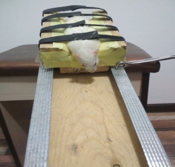 | Figure 1. The horizontal arrangement of the experimental object before the infliction of traumatic brain injury |
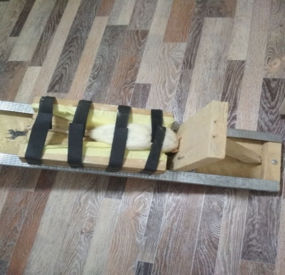 | Figure 2. The horizontal arrangement of the experimental object after traumatic brain injury |
3. Results and Discussions
- At the time of observation, the kidneys of the rat are smooth, one papillary, bean-shaped, red-brown in color. The weight of each of the kidneys is approximately 0.45-0.5 g, together 0.95-1.0 g. The size of the kidneys is variable. The kidneys of a 3-month-old rat averaged 14-16 mm long, 5-8 mm wide and 3-5 mm thick. The kidneys are located in the lumbar region. They are located in front of the twelfth thoracic - second lumbar segments (4-Fig). The lobulation of the kidneys in these laboratory rats is poorly visible, the cranial and caudal edges of the kidneys are dull. The kidney has a convex lateral and concave medial margin. In addition, the outside of the kidney is covered with dense fibrous connective tissue and poorly expressed fatty membranes, a serous membrane lying on the ventral surface of the organ (3-Fig). After the experiment, the kidney was removed for further study.
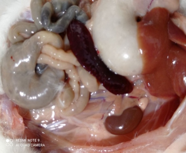 | Figure 3. Topographic picture of the kidneys (Top view) |
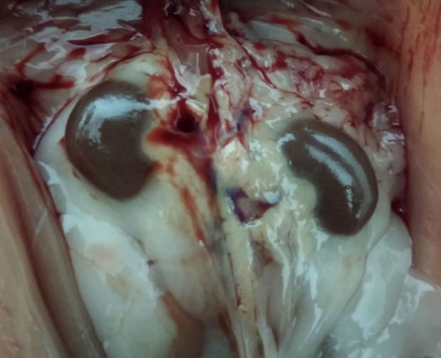 | Figure 4. The location of the kidneys in the lumbar region (top view) |
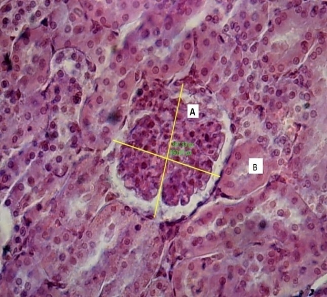 | Figure 5. Control group. Kidney: A - the glomerulus of the kidney, B - the tubule of the kidney. UV 60 x 80. Coloring by G.E. |
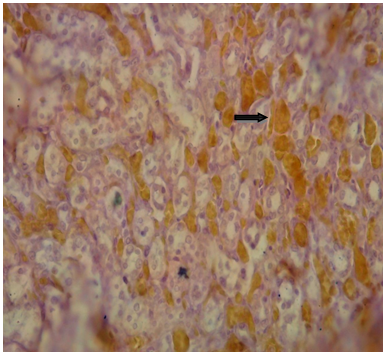 | Figure 6. Hemorrhage in the proximal and distal tubules. Coloring G.E. |