-
Paper Information
- Paper Submission
-
Journal Information
- About This Journal
- Editorial Board
- Current Issue
- Archive
- Author Guidelines
- Contact Us
American Journal of Medicine and Medical Sciences
p-ISSN: 2165-901X e-ISSN: 2165-9036
2021; 11(2): 148-153
doi:10.5923/j.ajmms.20211102.18
Received: Feb. 5, 2021; Accepted: Feb. 26, 2021; Published: Feb. 28, 2021

The Importance of Treatment Aimed at the Dynamics of Cartilage Oligomer Matrix Protein (COMP) in Patients with the Knee Joint Osteoarthritis
 Abstract
 Reference
 Full-Text PDF
 Full-text HTML
Sagdiana Buranova 1, Khalmurad Akhmedov 2, Feruza Razakova 3
1Assistant to Chair, Tashkent Medical Academy, Uzbekistan
2Doctor of Sciences, Professor, Tashkent Medical Academy, Uzbekistan
3National University of Uzbekistan Named after Mirzo Ulugbek, Uzbekistan
Correspondence to: Sagdiana Buranova , Assistant to Chair, Tashkent Medical Academy, Uzbekistan.
| Email: |  |
Copyright © 2021 The Author(s). Published by Scientific & Academic Publishing.
This work is licensed under the Creative Commons Attribution International License (CC BY).
http://creativecommons.org/licenses/by/4.0/

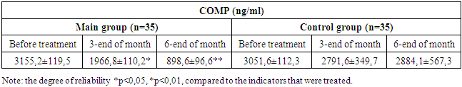
The study involved 50 patients aged 54.5±4.4 years with knee osteoarthritis and who accounted for 43.8% of the X-ray 0 stage of the disease, Phase I-40% and Phase II-16.2%, which were allocated to the main and control groups. It is implemented that for reducing the amount of cartilage oligomer matrix protein (COMP) in the primary group applied treatment and traditional treatment for control group. According to the results obtained, the application of the proposed method in the knee joint osteoarthritis (OA) differs from the traditional method by a decrease in the amount of COMP in the serum (blood) of patients, it plays an important role in improving and maintaining the functional activity of the joints. In this, treatment aimed at the dynamics of COMP differs from the traditional method by the fact that the cartilage absorbs the stress of decomposition processes.
Keywords: Osteoarthritis, Cartilage oligomer matrix protein, X-ray changes
Cite this paper: Sagdiana Buranova , Khalmurad Akhmedov , Feruza Razakova , The Importance of Treatment Aimed at the Dynamics of Cartilage Oligomer Matrix Protein (COMP) in Patients with the Knee Joint Osteoarthritis, American Journal of Medicine and Medical Sciences, Vol. 11 No. 2, 2021, pp. 148-153. doi: 10.5923/j.ajmms.20211102.18.
1. Introduction
- Currently, despite the rapid development of theoretical and practical medicine, chronic diseases of the musculoskeletal system in the activity of doctor cause great difficulties.In particular, on the basis of which cartilage tissue fragmentation, bone structure remodeling, osteophytosis, and inflammatory processes, lying osteoarthritis (OA) is characterized by its clinical appearance and features due to anatomo-physiological disorders in the joints, which determine the social significance and relevance of the problem, especially in middle-aged patients with early onset of malnutrition [1,3]. Moreover, more than half of the population of developed countries with more than 50 years of age and more than 60% of the population with more than 65 years of age suffer from these diseases, which in turn is a serious problem that must be solved in the health system of countries.According to information presented in the existing literature of recent years [3,4,7], inflammation on the basis of OA pathogenesis occurs irreversible erosions in the bones under the aggressive influence of cytokines, namely IL-1, IL-6 and FNO-α, and this in turn causes the emergence of degenerative changes in the cartilage. This process stimulates the synthesis of enzymes of collagen and matrix metalloproteinase and causes the decomposition of 2-type collagen, one of the biochemical properties of OA [4,7,13]. According to scientific research in recent years [5,6,12], cartilage oligomer matrix protein (COMP) provides important information on metabolic changes occurring under the influence of the above-mentioned ferments on the cartilage matrix. Indeed, the increase in COMP blood serum causes the formation of thoughts about how to serve as a biomarker in the early detection of this disease. It remains to be noted that the study of dynamic changes in COMP blood serum in relation to the clinical course of OA is of scientific and practical interest.Currently, despite the fact that important pathogenetic processes in OA is being studied, doctors have many difficulties in the treatment of this disease in practice. As practice experience shows, nonsteroidal anti-inflammatory drugs and chondroprotic agents, which are widely used as OA treatment methods, do not solve all problems. Indeed, in the literature data [6,8] there are conflicting views on these methods of treatment. These methods have a certain positive effect on the clinical improvement of the patient and the quality of his or her life. According to many authors [7,14], the level of pain syndrome in OA may not be correlated with the radiographic image and stage of the disease: in 74% of patients with the initial stage of OA of the knee joint, the level of pain syndrome and functional disorders is observed, just as in the last stage of OA, the existing method of treatment more often serves to reduce pain in the joints and to a certain extent improve its functional state [8,10,11]. On the other hand, existing methods are characterized by a lot of cost on the patient side and do not affect the reduction of indications for endoprosthesis in patients [9,13]. This condition, in turn, attracts attention to its important aspects in the pathogenesis of the disease. However, it is good to reduce the level of COMP in the coordination of cytokines, collagenase and matrix metalloproteinases in the development of the pathological process. In present-day literature, this problem is not widely covered, but some sources [10,12,15] describe the experimental results of some of the tools that affect matrix metalloproteinase. However, this can’t fully mean the solution of the problem. Therefore, a complete study of the effectiveness of the clinical course of the disease in the treatment aimed at reducing the amount of COMP from a pathogenetic point of view is of great scientific and practical interest.The purpose of the study: Determining efficacy the clinical course of the treatment method, aimed at the dynamics of cartilage oligomer matrix protein (COMP) in patients with knee joint OA.
2. Materials and Methods
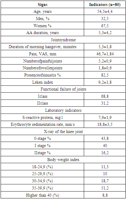
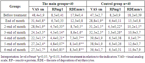
- The study involved 50 patients with primary OA of the knee joint, aged 41-65 (average 54.5±4.4), with an average duration of 5.3±4.2 years. Laboratory examination of all patients was performed with standard X-ray of the knee joint in the right (anterior posterior) projection and Kellgren modification method was used to determine the Radiological stage of the OA. In this there was 43.8% of OA X-ray 0, stage I – 40% and Stage II – 16.2% of patients. All patients with OA were divided into 2 groups according to the method of treatment and they were mutually matched by age and gender.The main group (n=40) - consisted of 56,4±3.9 years old patients with OA treated according to the above-mentioned COMP dynamics. The following measures were taken to them:- Physical education therapy- Measure to reduce body weight index- Medicamentous treatment – the use of garpagofitum (Sustavin) on the basis of chondroprotaes.3 exercises are recommended. These exercises are aimed at increasing the formation and volume of muscles located around the knee joint and not actively involved in the movement of a person. All patients were recommended a hypocaloric diet with a fat content of <30%, carbohydrates 50-55%, proteins - 15-20%, with a calorie deficit of 500-600 kcal compared to the calculated indicator. The drug garpagofitum (Sustavin), which is produced in Uzbekistan and has a significant role in reducing the synthesis of matrix metalloproteinase, was used in the formation of treatment aimed at reducing the amount of COMP. In this OA in the X-ray 0-stage: in a situation where COMP blood serum is more than 1000 n/ml, patients under the age of 50 take the same drug 250 mg for 3 months per day (if the amount of COMP is 1000 n/ml. up to 6 months); in patients aged 50-65 years in a situation with an excess of 1500 n/ml, 500 mg was administered daily for 6 months, and in adults older than 65 years with an excess of 2000 n/ml, 750 mg was administered daily for 6 months. In turn, in the radiographic I and II stages of OA, all patients were recommended 750 mg per day for 6 months, 750 mg per day for 6 months. Alternatively, all patients received chondroitin sulfate from 500 mg 2 times a day for 3 months.Control group (n=40) – 53.1±6.4 patients with OA treated by conventional method. The following measures were taken to them:- maintaining the right lifestyle, make the diet and weight, complex treatment to adhere to the physical exercises.- reception of non-steroidal anti-inflammatory drugs (nimesulide from 14 mg 2 times a day for 2 days and on subsequent days, depending on the need).- reception of chondroprotins per os for 6 months (chondroitin sulfate 500 mg 2 times a day).Criteria for attracting patients to the study:1. Primary OA diagnosis based on EULAR/ACR criteria;2. Patients older than 40 years;3. Radiographic stages of knee joint 0, I and II;4. I – III functional class;Criteria for exclusion of patients for the study:1. the fact that OA was not treated using surgical method before the scientific research work and during the examination;2. secondary AA;3. severe concomitant pathology (kidney, liver, heart failure, high levels of uncontrollable silver (AG), decompensated diabetes and etc.); injuries, malignant tumors, alcohol abuse, mental disorders, as well as cases of dementia and cognitive impairment;4. the presence of allergic reactions to individual components of the drug sustavin;5. knee joint III-IV radiographic stages;6. the fact that he took anti-inflammatory or painkillers in the interval of 10 days before the study.Determination of the effectiveness of the methods used was carried out for 6 months, and the following indicators were taken into account in this:- Evaluation of knee joint syndrome dynamics based on VAS (visual analog scale);- Leken algo-functional index; СОМР миқдори динамикаси.- Radiography of the knee joint.- The level of oligomer matrix protein of cartilage was studied using the immunoferment analysis method (ELISA, Russia).The results obtained from the research were statistically processed using the STATISTICA 6.0 software package as well as using the built-in statistical processing methods.
3. Results and Discussion
- The main share among the patients with OA who were examined was 54 (67.5%) women, and those who were ill for less than 5 years accounted for 61 (76.2%), those who were more than 5 years accounted for 19 (23.8%). In addition to the syndrome of joints, symptoms such as general malaise (31,3%), irritability, sleep and attention impairment (52,5%), irritability and fear (43,8%) are noted. Alternatively, in 87,5% of patients, level I anemia was detected.According to the analysis of the data on the disease Anamnesis, the average age of patients was 41,3±2,8 at the time of occurrence of the first symptoms of OA (early stage). The average period before this diagnosis with the appearance of the first symptoms was 24 months. During this time interval, patients were referred to various doctors up to 6±1.4 marotaba. Of these, 15% were diagnosed with OA within 6 months of the appearance of the first symptoms of the disease, every 5 - patients (20%) on average 10 months after the onset of the disease, 1/3 of them (31.3%) — 24 months, 34% knew about their diagnosis only after 3 years (36 months).Disorders of the functional state of the joints can also be associated with dynamic changes in the degenerative process in the cartilage. As can be seen from Table 1, 68.8% of the patients involved in the study consisted of I functional class and II of 31.2%. In turn, the leken algo-functional index consisted of 9,2±1,8. The main share of patients, that is, 51.2% obesity II degree.
|
|
|
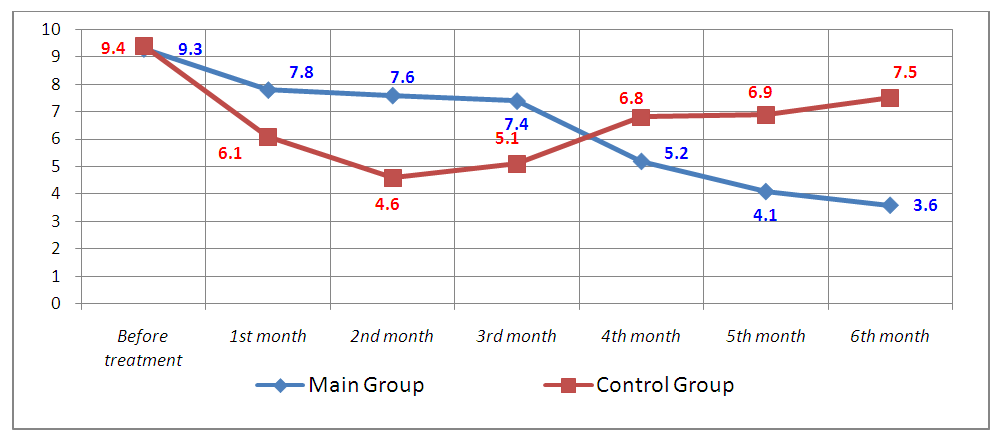 | Figure 1. Leken algo-change in the course of treatment of the functional index |
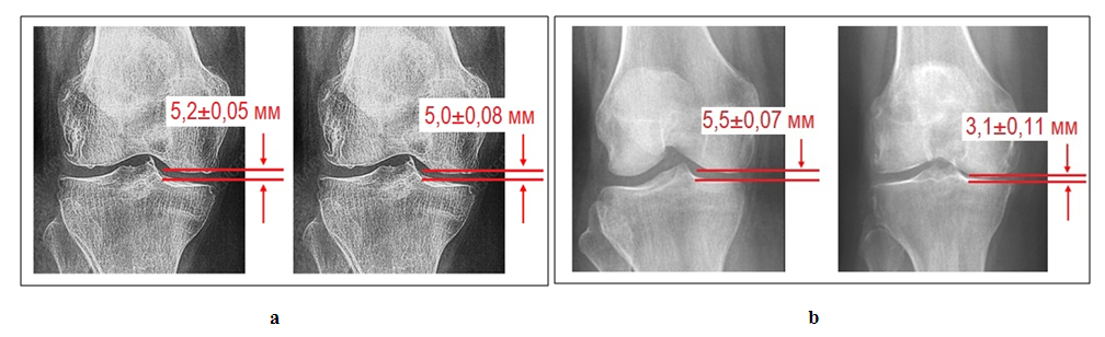 | Figure 2. In patients with knee joint OA, after a year in the course of treatment, an image of an X-ray (measurement of the width of the joint slit) is obtained. a – the main group and b –on the example of control group (on the left - before treatment, on the right – after a year) |
4. Conclusions
- The use of the proposed method in the knee joint OA differs from the traditional method by a decrease in the amount of COMP in the blood serum of patients, which plays an important role in improving and maintaining the functional activity of the joints. In thistreatment aimed at the dynamics of COMPdiffers from the traditional method by the fact that the cartilage absorbs the stress of decomposition processes. Alternatively, this method allows patients to reduce the need for non-steroidal anti-inflammatory drugs and reduce the appeal to the doctor on the treatment of the joints throughout the year.