I. K. Rustamova 1, S. A. Kasimova 2, N. K. Kayumova 2
1Associate Professor, Department of Neurology, Andijan State Medical Institute
2Assistant at the Department of Neurology, Andijan State Medical Institute
Correspondence to: I. K. Rustamova , Associate Professor, Department of Neurology, Andijan State Medical Institute.
| Email: |  |
Copyright © 2021 The Author(s). Published by Scientific & Academic Publishing.
This work is licensed under the Creative Commons Attribution International License (CC BY).
http://creativecommons.org/licenses/by/4.0/

Abstract
Ultrasonic assessment of blood flow in the cerebral arteries is the leading research method for ischemic stroke. The quality of life after a stroke largely depends on the patient's psycho-emotional state. We examined 112 patients with ischemic stroke. The relationship between hemodynamic parameters and the severity of anxiety and depression was pronounced. The results showed a relationship between depression and hemispheric lateralization of the lesion.
Keywords:
Ischemic stroke, Hemodynamics, Anxiety, Depression
Cite this paper: I. K. Rustamova , S. A. Kasimova , N. K. Kayumova , The State of Cerebral Hemodynamics and Emotional Disorders in Ischemic Brain Stroke, American Journal of Medicine and Medical Sciences, Vol. 11 No. 1, 2021, pp. 53-56. doi: 10.5923/j.ajmms.20211101.13.
1. Introduction
In Uzbekistan, more than 60 thousand cases of stroke are recorded annually (acute cerebrovascular accident). Moreover, disability after a stroke is 83.8%, and the percentage of hospital mortality is 17.3%. According to the World Health Organization, in economically developed countries, acute cerebrovascular accident takes third place among the causes of death, second only to coronary heart disease and cancer.The prognosis of recovery from a stroke is largely determined by the state of higher mental functions (Lincoln N.B. et al, 1989). The importance of neuropsychological studies after a stroke is emphasized in many works (A.N. Bogolepova, 2003, I. Fedin, 2002). D.R. Shtulman, 2002 considers that affective disturbances arising against the background of a developed catastrophe in the form of a stroke have an important influence on the patient’s condition [4].A comprehensive diagnosis of vascular diseases of the brain is based on the joint use of methods of radiation and ultrasound diagnostics. In this case, the leading place is taken by ultrasound assessment of blood flow in the cerebral arteries as the most mobile method for screening, dynamic observation and monitoring of patients with acute cerebrovascular insufficiency [1,2,3].
2. Purpose of the Study
Identify correlation relationships between hemodynamic parameters and the severity of anxiety and depression.
3. Research Methods
We observed 112 patients (mean age 63.8 + 7.2) who had an ischemic stroke with hemispheric lateralization of the lesion. Ultrasonic dplerographic research was carried out by the “Labodon” device of Naval Forces firm (France).We studied the blood flow characteristics of the vertebral, internal carotid and common carotid arteries in order to identify its disorders, and also studied the sonographic image of the arteries. In this case, the parameters of the dopplerographic spectrum were estimated. To identify anxiety and depression, we used the Hospital Anxiety and Depression Scale (Zigmond A.S., Shaith R.P., 1983), which was developed as a tool for identifying and assessing the severity of depression and anxiety in general medical practice.
4. The Results of the Study
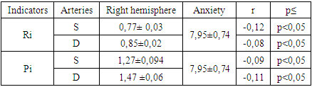
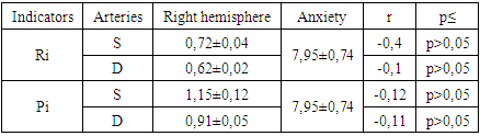
The results of the study showed that an important aspect of correlation relationships is the correlation between hemodynamic parameters and the degree of severity of anxiety. Table 1. Indicators of linear blood flow velocity (BFV) (according to S.E. Lelyuk, R.T. Lelyuk 1995) and anxiety in the common carotid artery (CCA) in case of a right hemisphere stroke
 |
| |
|
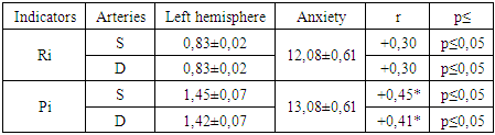
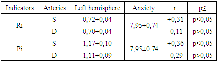
As can be seen from table №1, in case of a right hemisphere stroke, all the correlation relationships analyzed are negative and statistically significant both in the right and in the left carotid basin. In other words, the severity of increased vascular tone is inversely proportional to the severity of anxiety. That is, these data indicate that with damage to the right hemisphere, anxiety is realized against the background of statistically significantly lower values of the vascular tone indices, which is generally consistent with the analysis of correlation relationships between the linear velocity of blood flow in the common carotid artery and the tone of the autonomic nervous system. In this group there were reliable correlation relationships precisely in terms of severity of parasympathetic autonomic tone.The inverse correlation relationship between these indicators was revealed as can be seen from table No. 2 with a left hemisphere stroke.Table 2. Indicators of BFV (according to S.E. Lelyuk, R.T. Lelyuk 1995) and anxiety for the common carotid artery (CCA) in left hemisphere stroke
 |
| |
|
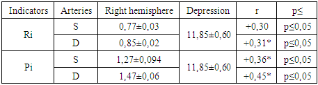
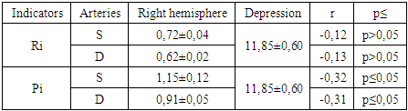
The results showed that all these correlation relationships are positive and statistically significant, in other words, damage to the left hemisphere implements anxiety against the background of increased vascular tone, which is generally consistent with the results of the analysis of the correlation relationship between hemodynamic parameters and autonomic tone. In this group, these relationships were inversely related. Table 3. Indicators of BFV (according to S.E. Lelyuk, R.T. Lelyuk 1995) and depression in the common carotid artery (CCA) in case of a right hemisphere stroke
 |
| |
|
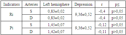
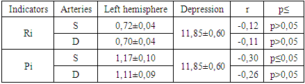
An analysis of the correlation relationships between hemodynamic parameters and the severity of depression, as can be seen from table №3, showed that in case of a right hemisphere stroke all these correlation relationships were positive and statistically significant, which indicates that, on the whole, there is a difficulty in cerebral blood flow both in the right and in the left hemisphere is reliably associated with the severity of depression.Interestingly, the reverse relationship was found in patients with left hemisphere stroke. These data are presented in table №4, from which it is seen that all correlation relationships between the studied indicators are negative.Table 4. Indicators of BFV (according to S.E. Lelyuk, R.T. Lelyuk 1995) and depression in the common carotid artery (CCA) with left hemisphere stroke
 |
| |
|
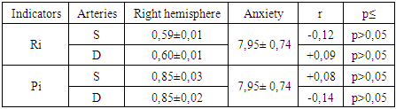
However, none of these indicators showed reliable correlation relationships. This suggests that with damage to the left hemisphere, the severity of depression is independent of cerebral hemodynamics.Table 5. Indicators of BFV (according to S.E. Lelyuk, R.T. Lelyuk 1995) and anxiety for the internal carotid artery (ICA) in case of a right hemisphere stroke
 |
| |
|
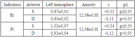
An analysis of the correlation relationships between hemodynamic parameters and the degree of severity of anxiety as shown in table №5 revealed that, with a right hemisphere stroke, hemodynamic indicators had generally negative correlation relationships with anxiety. However, in no case did they achieve a degree of statistical certainty, this indicates that anxiety in a right-hemisphere stroke is relatively independent of the state of cerebral hemodynamics.Table 6. Indicators of BFV (according to S.E. Lelyuk, R.T. Lelyuk 1995) and anxiety in the internal carotid artery (ICA) in left hemisphere stroke
 |
| |
|
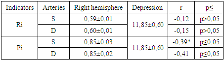
At the same time, as can be seen from table №6, the assessment of dynamic indices of the internal carotid artery in case of left hemisphere stroke revealed mainly positive correlation relationships reaching a degree of statistical reliability. In other words, with damage to the left hemisphere, anxiety was significantly correlated with the severity of increased tonus vessels in the basin of the internal carotid artery.Table 7. Indicators of BFV (according to S.E. Lelyuk, R.T. Lelyuk 1995) and depression of the internal carotid artery (ICA) in case of a right hemisphere stroke
 |
| |
|
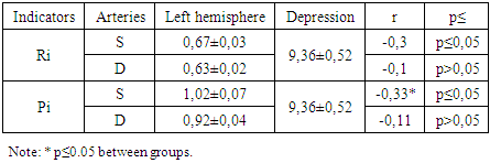
Table 8. Indicators of BFV (according to S.E. Lelyuk, R.T. Lelyuk 1995) and depression along the internal carotid artery (ICA) with left hemisphere stroke
 |
| |
|
The results of a correlation analysis of the severity of depression with hemodynamic indicators revealed that with right-hemisphere stroke, as can be seen from table №8, these relationships were negative, which indicates that an increase in vascular tone is associated with a lesser degree of severity of depression. Moreover, according to the indicator Pi, there are reliable correlation relationships. With a left hemisphere stroke (table No. 8), similar correlation relationships as a whole were also negative, but there was less pronounced tendency to reliability. Only in terms of the indicators of the left basin of the internal carotid artery and indicators of depression was there a statistically significant correlation, which indicates that, in general, hemodynamic parameters behave relatively independently in both right-hemispheric and left-hemispheric strokes with respect to depression.Table 9. Indicators of BFV (according to S.E. Lelyuk, R.T. Lelyuk 1995) and anxiety in the vertebral artery (VA) in case of a right hemisphere stroke
 |
| |
|
An analysis of the severity of anxiety in terms of linear blood flow velocity indices in Table №9 revealed that in case of a right hemisphere stroke, these correlation relationships, both in the left and right vertebrobasilar arteries, were ambiguous and the correlation relationships did not reach the degree of statistical significance. In other words, anxiety in a right hemisphere stroke behaved independently, as indicated above in the study of similar relationships in the carotid basin compared with hemodynamic parameters, that is, it was independent in relation to hemodynamic parameters.Table 10. Indicators of BFV (according to S.E. Lelyuk, R.T. Lelyuk 1995) and anxiety in the vertebral artery (VA) for left hemisphere stroke
 |
| |
|
At the same time, as can be seen from table No. 10, a comparison of linear blood flow velocity and anxiety in case of damage to the left hemisphere revealed individual statistically significant differences in the positive orientation.Moreover, in both cases, these differences were significant, when assessing the relationship between hemodynamics and anxiety in the vertebrobasilar basin on the left, on the right, these indicators had relative independence.Table 11. Indicators of BFV (according to S.E. Lelyuk, R.T. Lelyuk 1995) and depression along the vertebral artery (VA) in case of a right hemisphere stroke
 |
| |
|
Table 12. Indicators of BFV (according to S.E. Lelyuk, R.T. Lelyuk 1995) and depression along the vertebral artery (ICA) with left hemisphere stroke
 |
| |
|
Evaluation of the correlation relationships between the hemodynamic parameters of the vertebral artery and the degree of severity of the depression table (№ 11 and № 12) showed that in case of a right hemisphere stroke all these correlation relationships were negative and, according to individual indicators, in particular, Pi were statistically significant both in the right and left vertebrobasilar pool, in other words, with a right hemisphere stroke, a certain dependence of the severity of depression on the hemodynamic parameters was found, while it was found that the higher the severity of the linear velocity of blood flow, the less pronounced the degree of depression. Similar negative correlation relationships were identified in the analysis of hemodynamic parameters with the severity of depression in patients with left hemisphere stroke. However, here the reliability of correlation relationships was revealed in the left vertebrobasilar basin, which indicates that hemodynamics and depression are to some extent interdependent precisely in patients with left hemisphere stroke.
5. Conclusions
The results of the study showed that hemodynamic and depression indicators are to some extent interdependent precisely in patients with left hemisphere stroke, while anxiety in right hemisphere stroke is relatively independent of cerebral hemodynamics.
References
| [1] | Bahadirova M.A. Indices of cerebral hemodynamics in the examination and rehabilitation of patients with the consequences of a stroke. Abstract. Dis .... kmn. Tashkent: 2002. –p. 20. |
| [2] | Borisenko V.V., Vereshchagin N.V. Transcranial Doppler. |
| [3] | Wozniuk I.A, A.Yu. Polushin A.Yu, Belyasnik A.S, Zabirov S.Sh, Morozova E.M. “Ultrasound Doppler ultrasonography in acute cerebral ischemia”. |
| [4] | Gusev E.I, Gekht A.P, Bogolepova A, Sorokina I. Features of the depressive syndrome in patients after ischemic stroke. // App. To Journal. nevrol. and psychiatrist. 2001. -No3. -pp. 28-32. |

 Abstract
 Reference
 Full-Text PDF
 Full-text HTML