American Journal of Medicine and Medical Sciences

p-ISSN: 2165-901X    e-ISSN: 2165-9036

2021;  11(1): 46-52

doi:10.5923/j.ajmms.20211101.12

Received: Dec. 7, 2020; Accepted: Jan. 15, 2021; Published: Jan. 25, 2021


Studying of the Remote Results in the Application of Intrauterine Contraception after Cesarean Section

Nasirova Zebiniso Azizovna

Samarkand State Medical Institute, Uzbekistan

Correspondence to: Nasirova Zebiniso Azizovna, Samarkand State Medical Institute, Uzbekistan.

Email:

Copyright © 2021 The Author(s). Published by Scientific & Academic Publishing.

This work is licensed under the Creative Commons Attribution International License (CC BY).
http://creativecommons.org/licenses/by/4.0/

Abstract

Contraception for women in the postpartum period must be highly effective, safe (allowed for use in the postpartum period, do not contain estrogens) and acceptable (prolonged). The method of long – term reversible contraception-intrauterine contraception (IU) – is a safe method that is suitable for most women. To study the effect of intrauterine contraception on the state of the reproductive system, ultrasound examination of the pelvic organs in dynamics in the postoperative period for 12 months in patients with IUD was performed. When using IUD intraoperatively and at intervals throughout the year, the echographic picture of the reproductive system does not differ significantly from those of women who did not use contraception. It was found that the localization of the IUD inside the uterus may change during the first three to six months after administration. This may be due to bleeding, pain, and expulsions.

Keywords: Cesarean section, Intraoperative, IUD, Echographic parameters, Contraception, Expulsion

Cite this paper: Nasirova Zebiniso Azizovna, Studying of the Remote Results in the Application of Intrauterine Contraception after Cesarean Section, American Journal of Medicine and Medical Sciences, Vol. 11 No. 1, 2021, pp. 46-52. doi: 10.5923/j.ajmms.20211101.12.

1. Introduction

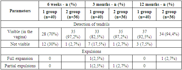
High frequency of abdominal delivery is a distinctive feature of modern obstetrics. Currently, the frequency of this operation is steadily increasing. In Uzbekistan, over the past 10-15 years, its frequency has more than doubled, and according to the statistical Department of the RSNPMC A and G and the ROC in 2017, it was 18%, reaching up to 35-40% in some institutions [21].
The increase in the frequency of operative delivery creates a new problem - an individual approach to choosing the optimal method of contraception in women with a uterine scar after cesarean section in order to best observe the interval between deliveries.
The most important factor in maintaining the reproductive health of women who have undergone cesarean section is to solve the problem of unwanted pregnancy and to observe the intergenetic interval. Artificial abortion is a severe physical, psychological and hormonal stress that leads to functional and organic disorders in the reproductive system [18]. In this regard, the use of contraception is considered as one of the most important areas in the rehabilitation of women after operative delivery.
An essential element of preserving women's reproductive potential after CS surgery is ensuring an adequate interval between pregnancies and childbirth. The most optimal interval between childbirth is from 3 to 5 years. Pregnancy earlier than 2 years, even after natural birth, is associated with a high risk of complications: miscarriage, bleeding, postpartum and inflammatory diseases, uterine ruptures, and maternal mortality [11,20]. However, ensuring an adequate interval between pregnancy and delivery is especially important for women with uterine scarring due to the increased risk of pregnancy and delivery complications. This becomes possible only with the use of effective planned contraception. Contraception for women in the postpartum period must be highly effective, safe (allowed for use in the postpartum period, do not contain estrogens) and acceptable (prolonged). The method of long – term reversible contraception-intrauterine devices (IUDs)– is a safe method that is suitable for most women.
The main advantage of long-term reversible contraception methods is that they do not require constant effort on the part of the user. In addition, after removal of the contraceptive, fertility is rapidly restored [21,26].
The period immediately after delivery is particularly well suited for the introduction of an IUD. Women who have just given birth to a child are usually highly motivated to use contraception; it is safe to rule out pregnancy, and the introduction of a contraceptive during a hospital stay is convenient for both patients and medical professionals. In addition, there is a risk of unwanted pregnancy in the period immediately after delivery [17].
It is the postpartum period that is an important time to start using an effective method of contraception in order to increase the interval between pregnancies. This is especially important for women after CS surgery.
Thus, the postpartum period is an important time to start using an effective method of contraception in order to increase the interval between pregnancies, which is especially important for women after CS surgery. One of the most suitable solutions is the use of prolonged reversible contraception-IUD-highly effective and long-acting contraceptives. The use of these methods of contraception allows you to ensure an adequate interval between pregnancies and childbirth and thereby preserve the reproductive potential of women after CS surgery.
All of the above determines the need to study the reproductive behavior of women after cesarean section and optimize the use of highly effective methods of contraception in women with a scar on the uterus.
The location of the IUD inside the uterus may change during the first three to six months after administration. This may be due to bleeding, pain, and expulsions. Some authors have found that after 6 weeks, the distance from the upper edge of the IUD to the bottom of the uterus of more than 10 mm will lead to expulsions after 24 weeks with interval IUD administration [9,12]. Among women with interval IUDs, expulsions occurred at a lower position inside the uterus, while women did not have the desire to remove the IUDs due to side effects. An IUD administered post-placentally will have a different localization inside the uterus compared to an interval injection [10,16].
The main goal of the study was to study the long-term results of using IUDs post-placentally and at intervals (after 6 weeks) in women who underwent cesarean section.

2. Method of Research

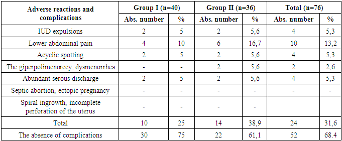
A prospective study was conducted in the city Maternity hospital No. 2 in Samarkand to assess the long-term results of post-placental and interval administration of CU T 380 A IUD in women who underwent cesarean section. The study included 76 women who were delivered by caesarean section and wanted to use the CU T 380 A IUD, who met the who standard medical criteria for IUD administration and were willing to follow the study Protocol. The exclusion criteria were: women who do not meet the who medical eligibility criteria for the use of IUDs.
Women were consulted about post-placental and interval IUD administration during prenatal visits and / or after hospitalization. The women were told in detail about the study, including the advantages and limitations of different methods, and repeated counseling was performed before the caesarean section. Written informed consent was obtained from women who were willing to participate in the study and comply with the study Protocol.
Post-placental insertion of the IUD was performed after removal of the placenta using Kelly forceps / manually through a uterine incision, and the device was placed on the bottom of the uterus. No attempt was made to direct the IUD tendrils into the vagina. Antibiotics were administered according to the Protocol of the maternity hospital for caesarean section. The women were monitored daily for postpartum bleeding, as well as any other complaints, throughout their hospital stay. Interval administration of IUDs was administered in the usual sterile manner, as described in the manufacturer's instructions for a 6-week postpartum visit. Before discharge, patients were indicated on the exchange card: VMC type, date of introduction, and expiration date. Participants were asked to return for scheduled follow-up visits in 6 weeks, 3 months, 6 months, and 12 months.
Outcomes and side effects the use of IUDs after 6 months was determined by confirming the presence of IUDs in the uterus during physical examination or ultrasound. Expulsions were defined as the complete passage of the device through the cervix or the presence of an IUD in the cervical canal. Removal of the IUD was determined by the patient's report or removal documentation. Infection of the pelvic and cervical organs was thought to be present in women with purulent discharge, soreness of the cervix, appendages, or uterus, with or without fever. Patient satisfaction was determined by the patient report. Secondary outcomes included removal, expulsions, satisfaction, and adverse events such as infection, perforation, and pregnancy.
To study the effect of intrauterine contraception on the state of the reproductive system, an ultrasound study of the pelvic organs was performed in dynamics in the postoperative period for 12 months in 76 patients. Echographic indicators were obtained using a real-time ultrasound detector from Toshiba (Xario SSA-660A).
Ultrasound biometrics included changing three dimensions of the uterus (length, anterior-posterior size, and width), measuring M-echo, measuring the distance between the IUD and the bottom of the uterus, and measuring the distance between the IUD and the upper endometrial layer.
The length of the uterine body was measured from the internal pharynx to the farthest point of the bottom in the longitudinal section. The anterior-posterior size of the uterine body was determined perpendicular to the previous measurement by the external contours of the anterior and posterior walls in the widest part of the uterine body. The width of the uterine body was measured by cross-scanning in the widest part from the right to the left contour of the side walls of the uterine body.
Ultrasound of the endometrium assessed its thickness, structure, and compliance with the phase of the menstrual cycle. To assess the thickness of the endometrium, anterior-posterior m-echo measurement was used for longitudinal scanning of the uterus.
The thickness of the anterior wall was measured in the projection of the scar, and attention was paid to the state of the ovaries.
The first assessment of the state of the reproductive system was carried out 6-8 weeks after the start of contraceptive use, and then 3-6-12 months later. Particular attention was paid to the localization of the IUD, changes in values from the upper edge of the IUD to the bottom of the uterus, and the distance between the IUD and the upper endometrial layer.
The conducted studies showed that the echographic parameters changed in the dynamics of the postoperative period and were in a certain dependence on the contraceptive used.

3. Results and Discussion

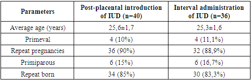
A prospective study was conducted in the city Maternity hospital No. 2 in the city of Samarkand. To assess the echographic parameters of post-placental and interval administration of CU T 380 A IUDs in women who underwent cesarean section, the study included 76 women who were delivered by caesarean section and wanted to use CU T 380 a IUDs. All women met the who standard medical criteria for IUD administration and were prepared to comply with the study Protocol.
The majority of women were between 21 and 30 years old (77.89%). Two women (4%) were over 36 years old and only one woman was under 20 years old (table 1).
Table 1. Characteristics of women using IUD Cu T 380 A intraoperatively (n =40) and interval (n =36)
     
The age of women subject to postpartum contraception in both groups varied from 18 to 40 years. The average age in the first group was 25.6±1.7 years, in the second group 25.3 ±1.6 years. First-time pregnancies in group 1 were 25% (10 women), in the second group 25.8% (10 women). 6 women had spontaneous miscarriages before the upcoming birth. The remaining women in both groups were re-pregnant and re-giving birth. The health index of the examined women for extragenital diseases was relatively satisfactory. The most common diseases of the visual organs were -10% in women with post-placental IUD administration and 8.3% in women with interval IUD administration. The most common indications for cesarean section were the failure of the scar and the scar itself (24.2%), extragenital pathology (9.4%), and the pelvic position of the fetus (8.7%). Thus, the clinical characteristics of the women of both groups observed by us indicate the homogeneity of the contingent.
It is characteristic that 68.4% of women using IUDs did not experience any side effects when using this contraceptive post-placental and interval. The presented table 2 shows certain complications of IUD.
Table 2. Frequency of adverse reactions in women who have had a caesarean section when using IUDs
     
The most common complication when using IUD was pain felt by women in the lower abdomen, which varied in duration and severity. In most women, they were aching, giving in the lower back, in some-cramping in the first days of the introduction of the IUD or during menstruation.
On average, the frequency of pain in the group was 13.2%, however, in women with interval administration, pain was observed 6.7% more often than in women with intraoperative IUD administration. Pain was analyzed on the VASH rating scale.
Visual-analog scale (VAS)-represents a vertical or horizontal straight line segment with a length of 10 cm (100 mm) (figure 1). The patient was asked to make a mark on it corresponding to the intensity of the pain they were experiencing, where 0 points is the absence of pain, and 10 points (10 cm on the scale) is unbearable pain.
Figure 1. VASH Scale
It should be noted that in 2 women with post-placental IUD administration, pain was characterized as insignificant, 22 of these patients were primiparous and primiparous, pain syndrome was observed in them for the first 2 weeks after cesarean section with post-placental IUD administration and was stopped by NSAIDs (paracetamol 500 mg). The rest of the time, contraception was not resumed. One repeat-giving woman had mild aching pains only in the first 2 days after cesarean section and disappeared without medication. A fifth woman had pain before and during menstruation during the first two menstrual cycles after contraception. Pain was relieved by prescribing antispasmodics and NSAIDs (no-shpa, paracetamol 500 mg). All subsequent months of contraception during lactation and after its completion, women were not bothered by pain.
Of the women who had a cesarean section with an interval introduction of IUD, 4 patients complained of pain only in the first 2 days after contraception, they were aching, insignificant in nature and disappeared on the 3rd day without medication. In one woman, the pain persisted throughout the first month and increased during several periods. Medical treatment was effective, the pain was stopped and subsequently did not resume. It should be noted that this patient had a history of an artificial abortion.
Another woman had a history of algodismenorrhea, and after the introduction of the IUD, the pain gradually increased, and at the request of the woman, the IUD was removed in the fourth month of use. Later, the woman was offered another contraception.
The second place in the frequency of complications when using IUDs was occupied by their expulsions (5.3%), and in women with post-placental IUDs only in 2 cases (5%), and in women with interval IUDs, this complication was noted much more often (5.8%). During the first control visit after 6 weeks, there were no cases of IUD expulsions in both groups. During the second control visit at the 3rd month, IUDs were partially expulsed in women with post-placental administration in 1 (2.5%) case, which led to the need to remove the IUDs, and in women with interval administration also in one case. Further, 1 more woman requested the removal of the IUD due to the requirement of a sexual partner. During the third control visit, 6 months later, there was one case of complete IUD cu T 380 a expulsions, and in 1 case Cu T was removed at the request of a woman in the first group, and cases of expulsions in the second group. Thus, at the end of the study, after 12 months of postpartum follow-up, there were 2 cases (5%) of expulsions and 2 cases (5%) of removal at the request of the patient in the group of women with intraoperative IUD administration and 2 cases of expulsions and 3 cases of removal in women with interval IUD administration.
At the first control visit after 6 weeks, when viewed in mirrors, the antennae were visible in 70% (28/40) of women in the first group and in 82.5% (33/36) of women in the second group. However, the antennae were not visible in 12 (30%) women of the first group during the first control visit. This is due to the fact that during intraoperative introduction of the IUD, the device's antennae are only directed to the internal pharynx, as the uterus contracts and lochia secretions, the antennae themselves are brought out (table 3).
Table 3. Detection of tendrils and IUD expulsions
     
It is noteworthy that all women with expulsions were multi-pronged (3-4 births), two of them had IUD expulsions after 2 months, and one-at the 4th month of contraceptive administration.
It should be noted that in the group with interval IUD administration, expulsions also occurred in women with a burdened history. So, four women had a history of 3-4 medical abortions. In this group, women had partial expulsions in the first month after IUD administration, while other women had full expulsions 3 months after administration.
The third most frequent complication was menstrual function disorders, which occurred in only 6 out of 76 women who used IUD (7.9%). the frequency of spotting in both groups was almost the same (7.5% and 8.3%, respectively), which coincides with the opinions of Agarwal K. et al. [1].
As a rule, these disorders were manifested in the form of acyclic bloody discharge and in most cases were stopped in a short time without the appointment of medication for hemostatic therapy. It should be noted that the majority of women with acyclic bleeding had gynecological diseases and a burdened obstetric history, the same data is given by Mohamed A. I., Elati I. H. A., Ramadan E. N. [20].
Thus, of the two women who had a caesarean section with post-placental IUD administration, one of them had a broken menstrual cycle before pregnancy and delivery, characterized by irregular menstruation with periodic anovulatory cycles. After restoring menstrual function in the 2nd month of the postoperative period, acyclic spotting stopped.
From the group of women who had a cesarean section with an interval introduction of the IUD, there were also spotting spotting that lasted 8 days, stopped on their own and did not resume later. Two women who had a cesarean section with an interval introduction of IUDs, who had a history of PID, had intermenstrual bleeding in the first 2 menstrual cycles after the introduction of IUDs, and after the appointment of hemostatic therapy in the following months, there was no intermenstrual discharge. In the fourth patient, who had a history of 2 births and 3 abortions, after the introduction of IUD, spotting was constant for 2 months, anemia developed, hemoglobin decreased to 68.0 g / l, conservative therapy did not have an effect, and therefore the spiral was removed and pure progestin contraception was prescribed.
In women who had a caesarean section and used intrauterine devices for contraception, the echographic indicators were slightly different from those of women who did not use contraception. In these women, we measured the distance from the upper edge of the spiral to the bottom of the uterus, as well as from the device to the upper layer of the endometrium. According to some authors [16], these changes may anticipate expulsions and the occurrence of side effects, such as pain and bleeding. Our goal was to determine whether these changes in distance are associated with the risk of IUD expulsions or removal due to side effects. Comparison of echographic data is presented in table 4.
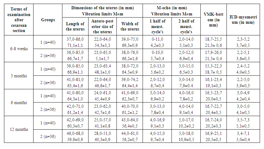
Table 4. Echographic parameters in the dynamics of women using the IUD
During the use of IUD post-placental and interval, the size of the uterus decreased by the 12th month, almost equally, which is reflected in table 4. it is Characteristic that as the postoperative period increases, all the sizes of the uterus in both women of the first group and the second group of women decreased gradually, reaching the minimum values by the 12th month. Analysis of the m-echo value in women with IUDs showed that there were no significant differences in endometrial changes in women with post-placental IUDs and with interval IUDs. As the postoperative period increased, the average thickness of the M-echo increased in all women. The scar area was also without features in all the women's visits.
Our next step was to measure the distance between the IUD and the uterine floor and the IUD and myometrium. Six weeks and three months after delivery were within the measurement range described for IUDs established outside the postpartum period, and similar between patients who had or had not had IUDs expulsions and removal due to symptoms within one year. The median IUD measurements at the uterine floor for the entire population were 21.3 mm (18.7–25.5 mm) at 6 weeks, 18.7 mm (15.3–22.3 mm) at 3 months, and 20.2 (16.7–24.0 mm) at 1 year. The median measurement of IUD-myometrium was 3.7 mm (2.3–5.2 mm) at 6 weeks, 4.0 mm (2.4–5.2 mm) at 3 months, and 5.1 mm (3.5–7.3 mm) at 1 year.
Our results are consistent with the recommendations of Gonzalez J. et al. [9], according to which ultrasound measurements can be clinically useful for observation.
As an additional limitation, we did not evaluate other potential ultrasound results, such as incorrect rotation within the endometrial cavity or the distance of the IUD from the internal pharynx. We selected fundal and myometrial measurements so that they can be easily identified using ultrasound, as well as so that they can be used by practitioners.

4. Conclusions

Thus, properly selected contraception provides high efficiency and acceptability and thus plays a crucial role in maintaining women's reproductive health. Increasing access to contraception is a way to preserve the health of women and children and reduce maternal and infant mortality. Ensuring satisfaction with obstetric and perinatal care and family planning will reduce: maternal mortality by more than 2/3 (from 358,000 to 105,000); neonatal mortality by more than half (from 3.2 to 1.5 million); the number of unwanted pregnancies increased by more than 2/3 (from 75 to 22 million); the number of unsafe abortions - from 20 to 5.5 million; the death rate from unsafe abortions-by more than 4/5 (from 46,000 to 8,000). Every dollar spent on contraception saves $ 1.40 of costs for obstetric and perinatal care [11,18].
Our results indicate that for the majority of women who have had a cesarean section, IUD does not have an adverse effect on their health and is a completely acceptable method of contraception, provided that its technology is followed. Adverse reactions in most women were transient and disappeared independently during the first 3-4 months of contraception.
When using copper-containing intrauterine contraceptives administered to patients during cesarean section, when observed for 12 months, the echographic indicators of the reproductive system do not differ significantly from women who did not use contraception. The practice of installing a post-placental IUD during cesarean section is safe, but follow-up may differ from that of patients with normal interval management of IUD. It was also found that after 6 weeks, a distance of less than 10 mm from the upper edge of the IUD to the bottom of the uterus can lead to expulsions after some time of using the IUD.
The effectiveness of contraception, short duration and low severity of adverse reactions allow the use of IUDs in women after abdominal delivery post-placental and interval.
It should be noted that in our study there were no cases of complications in the form of uterine perforation and common inflammatory diseases (endometritis, peritonitis).
During the use of intrauterine contraception, involutive processes in the uterus at 12 months were 60.3±0.7 mm in length, 41.1±0.8 mm in thickness, 56.4±0.5 mm in width in women with intraoperative IUD administration and 59.9±0.9 mm in length, 40.3±0.9 mm in thickness, 56.2±0.7 mm in width in women with interval IUD administration (p<0.05).
The results do not indicate any differences in changes in the morphology of the uterus and endometrium in women who underwent cesarean section, who used IUD post-placental and interval. Echographic indicators of ovarian size in women using IUD post-placental and interval did not actually differ. The average thickness of the anterior wall in the projection of the scar was 9.1±1.8 mm in women who underwent cesarean section and used IUD post-placental and interval.
Thus, changes in the echographic indices of the uterus and ovaries, the average thickness of the front wall in the projection of the scar in women undergoing cesarean section, primedic intrauterine devices intraoperatively and interval, after 6 weeks suggest that depending on the reproductive system, from the hypothalamic - pituitary activity in the postoperative period and different for involutive processes and the recovery mechanism of the reproductive system under the influence of contraceptive, but these data do not differ from the time of introduction of intrauterine system.
In conclusion, I would like to say that the method of long - term reversible contraception-IUD, based on their effectiveness, are high-class contraceptives, since with their ideal and typical use, the incidence of pregnancy is less than 1% per year. These drugs are characterized by the highest frequency of satisfaction and long-term use of all methods of reversible contraception.

References

[1]  Agarwal K. et al. Visibility of strings after postplacental intracesarean insertion of CuT380A and Cu375 intrauterine contraceptive device: a randomized comparative study // The Journal of Obstetrics and Gynecology of India. – 2017. – Т. 67. – №. 5. – С. 324-329.
[2]  An A. V., Pakhomovа, E. J. the Critical state for obstetric hemorrhage //Questions of gynecology, obstetrics and Perinatology. - 2010. - Vol. 9. - No. 4. - P. 33-37.
[3]  Atakhodzhayeva F. A., Fazliddinova F. N. The use of intrauterine contraception in the postpartum period // Problems of biology and medicine. -2013. - No. 4. - page 18.
[4]  Ayupova F. M., Khamdamova M. T. Clinical aspects of the use of copper-containing intrauterine contraceptives // Uzbekistan tibbet journal. -2010. № 6. 59-64.
[5]  Bayoumi Y. A. et al. Post-placental intrauterine device insertion vs puerperal insertion in women undergoing caesarean delivery in Egypt: a 1 year randomised controlled trial // The European Journal of Contraception & Reproductive Health Care. – 2020. – С. 1-6.
[6]  Boydell N. et al. Women’s experiences of accessing postpartum intrauterine contraception in a public maternity setting: a qualitative service evaluation //The European Journal of Contraception & Reproductive Health Care. – 2020. – С. 1-9.
[7]  Çelen Ş. et al. Immediate postplacental insertion of an intrauterine contraceptive device during cesarean section // Contraception. – 2011. – Т. 84. – №. 3. – С. 240-243.
[8]  Colwill A. C. et al. Six-week retention after postplacental copper intrauterine device placement //Contraception. – 2018. – Т. 97. – №. 3. – С. 215-218.
[9]  Curtis K. M., Peipert J. F. Long-acting reversible contraception //New England Journal of Medicine. – 2017. – Т. 376. – №. 5. – С. 461-468.
[10]  Dall’Asta A., Ghi T., Frusca T. Doppler Diagnosis //Fetal Growth Restriction. – Springer, Cham, 2019. – С. 139-170.
[11]  Dubossarskaya Z. M. analysis of the application of modern methods of postpartum contraception //Reproductive health. Eastern Europe, 2018, Pp. 32-34.
[12]  Elsokary A., Elkhyat A., Elshwaikh S. Evaluation of Post-Placental IUD Insertion during Cesarean Section at a Tertiary Care Hospital in Egypt // Open Journal of Obstetrics and Gynecology. – 2020. – Т. 10. – №. 4. – С. 516-525.
[13]  Emarah M. A. et al. Predicting successful use of post-placental intrauterine contraceptive device by ultrasound //Menoufia Medical Journal. – 2020. – Т. 33. – №. 2. – С. 440.
[14]  Hochmuller J. T. et al. Expulsion rate of intrauterine device: mediate vs. immediate puerperium period // Journal of the Turkish German Gynecological Association. – 2020. – Т. 21. – №. 3. – С. 143.
[15]  Goldstuck N. D., Steyn P. S. Insertion of intrauterine devices after cesarean section: a systematic review update // International journal of women's health. – 2017. – Т. 9. – С. 205.
[16]  Gonzalez J. et al. Ultrasound location of intrauterine devices placed at cesarean section over the first year postpartum // Contraception. – 2020.
[17]  Lester F. et al. Intracesarean insertion of the Copper T380A versus 6 weeks postcesarean: a randomized clinical trial // Contraception. – 2015. – Т. 91. – №. 3. – С. 198-203.
[18]  Maltseva L. I. the choice of contraception after cesarean section // Questions of gynecology, obstetrics and Perinatology. - 2014. - Vol. 13. - No. 2. - Pp. 88-94.
[19]  Mikova V. N., Sandakova E. A., Кolomoitseva T. N. Features of the echographic picture of the reproductive system when using various types of intrauterine contraception //Perm medical journal, 2009, Vol. 26, no. 5.
[20]  Mohamed A. I., Elati I. H. A., Ramadan E. N. Assessment effect of Post Puerperal and Immediate Post Placental Intrauterine Contraceptive Devices Insertion after Cesarean Delivery on Women’s Health // IOSRJNHS. – 2015. – Т. 4. – №. 3. – С. 1-7.
[21]  Nasirova Z. A., agababyan L. R. post-Placental introduction of intrauterine spirals in women delivered by abdominal route // Problem Reproduktsii. - 2017. - Vol. 23. - no. 2.
[22]  Ovsepyan L. S. the Role of three-dimensional sonography in predicting the risk of Levonorgestrel-releasing intrauterine system Mirena: dis. - Yerevan state medical University named after M. Heratsi, 2018.
[23]  Tjahjanto H., Haryuni I. T. Hang-up IUD, new technique for suturing CuT-380A IUD to uterine fundus in immediate postplacental insertion during cesarean delivery: twelve months follow up //Indonesian Journal of Obstetrics and Gynecology. – 2014. – С. 132-139.
[24]  Upmanyu P., Kanhere A. Acceptance of methods of family planning in patients undergoing repeat caesarean section //International Journal of Reproduction, Contraception, Obstetrics and Gynecology. – 2016. – Т. 5. – №. 4. – С. 976-9.
[25]  Verest A. et al. Intrauterine device (IUD) migration in cesarean delivery scar: What to do with the niche? //Facts, Views & Vision in Obgyn. – 2019. – Т. 11. – №. 3. – С. 251.
[26]  Whitaker A. K., Chen B. A. Society of Family Planning Guidelines: Postplacental insertion of intrauterine devices // Contraception. – 2018. – Т. 97. – №. 1. – С. 2-13.
[27]  Wildemeersch D., Goldstuck N. D., Hasskamp T. Current status of frameless anchored IUD for immediate intracesarean insertion //Dev Period Med. – 2016. – Т. 20. – №. 1. – С. 7-15.
[28]  Wildemeersch D. A., Goldstuck N. D., Janssens D. R. G. Immediate Post Placental Insertion of IUD-The Challenge to Solve the Expulsion Problem // Obstet Gynecol Int J. – 2014. – Т. 2. – №. 5. – С. 00052.
[29]  Zaconeta A. M. et al. Intrauterine Device Insertion during Cesarean Section in Women without Prenatal Contraception Counseling: Lessons from a Country with High Cesarean Rates // Revista Brasileira de Ginecologia e Obstetrícia. – 2019. – Т. 41. – №. 8. – С. 485-492.
[30]  Zhadan Yu. G. the Main aspects of rehabilitation of patients after caesarean section. – 2018.