American Journal of Medicine and Medical Sciences

p-ISSN: 2165-901X    e-ISSN: 2165-9036

2021;  11(1): 25-32

doi:10.5923/j.ajmms.20211101.07

Received: Dec. 3, 2020; Accepted: Dec. 31, 2020; Published: Jan. 15, 2021


USS Diagnostic Score in High-Altitude Trauma

A. M. Khadjibaev, J. A. Djuraev, P. K. Sultanov, Khadjayarov N. P.

Republican Research Centre of Emergency Medicine, Tashkent, Uzbekistan

Copyright © 2021 The Author(s). Published by Scientific & Academic Publishing.

This work is licensed under the Creative Commons Attribution International License (CC BY).
http://creativecommons.org/licenses/by/4.0/

Abstract

Introduction The frequency of abdominal organs and retroperitoneal space injuries at combined catatrauma remains high. Aim of research is to study the diagnostics methods at combined abdominal organs and retroperitoneal space injuries. Material and methods We analyzed diagnostics and treatment results of 180 victims of high-altitude trauma with combined injuries of the abdomen admitted to the surgical department of the Republican Research Centre of Emergency Medicine. Radiological and CT examinations, endosurgical interventions were performed to all patients. Results Combined injuries among patients with abdominal injuries were detected by instrumental methods in 94.9% and 88.3% of cases. The most frequently combined abdominal injuries were noted in patients with injuries of the chest organs (17.7% and 6.3%); head and extremities (10.1% and 2.7%); head + chest + pelvis + extremities (7.6% and 11.7%), head + chest + extremities (8.9% and 10.8%). Isolated abdominal injury was found in 5.1% and 11.7% of cases in the main and control groups. Injuries of abdominal organs at isolated or combined abdominal trauma during catatrauma were mainly manifested by ruptures of parenchymal organs with hemoperitoneum. Mortality among catatrauma victims with isolated and combined abdominal injuries was 17.7% in the main group and 27% in the control group. Conclusion It is very important to recognize simultaneously all the injuries immediately after the victim admits the hospital, while assessing the dominant ones. This need is imperative, vital, justified, urgent, as it is the motive for determining surgical tactics.

Keywords: Polytrauma, Combined injury, Catatrauma, Solated injury, Diagnostics, Abdominal injury

Cite this paper: A. M. Khadjibaev, J. A. Djuraev, P. K. Sultanov, Khadjayarov N. P., USS Diagnostic Score in High-Altitude Trauma, American Journal of Medicine and Medical Sciences, Vol. 11 No. 1, 2021, pp. 25-32. doi: 10.5923/j.ajmms.20211101.07.

1. Introduction

Polytrauma has been one of the most frequent problems of emergency surgery for recent years [1]. In peacetime, polytrauma most often occurs as a result of traffic accidents and when falling from a height [1-3]. They are characterized by severe combined injuries of various organs and systems [1-2]. Severe injuries of the brain and spinal cord internal organs of the chest and abdominal cavity dominate and become the direct cause of a victim’s death [4,2,5]. The combination of abdominal organs trauma with injuries of other anatomical areas of the body aggravates the condition of the victim, significantly complicates the diagnostics and worsens the prognosis [4,2,5]. Diagnostics of the abdomen injuries with combined catatrauma is significantly difficult due to the presence of a coma in most victims as a result of a traumatic brain injury (TBI) [6-9]. The presence of the brain compression, massive internal bleeding, multiple fractures of the ribs and pelvic bones, the development of fat embolism severely limit, and sometimes exclude the use of special investigation methods (radiological, contrast, instrumental, etc.) [10-11,3,12]. The clinical picture characterized by a large polymorphism, as a rule, does not correspond to the nature and severity of injury [5,13]. In addition, there are various factors of catatrauma to be the cause of it: the heterogeneous nature of the traumatic forces and places of their application, sometimes significantly remote from the projection of the injured organ; the different nature and degree of injury at the same traumatic force [14-16]. Mortality of catatrauma victims with combined injuries of the abdominal cavity and retroperitoneal space remains high and amounts 40-60% [14,17-18].
The aim of our study was to improve the diagnostics of abdominal organs and retroperitoneal space injuries at combined catatrauma.

2. Materials and Methods

We analyzed the diagnostics and treatment results of 190 victims of high-altitude trauma with combined injuries of the abdomen from 2010-2015, who were hospitalized to the Republican Research Center of Emergency Medicine. There were 158 (83.2%) males and 32 (16.8%) females. The age of patients varied from 15 to 80 years (mean age made up 35.83 years).
The patients were divided into two groups: main group (79) and control group (111). The difference between the main and control groups of patients was in the ultrasonic gradation of free fluid volume in the abdominal cavity and, accordingly, in the tactics of surgical treatment. Ultrasound was performed in dynamics after 1, 3 hours, 6 and 12 hours from the time of admission to clarify the ongoing bleeding.

3. Results

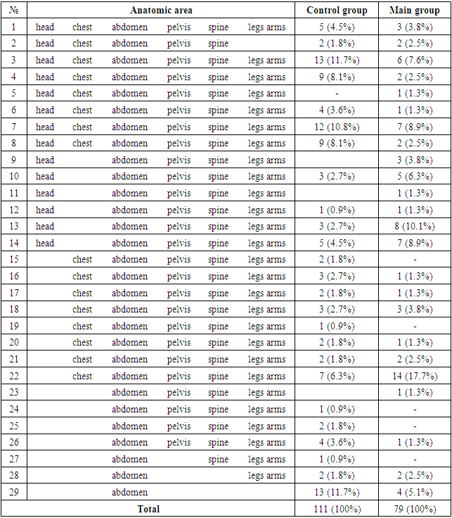
Сombined injuries were detected among patients with abdominal injuries in 94.9% and 88.3% of cases. Moreover, a combination of 6 areas was identified in 3.8% and 4.5% of cases; 5 areas - in 11.4% and 15.3% of cases; 4 areas - in 24.1% and 31.5% of the victims; 3 areas - in 25.3% and 20.7% and 2 areas - in 30.4% and 16.2% of cases, respectively, for the main and control groups (Tab. 1). The most frequently combined abdominal injuries were noted in patients with injuries of the chest organs (17.7% and 6.3%); head and extremities (10.1% and 2.7%); head + chest + pelvis + extremities (7.6% and 11.7%), head + chest + extremities (8.9% and 10.8%). Isolated abdominal injury was found in 5.1% and 11.7% of cases in the main and control groups.
Тable 1. The frequency of combined and isolated abdominal injuries
     
According to the ISS scale [19], injuries of critical (43.0% and 44.1%), severe (26.6% and 27.0%) and moderate (15.2% and 14.4%) degrees were observed in the main and control groups, respectively (Tab. 2).
Table 2. Severity score of combined catatrauma victims with abdominal injuries according to ISS scale
     
In 24 (30.4%) patients of the main and in 35 (31.5%) patients in the control groups the changes in neuro-reflex activity which prevented the collection of complaints and anamnesis were observed, as well as a distortion of the symptom complex of injuries. In addition, the timely and correct diagnostics in 18 (22.8%) cases of the main and in 31 (27.9%) patients of control group was impeded by the residual effect of treatment measures at the prehospital stage. In general, it stipulated the variability of the clinical picture due to the lack of absolute clinical signs. Thus, 28% of patients complained of pain in the abdomen both in the main and in the control groups, while 18.9% and 19.8% of the victims complained of spilled pain throughout the abdomen, and 22.8% and 20.7% of patients complained of local pain. 15.2% and 17.1% of the victims complained of nausea. Vomiting was observed only in 3.8% and 3.6% of patients in the main and control groups, respectively. However, such a clinical sign as dry mouth was observed in the majority of victims (Tab. 3).
Table 3. The frequency of clinical symptoms of abdominal organs injuries in patients with combined abdominal catatrauma
     
Pains of various intensities were noted in 33 (41.8%) and 45 (40.5%) patients on palpation examination of the victims, and only 8.9% and 12.6% of patients complained of sharp pains. Mostly local pain was observed in the areas of bruises (22.8% and 20.7% of cases), only 18.9% and 19.8% of patients had spilled pains throughout the abdomen. Tension of the anterior abdominal wall during palpation, limitation of its excursion in the act of breathing, atrophy in the sloping parts of the abdomen were noted in 10.1% and 11.7%; 7.6% and 7.2%; in 11.4% and in 25.2% patients of main and control groups respectively. Weak intestinal peristalsis was noted in 24.1% and 51.4% of the victims, while in 1.3 and 0.9% of patients it was not heard at all. A positive Shchetkin-Blumberg symptom was observed in 2.5% and 9.0% of patients in the main and control groups, respectively. The reliability of clinical signs according to our studies is low and makeup 45.9 ± 9.3% (P <0.01) at average. Evidence for this is that pain was not always associated with abdominal pathology, as spinal injury, fractures of the ribs and pelvic bones could be its cause. Nausea and vomiting were associated with TBI in most cases. Objective examination of the patient was difficult due to impaired consciousness of the patient in the form of lethargy or, conversely, by the excitation of the victim as a result of trauma and was always associated with the subjective sensations of the examining doctor. It was not possible to establish specific values of clinical and biochemical laboratory studies that could reliably judge the nature and severity of injuries at combined abdominal catatrauma in the early stages of injury. They could indicate indirectly the presence of blood loss and endogenous intoxication in the later stages from the moment of injury. All this required the use of special instrumental investigation methods.
X-ray studies were performed for all patients with catatrauma. Simultaneous survey radiography of the skull, spine, chest, abdomen (including lateography), pelvis and extremities (according to indications) made it possible to obtain a general idea of the presence of gross pathology for a short time and therefore was the basis for further clarification of the injury nature using more specific diagnostic tools. The diagnostic value of X-ray studies ranged from 41.2% to 66.4%, depending on the number of injured anatomical areas and the nature of their combinations. Contrast radiography of the urinary tract significantly improved the quality of diagnostics of their injuries. So, during X-ray analysis of the abdominal organs free gas in the abdominal cavity was revealed only in 3 (3.8%) cases of the main and in 2 (1.8%) cases of the control group.
Computed tomography was performed in 34 (43.0%) patients with combined abdominal catatrauma of the main group and in 46 (41.4%) cases of the control group. It turned out to be a highly informative diagnostic research method, since the reliability of the information was 65 ± 4.6%. Basically, CT was performed in severe patients with combined injury of the brain and spinal cord, chest and abdominal organs, pelvic bones. Moreover, MSCT of the following organs was performed: head and neck in 18 (22.8) cases of the main and in 28 (25.2%) patients of the control group, chest - 13 (16.5%) and 22 (19.8%), abdomen - 10 (12.7%) and 14 (12.6%), the pelvis - 8 (10.1%) and 10 (9.0%). In 4 (5.1%) cases of the main and 4 (3.6%) of the control groups total MSCT from the head to the upper third of both thigh bones was performed; in 3 (3.8%) and in 3 (2.7%) cases, they were simultaneously visualized chest with abdomen, in 1 (1.3%) and in 9 (8.1%) cases MSCT of the head and chest was performed. As it can be seen, a CT scan of the abdomen was conducted in 13% of patients. Basically, hemoperitoneuyum, retroperitoneal hematomas, ruptures of liver, spleen, the kidneys and bladder (with contrast) were revealed.
Ultrasound diagnostics of internal organs injuries was carried out by the FAST method, according to which an ultrasound scan is performed in the sub-hyphoid region (search for fluid in the pericardium), the upper right quadrant (search for fluid in the hepatorenal pocket and Morison’s space), the upper left quadrant (search for fluid in the splenorenal pocket) , suprapubic region (fluid search in the small pelvis), as well as the right and left lateral canals, intestinal space and pleural cavities [20]. Its use in all victims revealed its high information content (91.6 ± 5.3%). Fluids in various amounts were detected mainly around injured organs, in the pelvis and lateral canals. A large amount of fluid was more often detected at injury of the liver (Fig. 1) and spleen (Fig. 2).
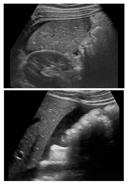
Figure 1. Ultrasound of the abdominal cavity of patient K. with catatrauma. Free fluid around the liver
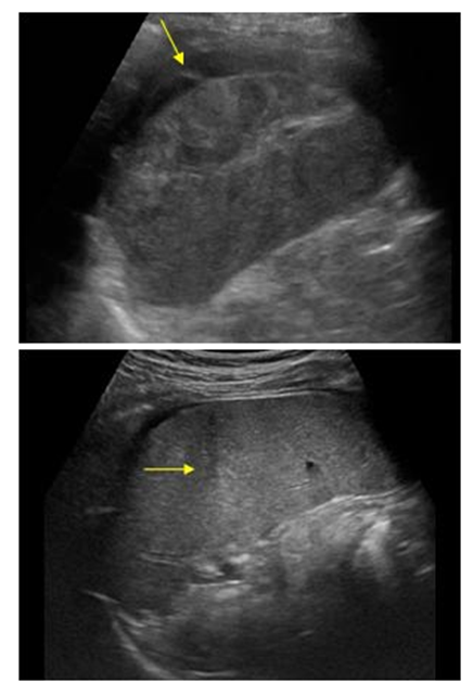
Figure 2. Ultrasound of the abdominal cavity of patient B. with catatrauma. Free fluid around the spleen
In some patients an echogenic region with uneven contours of various sizes and signs of rupture of the liver and spleen were determined in the parenchyma of the liver and spleen. Signs of kidney ruptures with retroperitoneal hematomas were also revealed besides detecting free fluid in the abdominal cavity patients with combined abdominal catatrauma. Along with this, ultrasound in patients revealed concomitant diseases of the abdominal cavity and retroperitoneal space: compaction of the liver parenchyma, cirrhosis, chronic pyelonephritis, fatty hepatosis of various degrees, cholelithiasis, etc. We evaluated in the control group the volume of free fluid according to the traditional gradation: free fluid in various parts of the abdominal cavity in the form of a layer, in small, moderate and large quantities. Free fluid was not detected in 46 (41.4%) cases of the control group according to the ultrasound data. A layer of free fluid was visualized around the spleen or liver, as well as between the loops of the small intestine in 32 (28.8%) cases. A small or moderate quantity of free fluid in the abdominal cavity was also visualized in 12 (10.8%) patients. In 21 (18.9%) cases there was a large quantity of free fluid in the abdominal cavity with ultrasonic signs of the spleen or liver injuries. To compare the traditional and quantitative estimation of free fluid volume in the abdominal cavity on the basis of clinical material, we retrospectively calculated the USS score in the control group of patients (Tab. 4).
Table 4. Comparison of the ratios of traditional and quantitative estimation of free fluid volume in the abdominal cavity according to ultrasound data
     
The quantitative assessment of the ultrasonic gradation of free fluid in the abdominal cavity is more informative, since it adds up the volume of free fluid in several regions of the abdomen and allows determining the tactics of surgical intervention on the abdominal organs based on a scoring. Considering this, we conducted a retrospective analysis of the control group of patients, comparing the USS score with the performed surgical interventions (Tab. 5).
Table 5. Comparative evaluation of ultrasound data and surgical interventions on the abdominal organs
     

4. Discussions

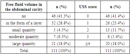
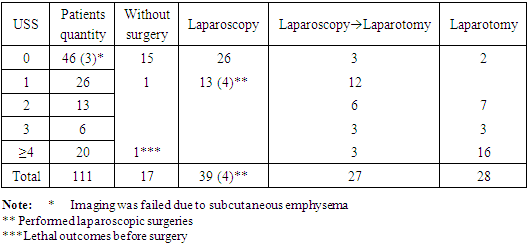
Ultrasound diagnostics is the most common non-invasive method for diagnosing injury of internal organs during polytrauma [20,12]. Its advantage is portability, accuracy and quick investigation. The main task of ultrasound in our research was to detect free fluid (blood) in the cavities. The nature of the parenchymal organ injury was judged by the change of the size and configuration of the organ and the presence of increased echogenicity zones [21]. The method deserves serious attention for the diagnostics of internal organs injuries in patients with combined abdominal catatrauma. It is simple and accessible to use. It can be used both in the emergency room, in the intensive care room, and in the operating room, including intraoperative diagnostics. In the 90s, they offered a ball score for determining the amount of fluid in the abdominal cavity by its thickness (Ultrasound Score). So, in 1994, Huang M. introduced physiological saline during diagnostic peritoneal lavage and he measured the separation of the peritoneum leaves by ultrasound examination. The result of his research was the proposal to equate every 2 mm of free fluid in the abdominal cavity with one point and to carry out surgical treatment at a score of ≥3. Mc Kenney K.L. in 1996, examining 4 sections of the abdomen (subphrenic, subhepatic, periolane and pelvis) proposed a counting system based on the estimation of the antero-posterior maximum fluid size detected in the abdominal cavity. Mc Kenney K.L. offered surgery in the presence of free fluid ≥2cm in the abdominal cavity. In 2001, Sirlin C.B. suggested ranking by one point in the presence of free fluid in each of the 7 areas of the abdominal cavity [22]. Moreover, every 10 mm of free fluid in one area of the abdominal cavity amounts one point. For example, up to 10 mm of free fluid in the subhepatic region is equal to one point, up to 20 mm - two points, etc. If there is free fluid up to 10 mm in the subhepatic region, in the right lateral canal and in the small pelvis, then it will be evaluated at 3 points. The higher the score, the higher is the likelihood of the abdominal organs injury and the need for surgical intervention.
According to our studies results, abdominal organs requiring emergency surgery were revealed in 10.9% of cases in patients with a “0” USS hemoperitoneum score. In 2 patients during the examination free gas was revealed in the abdominal cavity due to rupture of the walls of the stomach and small intestine, which required an urgent laparotomy. In 3 patients laparoscopy revealed injury of the spleen (in one case) and liver (in two cases): in the first case 400 ml of fresh blood with clots were detected intraoperatively, in the second and third ones - 100 ml and 200 ml, respectively. It is, probably, connected with poor ultrasound imaging of the abdominal organs due to the evident subcutaneous emphysema of the anterior abdominal wall as the result of combined injury of the chest organs. It should be said that at high-altitude trauma, the mechanogenesis of organ injury has its own characteristics, in particular, the heterogeneous nature of the traumatic forces and their application [14,16]. Obviously, it determined the two-moment rupture of the parenchymal organs and the absence of a sufficient quantity of free fluid in the abdominal cavity at the time of injured persons’ admission to the hospital. In our opinion, in this case, it is necessary to observe a patient with mandatory dynamic ultrasound monitoring of the abdominal cavity.
Patients with a score "1" in 61.5% of cases had injuries of the abdominal parenchymal organs of various degrees. In 4 cases injuries in the form of tears of the liver and spleen capsules (I degree according to AIS) [23] became possible to be eliminated by laparoscopic means. And in 12 cases, conversion laparotomy was performed as a result of ruptures of the liver and / or spleen requiring closure of the liver rupture or splenectomy. In such situations, laparoscopy is necessary for the timely detection of internal organs injuries.
Victims with a USS score of equal to or more than “2” in 100% of cases needed emergency laparotomy, as laparoscopy revealed 8 cases of splenic rupture of the II and III degree requiring laparotomy. In one of them, spleen damage was combined with rupture of the kidney, in one - with rupture of the bladder, in 2 cases - with ruptures of the liver. Victims with a USS score of equal to or more than “2” in 100% of cases needed emergency laparotomy, as laparoscopy revealed 8 cases of splenic rupture of the II and III degree requiring laparotomy. In one of them, spleen injury was combined with the kidney rupture, in one - with rupture of the bladder, in 2 cases - with ruptures of the liver. To evaluate the quantity of free fluid in the abdominal cavity in the main group we have already used USS ultrasonic gradation (Fig. 3).
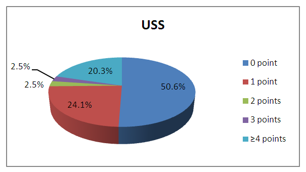
Figure 3. The volume of free fluid in the abdominal cavity according to USS data in the main group
Instrumental research methods occupy a central place in the diagnostics of abdominal organs injuries and in the definition of treatment tactics. The unclear clinical picture, the fuzzy data of the X-ray examination and ultrasound were indications for performing diagnostic laparoscopy [24-25]. The diagnostic value of laparoscopy performed in 44 (55.7%) patients of the main group and in 66 (59.5%) patients of the control group was 94.9 ± 4.1%.Despite the high information content of the method, which allows not only to diagnose damage, but also to determine the possible volume of surgical intervention, it has serious contraindications for use in patients with combined abdominal catatrauma. The main of them are: severe TBI with signs of the brain compression, suspicion of the diaphragm rupture, severe respiratory failure, unstable hemodynamics, severe bloating, numerous postoperative scars of the anterior abdominal wall, etc. The nature of abdominal injuries was very diverse (Tab. 6). Injuries of the parenchymal organs were mainly noted, including spleen ruptures (16.5% and 27%), liver injury (20.3% and 23.4%), hollow organ ruptures, mainly of the small and large intestines (13.9% and 13.5%) followed by peritonitis. In 43.0% of patients in the main group and in 36.9% of cases in the control group, a bruise or hematoma of the anterior abdominal wall was noted.
The main source of hemoperitoneum in our observations were injuries of the liver and spleen. According to the classification of parenchymal organs trauma of the American Association of Trauma Surgery, 1994 (AATS), the basis of which is the Abbreviation Injury Scale (AIS), where 5 degrees of severity are distinguished [19,23], we identified the following injuries: the spleen injury of I degree was noted in 1 case (3.3%) of the control group; the spleen injury of the II degree was observed in 1 (7.7%) patient in the main group and in 2 (6.7%) patients in the control one; III degree - in 5 (38.5%) and in11 (36.7%) patients; IV degree - in 7 (53.8%) and in 16 (53.3%) patients, respectively. The spleen injuries of the V degree were not noted. The liver injury of I degree was detected in 2 (11.7%) patients of the main and in 5 (17.8%) patients of the control group; II degree - in 4 (23.5%) and in 8 (28.6%); III - in 7 (41.2%) and in 11 (39.3%); IV– in 3 (17.6%) and in 4 (14.3%). The V degree of liver injury in our observations with catatrauma was not detected.
Table 6. The frequency of the abdominal organs injuries at catatrauma
     

5. Conclusions

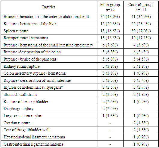
Thus, injuries of the abdominal organs at an isolated or combined abdominal trauma during catatrauma had a multiple nature and they were mainly manifested by ruptures of parenchymal organs with hemoperitoneum. Mortality among catatrauma patients with isolated and combined abdominal injuries was 17.7% in the main group and 27% in the control group. Our experience allows us to consider that the structure of the diagnostic search for combined abdominal catatrauma can be represented as follows: primary determination of the patient’s severity according to the clinical examination and the ascertainment of the need for resuscitation aid, the identification of life-threatening complications that need to be eliminated immediately (cardiac and respiratory arrest, asphyxiation, external and internal bleeding), the identification of the dominant damage and its role in the severity of the patient’s condition according to clinical and para-clinical examination methods data (traumatogenesis); diagnostics of injuries aggravating (non-aggravating) the course of the leading trauma.
The below mentioned principles should be subject to a diagnostic process that is strictly regulated and corresponding, in our opinion, to the following requirements:
1) Simultaneous recognition of all available injuries immediately after the patient arrives to the hospital, while assessing the dominant ones. This need is imperative, vital, justified, urgent, as it is the motive for determining surgical tactics.
2) Carrying out diagnostic measures in the anti-shock zone of the admission department against the background of intensive care and resuscitation aids aimed at eliminating life-threatening injuries, which requires determining the priority of diagnostic measures.
3) To assess the ultrasonic gradation of free fluid in the abdominal cavity, it is necessary to use a USS score based on which the treatment tactics of the abdominal organs injuries is determined.

References

[1]  Gumanenko E.K., Kozlova V.K. Polytrauma. M.: Geotar-Media. - 2008. -- P.608.
[2]  Bagnenko S.F., Shapot Yu.B., Tulupov A.N., Lapitsky A.V., Balabanova O.V. Medical care for mechanical trauma of the chest and abdomen at the prehospital stage // Bulletin of Surgery. - SPb. - 2007. - No. 2. - P. 47-50.
[3]  Chikaev V.F., Ibragimov R.A., Bondarev Yu.V. The principles of diagnostics and treatment of victims at combined trauma with injuries of the abdominal cavity parenchymal organs // Pract. medicine. Traumatology orthopedics. Surgery. - 2010. - No. 8. - P. 3-6.
[4]  Abakumov M.M., Galankina I.E., Vilk A.P. Possibilities of conservative treatment of spleen injuries with closed abdominal trauma. Jour. named after N.V. Sklifosovsky "Emergency medical care."2013; (4): 30-34.
[5]  Ivanov P.A. et al. Combined injuries of the liver, pancreas and duodenum with closed abdominal trauma // International Association of Surgeons-Hepatologists. - Moscow. - 2008. - Volume 13. - No. 1. - P.102-104.
[6]  Akilov H.A., Aminov U.S. Diagnostics and treatment of blunt abdominal injuries in children // Bulletin of emergency medicine. - Tashkent. - 2012. - No. 3. - P.12-14.
[7]  Kadirov Z.L., Makhmudov N.I., Akhrarov B.M. The role of laparocentesis and laparoscopy in the diagnosis of closed abdominal injuries // Bulletin of emergency medicine. - Tashkent. - 2010. - No. 2. - P.136-137.
[8]  Nazarova E.O., Karpov S.M., Apaguni A.E., Vyshlova I.A. A modern view on pathogenetic mechanisms // Bulletin of new medical technologies, electronic journal - 2018 - N 1. - P.126-130.
[9]  Khadjibaev A.M., Sultanov P.K., Rakhmanov R.O. The use of modern diagnostic and therapeutic technologies for breast injuries due to catatrauma at the Republican Scientific Center for Emergency Medical Care. “Emergency medical care named after N.V. Sklifosovsky.” 2015 No. 4. P. 44-51.
[10]  Kadyrov Z.L., Makhmudov N.I., Akhrarov B.M. Diagnostic standards for blunt abdominal injuries // Bulletin of emergency medicine. - 2010. - No. 2. - P.137.
[11]  Nishanov H.T., Muminov R.D., Dzhurabaev N.Z. Emergency laparoscopy in a comprehensive examination at abdominal injuries // Endoscopic surgery. - M. –2010. - No. 2. - P. 34-36.
[12]  Sirlin CB, Casola G, Brown MA, Patel NV, Bendavid EJ, Hoyt DB. Quantification of fluid on screening ultrasonography for blunt abdominal trauma: a simple scoring system to predict severity of injury //J. Ultrasound Med. – 2001. – Vol. 20. № 4. – P.359-364.
[13]  Brillantino A, Iacobellis F, Robustelli U, et al. Non operative management of blunt splenic trauma: a prospective evaluation of a 10 Journal of International Medical Research standardized treatment protocol // Eur J Trauma Emerg Surg. – 2016. – Vol. 42. - P593–598.
[14]  Khashimov F.E., Alimova H.P., Musaev T.S., Navruzov S.Yu., Shadmanov T.T. Organization of medical care for children injured in traffic accidents. // Bulletin of emergency medicine, 2011, No. 2. - P.32-35.
[15]  A.M. Khadjibaev, R.O. Rakhmanov, P.K. Sultanov, V.Kh. Sharipova. Diagnostics and surgical tactics in emergency conditions due to trauma and diseases of the chest cavity. General resuscitation. 2016 Volume 12 14 Pages 55-67. https://doi.org/10.15360/1813-9779-2016-4-57-67.
[16]  Ultrasonography for the Diagnosis of Intraperitoneal Free Air in Chest-Abdominal-Pelvic Blunt Trauma and Critical Acute Abdominal Pain // Arch Surg. 2009; 144(2): 137-141.
[17]  Koshelev V.N., Chalykh Yu.V. Causes of mortality in injuries of the liver and spleen // Bulletin of Surgery named after I.I. Grekov, Vol. 155. 1996, No. 2. - P.51-53.
[18]  Khadjibaev A.M., Sultanov P.K. Abdominal bleeding at catatrauma. Bulletin of Surgerynamed after I.I. Grekov. 2016 No.2. P.43-48.
[19]  Baker, S. P., O’Neill, B., Haddon, W., Jr., & Long, W. B. The injury severity score: A method for describing patients with multiple injuries and evaluating emergency care // Journal of Trauma. – 1974. – N14. – P187-196.
[20]  Becker A., Lin G., McKenney M.G. et al. Is the FAST exam reliable in severely injured patients? // Injury. – 2010. – Vol.41. – N.5. – P.479–483.
[21]  Da-wei Zhao, Meng Tian, Le-tian Zhang, Tao Li, Jie Bi, Jia-ying He, Ying-ying Zhang. Effectiveness of contrastenhanced ultrasound and serum liver enzyme measurement in detection and classification of blunt liver trauma // Journal of International Medical Research. – 2017. - Vol. 45(1). - P170–181.
[22]  Moore E.E., Shackford S.R., Pachter H.L. et al. Organ Injury Scaling Spleen, Liver and Kidney // J. Trauma. – 1989. – Vol. 2. – P. 1664-1666.
[23]  Khadjibaev A.M., Sultanov P.K., Kim Hoon. Features of Patients Associated with Falls from Heights Admitted to Republican Research Center of Emergency Medicine. Journal of Trauma and Injury (Seoul, South Korea). 2015 №28(4). P.223-294. DOI: 10.20408/jti.2015.28.4.248.
[24]  Puzanov S.Yu., Alishikhov A.A., Rutenberg G.M., Bogdanov D.Yu. The feasibility of using laparoscopy at traumatic injuries of the abdominal organs // Endoscopic surgery. - 2014. - No. 2. - P.14-17.
[25]  Alla Z., B. Todd Heniford, Ronald F. Sing, Diagnostic Laparoscopy in the Intensive Care Unit // Journal of Intensive Care Medicine. – 2015. – Vol.30. – N5. – P297-302.